. 2022 Feb 15;23(1):131.
doi: 10.1186/s12864-022-08347-5.

Whole-genome analyses reveal a novel prophage and cgSNPs-derived sublineages of Brachyspira hyodysenteriae ST196

Affiliations

Whole-genome analyses reveal a novel prophage and cgSNPs-derived sublineages of Brachyspira hyodysenteriae ST196

Ana Belén García-Martín et al. BMC Genomics. .

Abstract

Background: Brachyspira (B.) hyodysenteriae is a fastidious anaerobe spirochete that can cause swine dysentery, a severe mucohaemorragic colitis that affects pig production and animal welfare worldwide. In Switzerland, the population of B. hyodysenteriae is characterized by the predominance of macrolide-lincosamide-resistant B. hyodysenteriae isolates of sequence type (ST) ST196, prompting us to obtain deeper insights into the genomic structure and variability of ST196 using pangenome and whole genome variant analyses.

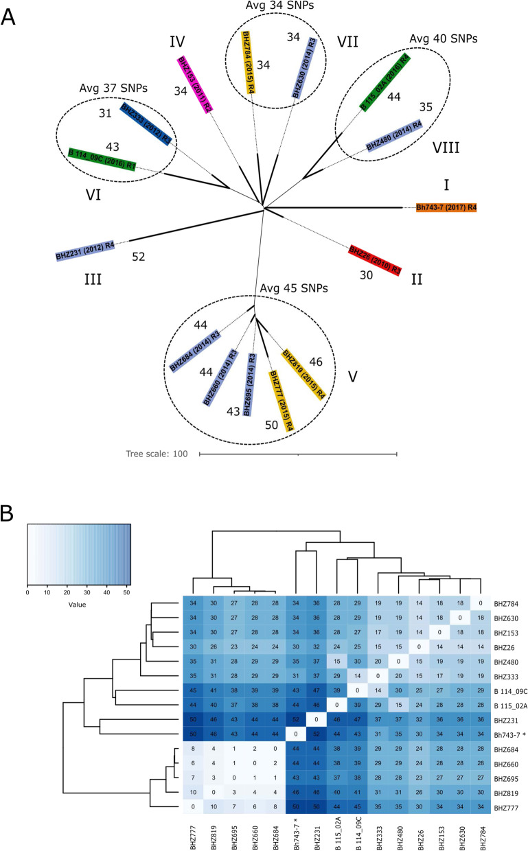
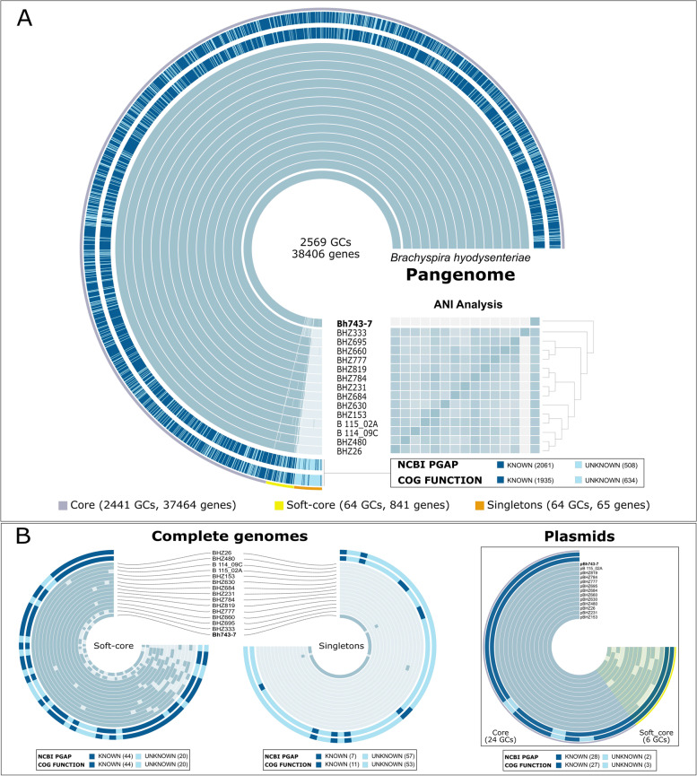
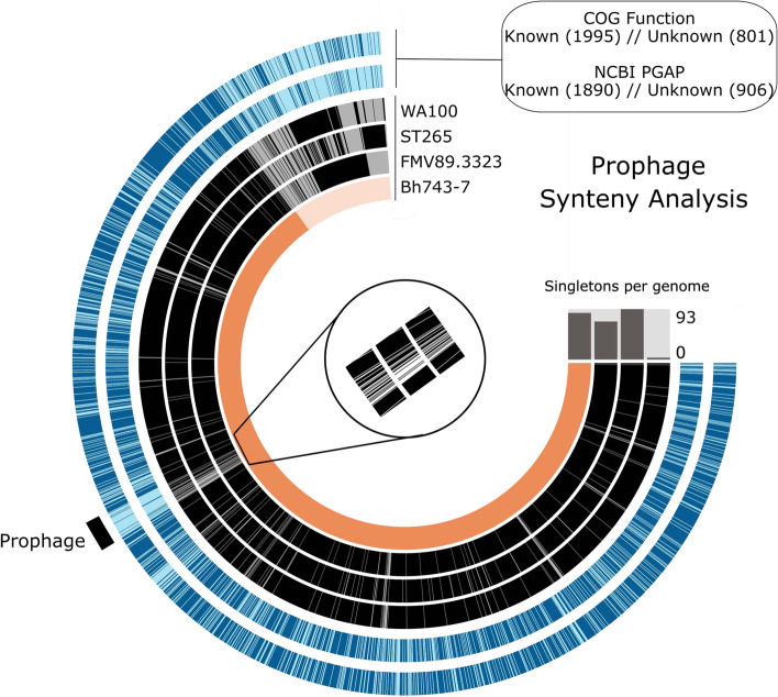
Results: The draft genome of 14 B. hyodysenteriae isolates of ST196, sampled during a 7-year period from geographically distant pig herds, was obtained by whole-genome sequencing (WGS) and compared to the complete genome of the B. hyodysenteriae isolate Bh743-7 of ST196 used as reference. Variability results revealed the existence of 30 to 52 single nucleotide polymorphisms (SNPs), resulting in eight sublineages of ST196. The pangenome analysis led to the identification of a novel prophage, pphBhCH20, of the Siphoviridae family in a single isolate of ST196, which suggests that horizontal gene transfer events may drive changes in genomic structure.

Conclusions: This study contributes to the catalogue of publicly available genomes and provides relevant bioinformatic tools and information for further comparative genomic analyses for B. hyodysenteriae. It reveals that Swiss B. hyodysenteriae isolates of the same ST may have evolved independently over time by point mutations and acquisition of larger genetic elements. In line with this, the third type of mobile genetic element described so far in B. hyodysenteriae, the novel prophage pphBhCH20, has been identified in a single isolate of B. hyodysenteriae of ST196.

Keywords: Bioinformatics; Horizontal-gene transfer; Pangenome; Singletons; Structural variations; Swine dysentery; WGS.


Conflict of interest statement

None of the authors of this paper have a financial or personal relationship with other people or organizations that could inappropriately influence or bias the content of the paper.

Figures

Fig. 1
Fig. 1
Core-genome SNP-based phylogeny of Brachyspira hyodysenteriae isolates of ST196. A The relationship among isolates of ST196 are shown according to the approximately-maximum-likelihood phylogenetic tree. Labels containing names of the isolates and regions (from Western to Eastern Switzerland: R1 to R4) of isolation are colored according to the years of isolation. Sublineages are indicated with roman numerals. Numbers of different non-recombinant cgSNPs identified respect to the reference genome are indicated for each isolate. Average (Avg) SNPs distance is also indicated for clusters V, VI, VII and VIII. B Isolates are clustered according to the distance matrix of pairwise differences calculated from the non-recombinant cgSNPs. Number of different cgSNPs used to identify genetic distances across all genomes are indicated in each cell of the heatmap
Fig. 2
Fig. 2
Pangenome analysis of Brachyspira hyodysenteriae isolates of the predominant ST196. Total number of genes as well as bin-specific gene clusters (GCs) are indicated. A The two outmost layers represent both functional annotations derived from the COGs database and NCBI-PGAP. Known and unknown functions are in dark and light blue, respectively. The number of either known or unknown functions are indicated in brackets. Each blue-colored layer represents the genome of a B. hyodysenteriae isolate of ST196. The inner one represents the complete genome of B. hyodysenteriae isolate Bh743-7 used as reference genome. Genomes are organized by average full nucleotide identity (ANI values: 0.995 − 1) and minimum number of genomes in which a certain gene is present. Core (grey), soft core (yellow) and singletons (orange) bins are shown for all the genomes (n = 15). B Soft-core and singleton bins are represented in more detailed at the complete genome level (n = 15), but also for the plasmids (n = 13), independently
Fig. 3
Fig. 3
Schematic gene map displaying the modular organization of the novel prophage and its integration into the chromosome of the prophage-carrying Brachyspira hyodysenteriae isolate Bh743-7 of ST196. Image was created using Easyfig v2.1 (Sullivan et al., 2011). Each arrow represents an open reading frame and its orientation. Phage-like genes annotations are as follows: 1: Phage recombination protein Bet; 2: Winged helix-turn-helix transcriptional regulator; 3: N-6 DNA-methylase; 4: DUF4406 domain-containing protein; 5: Phage tail protein; 6: PBSX family phage terminase large subunit; 7−10: Phage capsid protein; 11: Phage tail protein; 12: Damage-inducible protein D; 13−14: Phage tail proteins; 15: Glycoside hydrolase family 19 protein; 16: Integrase. Phage-like genes are color-coded according to their respective modules: DNA replication (light blue), packaging/capsid morphogenesis (dark blue), tail morphogenesis (orange), lysis (green), recombination/‌integration (yellow), other functions (purple), unknown functions (violet) and hypothetical proteins (grey). Direct comparison of the structures of the new prophage and the VSH-1 gene transfer agent of B. hyodysenteriae is also shown
Fig. 4
Fig. 4
Prophage synteny analysis. The two most outer layers represent both functional annotations derived from the COGs database and NCBP-PGAP. Number of genes with either known (dark blue) or unknown (light blue) annotations are also shown in brackets. The genomes of Brachyspira hyodysenteriae strains that share some of the phage-like genes found in the novel prophage of B. hyodysenteriae isolate Bh743-7 (in orange) are represented by the black inner layers. Although the novel prophage was not present in any of the analysed strains, homologous genes were found in those strains by forcing synteny as highlighted. In the centre, a zoom in barcode-like graph is showing the presence (black) /absence (white) of the homologous genes. Total number of singletons per genome is also indicated in the bar graph

References

    1. Hampson D, Lugsomya K, La T, Dale Phillips N, J. Trott D, Abraham S. Antimicrobial resistance in Brachyspira – An increasing problem for disease control. Vet Microbiol. 2019;229:59-71. - PubMed
    1. Card RM, La T, Burrough ER, Ellis RJ, Núñez-García J, Thomson JR, Mahu M, Phillips ND, Hampson DJ, Rohde J, et al. Weakly haemolytic variants of Brachyspira hyodysenteriae newly emerged in Europe belong to a distinct subclade with unique genetic properties. Vet Res. 2019;50(1):21. - PMC - PubMed
    1. Joerling J, Willems H, Ewers C, Herbst W. Differential expression of hemolysin genes in weakly and strongly hemolytic Brachyspira hyodysenteriae strains. BMC Vet Res. 2020;16(1):169. - PMC - PubMed
    1. De Luca S, Nicholson P, Magistrali CF, García-Martín AB, Rychener L, Zeeh F, Frey J, Perreten V. Corrigendum to “Transposon-associated lincosamide resistance lnu(C) gene identified in Brachyspira hyodysenteriae ST83” [Vet Microbiol. 214 (2018) 51–55]. Vet Microbiol. 2018;220:113. - PubMed
    1. De Luca S, Nicholson P, Magistrali CF, García-Martín AB, Rychener L, Zeeh F, Frey J, Perreten V. Transposon-associated lincosamide resistance lnu(C) gene identified in Brachyspira hyodysenteriae ST83. Vet Microbiol. 2018;214:51–5. - PubMed