. 2022 Feb 15;8(1):16.
doi: 10.1038/s41421-022-00381-7.

Broad ultra-potent neutralization of SARS-CoV-2 variants by monoclonal antibodies specific to the tip of RBD

Affiliations

Broad ultra-potent neutralization of SARS-CoV-2 variants by monoclonal antibodies specific to the tip of RBD

Hang Ma et al. Cell Discov. .

Abstract

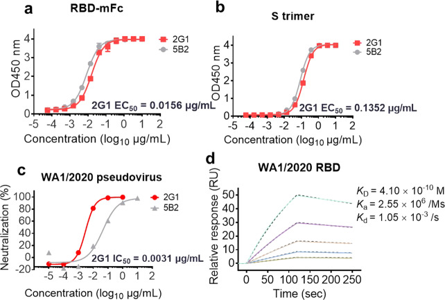
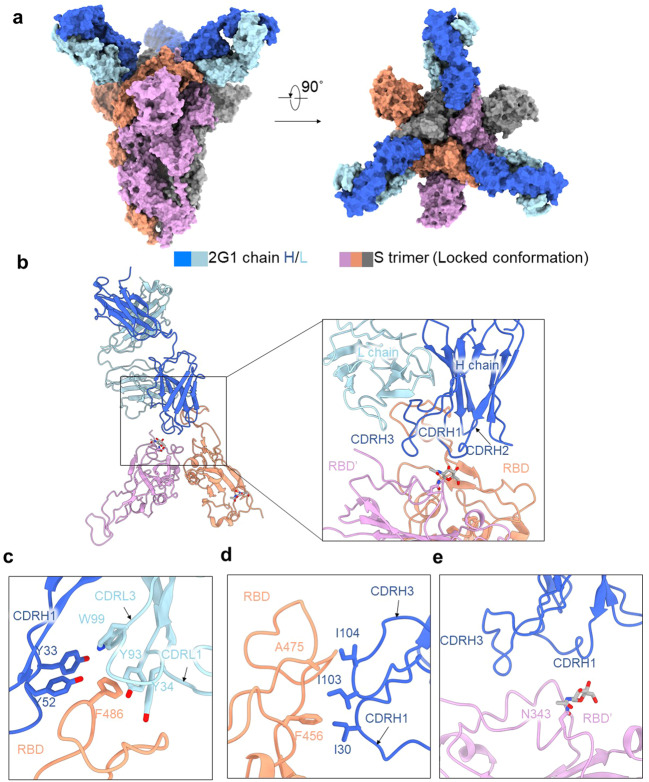
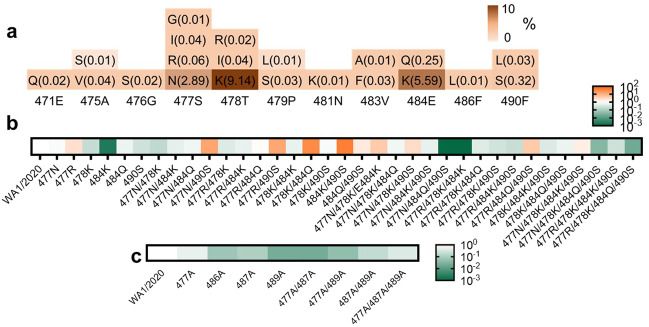
Severe acute respiratory syndrome coronavirus 2 (SARS-CoV-2) variants of concern (VOCs) continue to wreak havoc across the globe. Higher transmissibility and immunologic resistance of VOCs bring unprecedented challenges to epidemic extinguishment. Here we describe a monoclonal antibody, 2G1, that neutralizes all current VOCs and has surprising tolerance to mutations adjacent to or within its interaction epitope. Cryo-electron microscopy structure showed that 2G1 bound to the tip of receptor binding domain (RBD) of spike protein with small contact interface but strong hydrophobic effect, which resulted in nanomolar to sub-nanomolar affinities to spike proteins. The epitope of 2G1 on RBD partially overlaps with angiotensin converting enzyme 2 (ACE2) interface, which enables 2G1 to block interaction between RBD and ACE2. The narrow binding epitope but high affinity bestow outstanding therapeutic efficacy upon 2G1 that neutralized VOCs with sub-nanomolar half maximal inhibitory concentration in vitro. In SARS-CoV-2, Beta or Delta variant-challenged transgenic mice and rhesus macaque models, 2G1 protected animals from clinical illness and eliminated viral burden, without serious impact to animal safety. Mutagenesis experiments suggest that 2G1 is potentially capable of dealing with emerging SARS-CoV-2 variants in the future. This report characterized the therapeutic antibodies specific to the tip of spike against SARS-CoV-2 variants and highlights the potential clinical applications as well as for developing vaccine and cocktail therapy.


Conflict of interest statement

The authors declare no competing interests.

Figures

Fig. 1
Fig. 1. Cell isolation, antibody cloning, and candidate panning.
a Isolation strategy of highly potent neutralizing antibodies as depicted by a diagram. b RBD-specific B cells were isolated from convalescent subjects of SARS-CoV-2 infection by fluorescence-activated cell sorting. The 7ADD/CD19+/CD27+/IgG+/RBD+ gate is shown and highlighted in the boxes. c Statistics of the number of paired antibodies from each subject, as well as the number of kappa and lambda subtypes. d Binding scores of antibody candidates against SARS-CoV-2 RBD as measured by ELISA and scores higher than 2 are presented. 2G1 is highlighted in red. e Candidate panning using a WA1/2020 pseudovirus-based screening model. Antibodies were 10-fold serially diluted from 101 μg/mL to 104 μg/mL.
Fig. 2
Fig. 2. Characterization of 2G1 using WA1/2020 related S and RBD proteins and pseudovirus.
a, b 2G1 concentration-dependently binds to RBD-mFc (a) and S trimer (b) of SARS-CoV-2 in ELISA test. A neutralizing antibody 5B2 targeting SARS-CoV-2 RBD was used as control. Values from two replicates are shown as means ± SD. c Serial 10 fold-diluted 2G1 was incubated with SARS-CoV-2 WA1/2020 pseudovirus and used to infect 293T-ACE2 cells. After 48 h incubation, the infection was quantified using a fluorescence detection kit. d Binding kinetics of 2G1 to SARS-CoV-2 RBD in SPR. Serial dilutions of 2G1 Fab were flowed through a chip fixed with RBD recombinant protein. The kinetics data were fitted with results from different concentrations.
Fig. 3
Fig. 3. Binding, blocking, and extensive neutralization of 2G1 against SARS-CoV-2 variants.
a, b 2G1 competitively blocked the ACE2 binding to single point mutant RBD proteins (a) and VOC S trimers (b). c Affinity analysis of 2G1 bound to S trimers of SARS-CoV-2 WA1/2020, Alpha, Beta, Gamma, Kappa and Delta by SPR. Chips fixed with S trimers were loaded on a BIAcore 8 K system. 2G1 Fab varied from 1.250 μg/mL to 0.039 μg/mL were injected over the chips for measuring the real-time association and dissociation parameters. d Neutralization of 2G1 against diverse SARS-CoV-2 pseudoviruses. Pseudoviruses with active titer higher than 1 × 10⁷ TU/mL were employed in this study. Concentration-dependent neutralization of 2G1 was quantified by detecting the fluorescence from the luciferase reporter. Data in duplicate are displayed as means ± SD. e Live virus neutralization by 2G1. 100 TCID50 of SARS-CoV-2 (WA1/2020, Alpha, Beta, Gamma and Delta) were incubated with 3 fold-diluted 2G1 and then added to Vero E6 cells. After a 3-day incubation, cytopathic effect (CPE) was assessed by counting the plaque formation.
Fig. 4
Fig. 4. Therapeutic efficacy of 2G1 against SARS-CoV-2 variants in transgenic mice.
a High permissive AC70 human ACE2 transgenic mice were challenged with 100 LD50 of SARS-CoV-2 WA1/2020, Beta- or Delta- variants, followed by 20, 6.7, or 2.2 mg/kg of 2G1 treatment (n = 14). A 12-day clinical observation was implemented. b Body weight change of mice. c Clinical illness of mice was assessed based on a standardized 1 to 4 grading system that describes the clinical wellbeing of mice. d Mortality of mice. Mice were monitored until 12 dpi unless the designed endpoint was reached. e Viral load in lung and brain tissues. Data are shown as means ± SD. Vhcl vehicle control, p.i. post infection.
Fig. 5
Fig. 5. Therapeutic efficacy of 2G1 against SARS-CoV-2 variants in rhesus macaques.
a One male and one female rhesus macaques in each group were endotracheally challenged with 1 × 10⁵ TCID50 of SARS-CoV-2. 2G1 at 10 mg/kg or 50 mg/kg, or equal amount of PBS were intravenously given at 1 dpi. Throat and anal swabs were sampled daily until 7 dpi. b Viral load in throat swab. c Viral load in anal swab. d Viral load in lungs, tracheas, and bronchi. Data with duplications are shown as means ± SD. Vhcl vehicle control, p.i. post infection.
Fig. 6
Fig. 6. Cryo-EM structure of 2G1 and the complex with WA1/2020S protein.
a The domain-colored cryo-EM map of SARS-CoV-2S ectodomain trimer and 2G1 Fab fragments complex is shown, viewed along two perpendicular orientations. The heavy and light chains of 2G1 are colored blue and cyan, respectively. The three protomers of trimeric S protein are colored gray, orange and pink. be The binding interface between 2G1 and RBD and adjacent RBD’. RBD and 2G1 interact with each other mainly through hydrophobic interactions (c, d). 2G1 heavy chains (CDRH3 and CDRH1) lie above the adjacent RBD’ (e). Residues are numbered using the Kabat convention.
Fig. 7
Fig. 7. Analysis of different binding modes of 2G1, S2E12, B1-182.1, and REGN10933.
a The epitope surfaces of S2E12, B1-182.1, and REGN10933 on S protein are in red, orange, and green, respectively. b Comparison of binding modes of 2G1, S2E12, B1-182.1, and REGN10933. The epitope surface of 2G1 is in blue. The borderlines of ACE2-binding site, S2E12, B1-182.1 and REGN10933 are shown in black, red, orange and green respectively. The connecting lines between the center of 2G1 Fab and RBD are taken as the principal axis, and axis of Fab S2E12, B1-182.1 is rotated 6° and REGN10933 is rotated 13° approximately. c Mapping of S2E12, B1-182.1, and REGN10933 epitopes on RBD. d The neutralizing activity of 2G1, REGN10933, B1-182.1, and S2E12 was analyzed using WA1/2020 pseudotyped virus in parallel. Data in duplicate are displayed as means ± SD.
Fig. 8
Fig. 8. Identification of critical binding residues for 2G1.
a Statistics of mutation proportion in RBD residue 471Glu–490Phe where key for 2G1 epitope from GISAID database as of August 2021. b Identification of critical binding residues for 2G1. Spike genes with high frequency mutation sites between 471Glu and 490Phe (> 0.05%) were cloned and transiently expressed on the surface of 293T cells. The binding ability of 2G1 to these mutant S proteins was measured by flow cytometry. The fold change of binding ability was normalized by comparing to WA1/2020S protein. c Mutations in the key interaction sites of 2G1 that affect the binding ability of 2G1 to varying degrees.

References

    1. Volz E, et al. Assessing transmissibility of SARS-CoV-2 lineage B.1.1.7 in England. Nature. 2021;593:266–269. - PubMed
    1. Alpert T, et al. Early introductions and transmission of SARS-CoV-2 variant B.1.1.7 in the United States. Cell. 2021;184:2595–2604. - PMC - PubMed
    1. Liu J, et al. BNT162b2-elicited neutralization of B.1.617 and other SARS-CoV-2 variants. Nature. 2021;596:273–275. - PubMed
    1. Dejnirattisai W, et al. Antibody evasion by the P.1 strain of SARS-CoV-2. Cell. 2021;184:2939–2954. - PMC - PubMed
    1. Chen RE, et al. In vivo monoclonal antibody efficacy against SARS-CoV-2 variant strains. Nature. 2021;596:103–108. - PMC - PubMed