. 2022 Feb 15;13(1):871.
doi: 10.1038/s41467-022-28528-w.

Structural basis for SARS-CoV-2 Delta variant recognition of ACE2 receptor and broadly neutralizing antibodies

Affiliations

Structural basis for SARS-CoV-2 Delta variant recognition of ACE2 receptor and broadly neutralizing antibodies

Yifan Wang et al. Nat Commun. .

Abstract

The SARS-CoV-2 Delta variant is currently the dominant circulating strain in the world. Uncovering the structural basis of the enhanced transmission and altered immune sensitivity of Delta is particularly important. Here we present cryo-EM structures revealing two conformational states of Delta spike and S/ACE2 complex in four states. Our cryo-EM analysis suggests that RBD destabilizations lead to population shift towards the more RBD-up and S1 destabilized fusion-prone state, beneficial for engagement with ACE2 and shedding of S1. Noteworthy, we find the Delta T478K substitution plays a vital role in stabilizing and reshaping the RBM loop473-490, enhancing interaction with ACE2. Collectively, increased propensity for more RBD-up states and the affinity-enhancing T478K substitution together contribute to increased ACE2 binding, providing structural basis of rapid spread of Delta. Moreover, we identify a previously generated MAb 8D3 as a cross-variant broadly neutralizing antibody and reveal that 8D3 binding induces a large K478 side-chain orientation change, suggesting 8D3 may use an "induced-fit" mechanism to tolerate Delta T478K mutation. We also find that all five RBD-targeting MAbs tested remain effective on Delta, suggesting that Delta well preserves the neutralizing antigenic landscape in RBD. Our findings shed new lights on the pathogenicity and antibody neutralization of Delta.


Conflict of interest statement

Z.H. and C.Z. are listed as inventors on a pending patent application for MAb 8D3. The other authors declare no competing interests.

Figures

Fig. 1
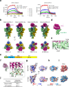
Fig. 1. Cryo-EM structures of the SARS-CoV-2 Delta variant S trimer.
a, b Cryo-EM maps of the Delta variant S-open a and S-transition b state. Protomer 1, 2, and 3 are shown in light green, royal blue, and gold, respectively, which color scheme was followed throughout. c Atomic model of the Delta S-open, and Delta mutations indicated by red sphere. d Population distribution of the Delta S-open and S-transition. e Overlaid RBD-1 from Delta S-open (light green) and S-transition (violet red), showing that the angle between the long axis of RBD and the horizontal plane of S trimer is reduced from S-open to S-transition. The RBD-1 of G614 S-open (PDB 7KRR, grey) also showed here as a comparison. f List of the angle between RBD-1 and the S-trimer plane in different states of Delta S, compared with that of the G614 variant,. g Top view of the overlaid NTDs of the Delta S-open (in color) and the G614 S-open (PDB 7KRR, gray), indicating a clockwise rotation/untwist of the Delta S relative to that of G614. h Side view of the overlaid protomer 1 of S-open, showing an outward tilt of NTD (~4.2 Å), associated with small outward shift of NTD loop290–303 and HR1 helix920–940, relative to that of G614 S-open (PDB 7KRR, gray). i In Delta S-open, all three FPs (blue) are disordered, the 630 loop in the RBD-up protomer 1 is disordered, and that in the RBD-down protomer 2/3 is partially disordered. j, k Two representative 3DVA motions of the Delta S-open dataset. The left two maps illustrate the two extremes in the motion, and the angular range and direction of the motion are displayed in the two views of the overlaid two extreme maps (right).
Fig. 2
Fig. 2. Structural basis of enhanced Delta variant S trimer/ACE2 interaction.
a Measurement of the binding affinity between ACE2 and the S trimer of the Delta (left) or G614 (right) variants using bio-layer interferometry (BLI). Association and dissociation steps were divided by dotted lines. ACE2 concentrations tested were shown. Raw sensor grams and fitting curves were shown in color and black, respectively. Source data are provided as a Source Data file. b Cryo-EM maps of the Delta S-ACE2 complex in four distinct conformational states. ACE2 is shown in violet red. This color scheme is followed throughout. c Population distribution of the Delta S-ACE2 conformers. d Density map of the focus-refined Delta RBD-1-ACE2. e Zoomed-in view of the S-ACE2 interaction interface, showing the side chain densities of the substituted L452R and T478K were well resolved. f The substituted K478 forms a new H-bond with N487, resulting in the formation of two new H-bonds with ACE2 (red dotted line), and a conformational change of loop473–490 (indicated by red arrow) relative to that in WT RBD-ACE2 (PDB 6M0J). g, h The electrostatic surface property g and hydrophilicity and hydrophobicity properties h of the Delta and WT RBDs, with the mutated residues indicated. i Similar surface properties for ACE2, with residues in proximity to Delta RBD-1 (< 4 Å) indicated. j Interaction interface areas between ACE2 and RBD of Delta or WT (PDB 6M0J) strain analyzed using PISA.
Fig. 3
Fig. 3. 3D variability analysis on the delta S-ACE2 complex.
ac Three representative 3DVA modes of the Delta S-ACE2 complex, with the left two maps showing the two extremes in the motion, and the angular range and direction of the motion displayed in the two views of the overlaid two extreme maps (right).
Fig. 4
Fig. 4. Neutralization breadth and binding properties of the MAbs (3C1, 2H2, 2G3, 3A2, and 8D3) against SARS-CoV-2 variants.
a Neutralization values and fold change in neutralization IC50 of the MAbs against the variant pseudoviruses, relative to the WT pseudovirus. A minus sign (-) denotes decrease. Orange shade, more than 10-fold decrease; red shade, more than 100-fold decrease. b Neutralization activity of the MAbs towards SARS-CoV-2 Beta, Kappa, and Delta variant pseudoviruses. The MAbs were serially diluted and tested for neutralization. Data are expressed as mean ± SEM of four replicate wells. c Binding properties of the MAbs with recombinant S trimers derived from the WT and Delta strains were determined by ELISA. Serial dilutions of S trimers were coated onto the wells. Data are expressed as mean ± SD of triplicate wells. d Binding properties of the MAbs with recombinant RBD proteins derived from the WT, Beta, and Delta strains were measured by ELISA. Serial dilutions of RBD were coated onto the wells. Data are expressed as mean ± SD of triplicate wells. Source data are provided as a Source Data file.
Fig. 5
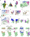
Fig. 5. Cryo-EM analyses on the Delta S-8D3 Fab complex.
a Side and top views of the cryo-EM map of the Delta S-8D3 complex, with the heavy and light chain of 8D3 Fab in royal blue and violet red, respectively. The color scheme was followed. b, c Model-map fitting of the focus-refined Delta RBD-1-8D3 structure b, and the zoomed-in view of the Delta RBD-1/8D3 interaction interface c. The sidechains of the key amino acids, including R452 and K478, are well resolved. d The RBD-1/8D3 interaction interface, with major involved structural elements and the two RBM mutations of Delta variant labeled. e ACE2 (coral, from our Delta RBD-1-ACE2 structure) would clash with the heavy chain of 8D3 Fab (dotted black circle). They share overlapping epitopes on the RBM loop473–490. f The side chain orientation alternation of K478 from Delta RBD-1-8D3 (in light green) relative to that from Delta RBD-1-ACE2 (coral). g Magnified view to show the H-bond interaction network formed between 8D3 Fab and the RBM loop473–490 of Delta variant. h Distinct binding of 2H2 (PDB 7DK4, blue) and 3C1 (PDB 7DCC, purple) Fab on the RBD (light green) of WT S. The Delta variant mutation sites (in red surface) are not located in the binding footprint of 2H2 and 3C1 on RBD. i The footprint (<4 Å contact) of ACE2 (coral, our Delta RBD-1-ACE2 structure), 8D3 (violet red), 2H2 (blue), and 3C1 (purple) Fab, respectively, on RBD. The second column shows the RBD with mutation sites of the Delta, Beta, and Kappa variants indicated in red.

References

    1. Wang Q, et al. Structural and functional basis of SARS-CoV-2 entry by using human ACE2. Cell. 2020;181:894–904 e899. - PMC - PubMed
    1. Tang T, Bidon M, Jaimes JA, Whittaker GR, Daniel S. Coronavirus membrane fusion mechanism offers a potential target for antiviral development. Antivir. Res. 2020;178:104792. - PMC - PubMed
    1. Shang J, et al. Structural basis of receptor recognition by SARS-CoV-2. Nature. 2020;581:221–224. - PMC - PubMed
    1. Rabaan AA, et al. SARS-CoV-2, SARS-CoV, and MERS-COV: a comparative overview. Infez. Med. 2020;28:174–184. - PubMed
    1. Lan J, et al. Structure of the SARS-CoV-2 spike receptor-binding domain bound to the ACE2 receptor. Nature. 2020;581:215–220. - PubMed

Publication types

MeSH terms

Substances

Supplementary concepts