. 2022 Feb 15;9(1):55.
doi: 10.1038/s41597-022-01157-0.

The Digital Brain Tumour Atlas, an open histopathology resource

Affiliations

The Digital Brain Tumour Atlas, an open histopathology resource

Thomas Roetzer-Pejrimovsky et al. Sci Data. .

Abstract

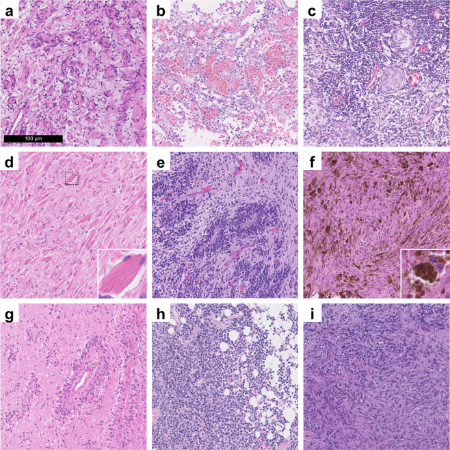
Currently, approximately 150 different brain tumour types are defined by the WHO. Recent endeavours to exploit machine learning and deep learning methods for supporting more precise diagnostics based on the histological tumour appearance have been hampered by the relative paucity of accessible digital histopathological datasets. While freely available datasets are relatively common in many medical specialties such as radiology and genomic medicine, there is still an unmet need regarding histopathological data. Thus, we digitized a significant portion of a large dedicated brain tumour bank based at the Division of Neuropathology and Neurochemistry of the Medical University of Vienna, covering brain tumour cases from 1995-2019. A total of 3,115 slides of 126 brain tumour types (including 47 control tissue slides) have been scanned. Additionally, complementary clinical annotations have been collected for each case. In the present manuscript, we thoroughly discuss this unique dataset and make it publicly available for potential use cases in machine learning and digital image analysis, teaching and as a reference for external validation.


Conflict of interest statement

The authors declare no competing interests.

Figures

Fig. 1
Fig. 1
Overview of the data acquisition and publication process. First, histological slides and clinical records of brain tumour patients were retrieved from the biobank of the Division of Neuropathology and Neurochemistry, Medical University of Vienna. Then, slides were digitized using a Hamamatsu slidescanner. Clinical data were translated into standardized annotations. At least two experienced neuropathologists checked each slide scan to ensure conformity of the diagnosis with the current revised 4th edition of the “WHO Classification of Tumours of the Central Nervous System” and sufficient scan quality. Ambiguous cases were excluded and WSIs of inferior quality were re-scanned. Finally, data were made available via EBRAINS to the international research community. (Brain illustration adapted from Meaghan Hendricks from the Noun Project).
Fig. 2
Fig. 2
Descriptive statistics of the ‘Digital Brain Tumour Atlas’ patient cohort (not including control patients). (a) The age distribution by sex shows a bimodal distribution with most patients belonging to the higher-age categories. Since some uncommon tumour types like medulloblastoma occur mainly in children and have been strategically over-sampled, there is also a peak in younger patients. (b) The distribution of the different WHO grades shows a slight predominance of grade I and grade IV tumours. Of note, some tumour entities are not assigned WHO grades (‘NA’) and very few tumour types are assigned intermediate grades II-III (a total of five cases, not shown in the figure). (c) Tumour distribution with colour-coded locations and ratio-specific circle sizes. (Brain illustration adapted from Patrick J. Lynch, wikimedia) (d) Distribution of the cell densities of all included tumour samples by tumour grade. Note that lower-grade tumours are not necessarily less cell dense (e.g., in the case of cellular schwannoma). (e) The distribution of the scanned tissue areas (per slide).
Fig. 3
Fig. 3
Exemplary images from exceedingly rare brain tumours, which are included in the DBTA. (a) Perineurioma component of a hybrid nerve sheath tumour. (b) Angiosarcoma. (c) Lymphoplasmacyte-rich meningioma. (d) Crystal-storing histiocytosis. (e) Embryonal tumour with multilayered rosettes. (f) Melanotic schwannoma. (g) Angiocentric glioma. (h) Cerebellar liponeurocytoma. (i) Pituicytoma.

Dataset use reported in

  • doi: 10.1038/s41591-018-0156-x

References

    1. Rouse C, Gittleman H, Ostrom QT, Kruchko C, Barnholtz-Sloan JS. Years of potential life lost for brain and CNS tumors relative to other cancers in adults in the United States, 2010. Neuro. Oncol. 2015;18:70–77. doi: 10.1093/neuonc/nov249. - DOI - PMC - PubMed
    1. Liu R, Page M, Solheim K, Fox S, Chang SM. Quality of life in adults with brain tumors: Current knowledge and future directions. Neuro. Oncol. 2009;11:330–339. doi: 10.1215/15228517-2008-093. - DOI - PMC - PubMed
    1. Ostrom QT, et al. CBTRUS Statistical Report: Primary Brain and Other Central Nervous System Tumors Diagnosed in the United States in 2013–2017. Neuro. Oncol. 2020;22:iv1–iv96. doi: 10.1093/neuonc/noaa200. - DOI - PMC - PubMed
    1. International Agency for Research on Cancer & Wiestler, O. D. WHO Classification of Tumours of the Central Nervous System. (International Agency for Research on Cancer, 2016).
    1. van den Bent MJ, et al. A clinical perspective on the 2016 WHO brain tumor classification and routine molecular diagnostics. Neuro. Oncol. 2017;19:614–624. doi: 10.1093/neuonc/now277. - DOI - PMC - PubMed

Publication types