Review
. 2022 Sep;22(9):550-566.
doi: 10.1038/s41577-022-00679-3. Epub 2022 Feb 15.

The role of chromatin loop extrusion in antibody diversification

Affiliations
Review

The role of chromatin loop extrusion in antibody diversification

Yu Zhang et al. Nat Rev Immunol. 2022 Sep.

Abstract

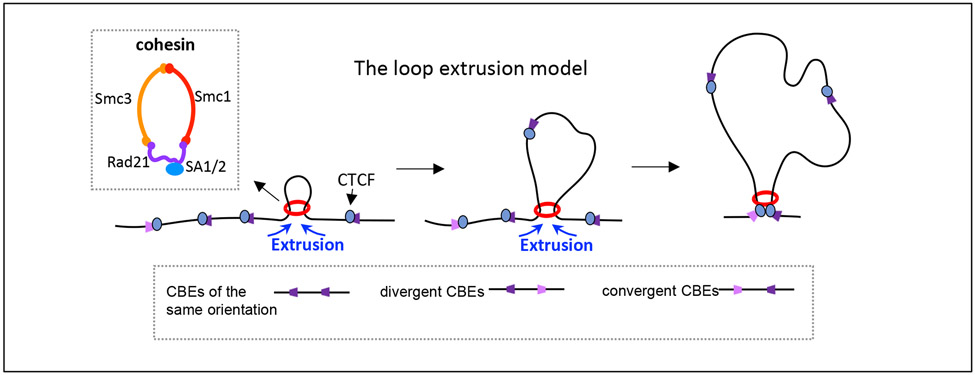
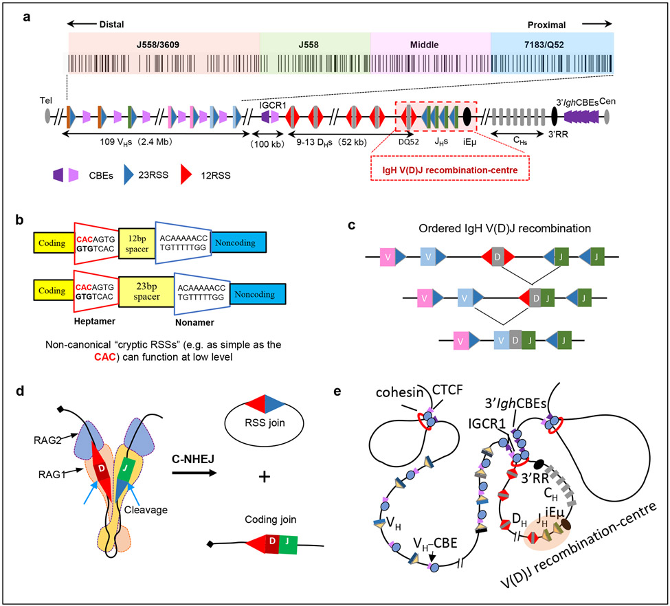
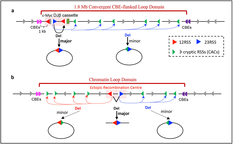
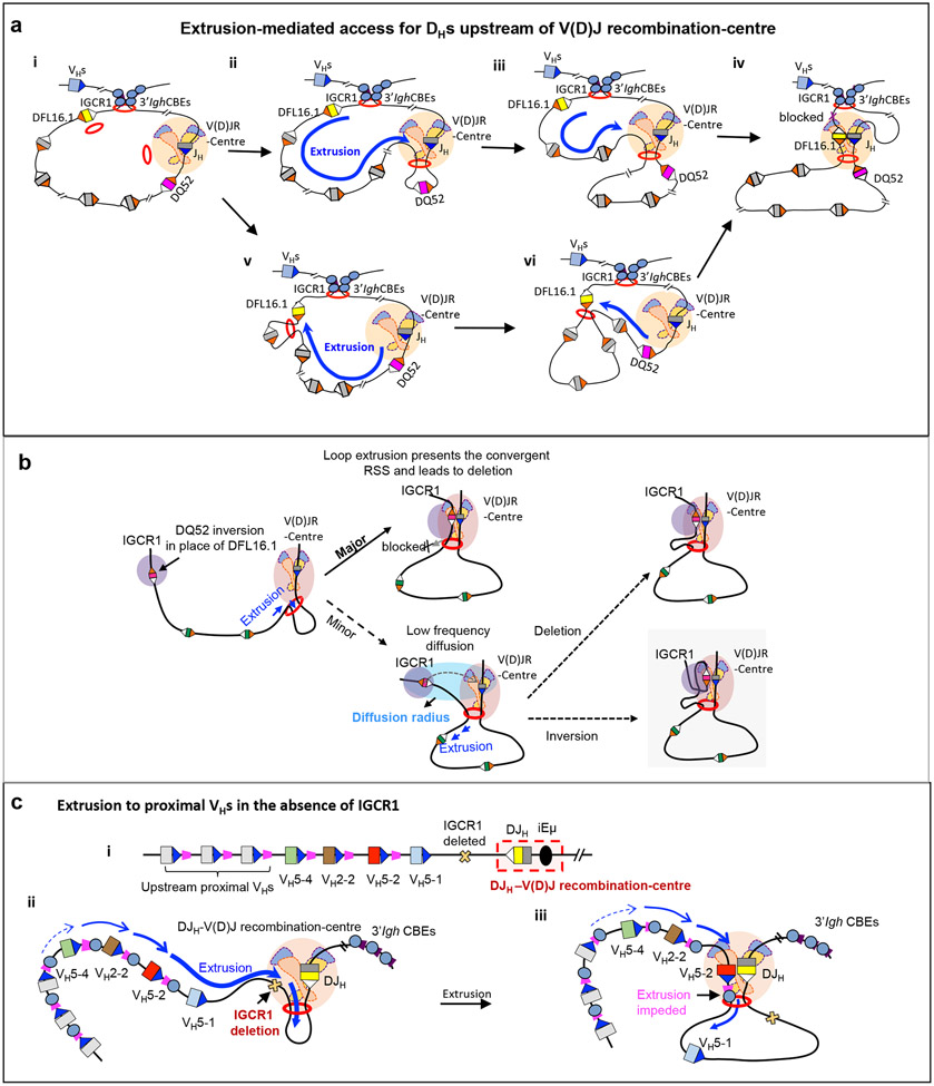
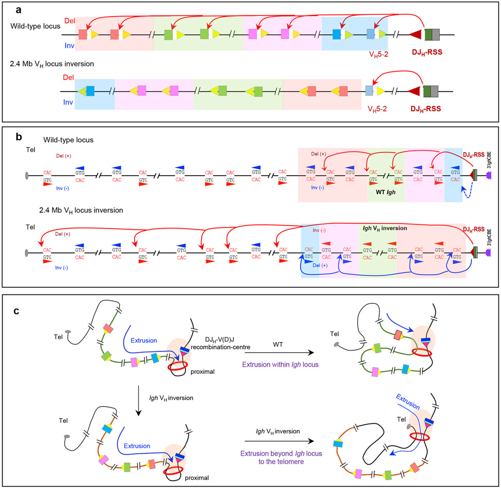
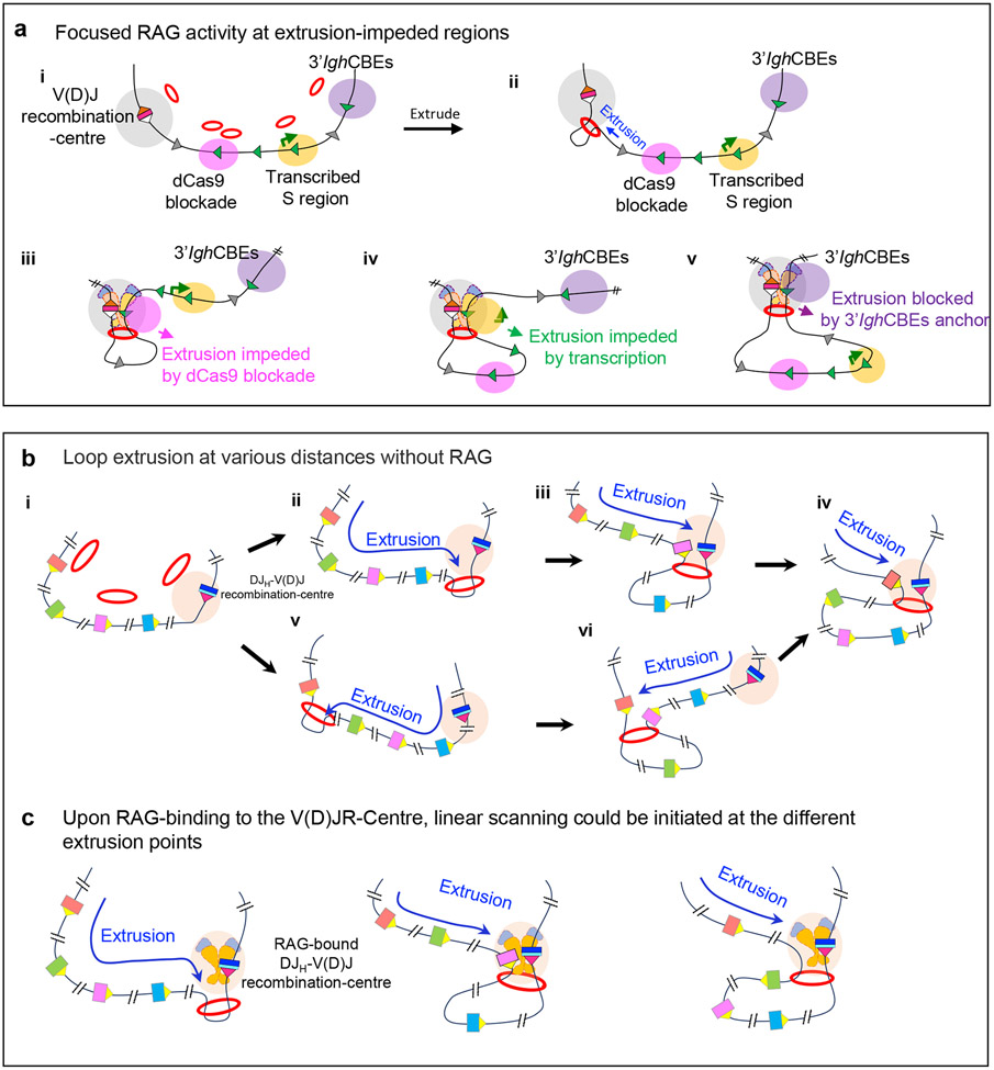
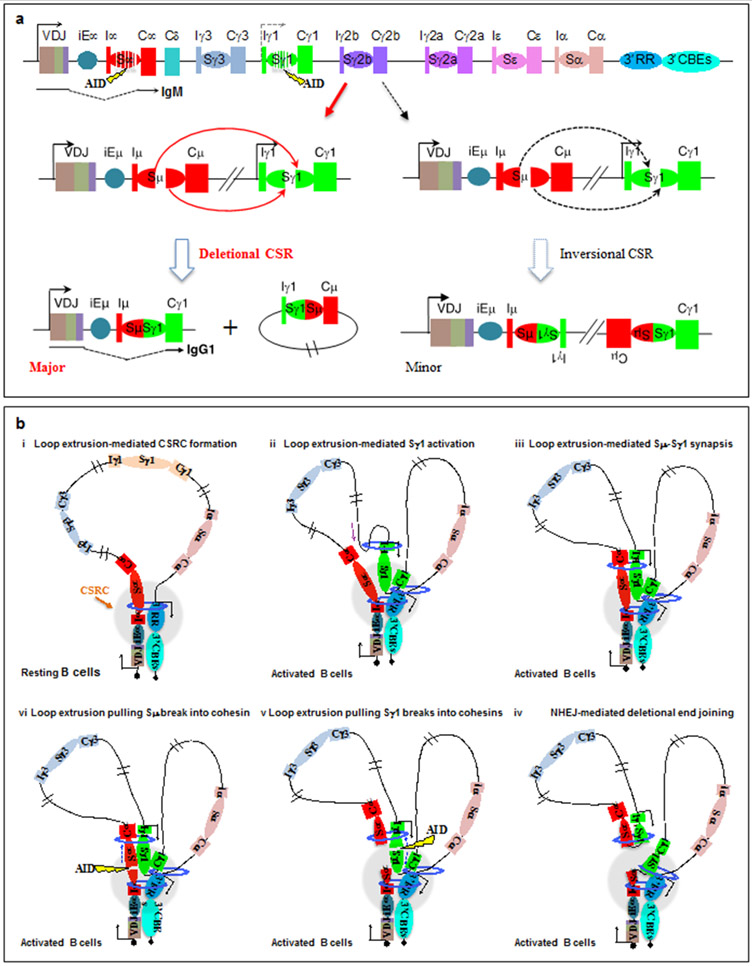
Cohesin mediates chromatin loop formation across the genome by extruding chromatin between convergently oriented CTCF-binding elements. Recent studies indicate that cohesin-mediated loop extrusion in developing B cells presents immunoglobulin heavy chain (Igh) variable (V), diversity (D) and joining (J) gene segments to RAG endonuclease through a process referred to as RAG chromatin scanning. RAG initiates V(D)J recombinational joining of these gene segments to generate the large number of different Igh variable region exons that are required for immune responses to diverse pathogens. Antigen-activated mature B cells also use chromatin loop extrusion to mediate the synapsis, breakage and end joining of switch regions flanking Igh constant region exons during class-switch recombination, which allows for the expression of different antibody constant region isotypes that optimize the functions of antigen-specific antibodies to eliminate pathogens. Here, we review recent advances in our understanding of chromatin loop extrusion during V(D)J recombination and class-switch recombination at the Igh locus.


Figures

Figure 1 ∣
Figure 1 ∣. The loop extrusion model.
A simplified version of the loop extrusion model is outlined (adapted with permission from REF. 53). The extrusion model suggests that progressive extrusion of chromatin by the cohesin complex (red ring) leads to the formation of chromatin loops anchored by the structural protein CTCF (CCCTC-binding factor) bound to convergent CTCF sites. CTCF sites can be found in the genome in three orientations relative to each other (same, divergent and convergent). The inset box shows a schematic of the highly conserved cohesin ring-shaped protein complex, which is composed of the core subunits SMC1, SMC3 and RAD21 associated with either SA1 or SA2 in somatic cells.
Figure 2 ∣
Figure 2 ∣. The Igh locus initiates V(D)J recombination at the V(D)J recombination-centre.
a ∣ The mouse 2.8 Mb immunoglobulin heavy chain (Igh) locus. Upstream of the Igh constant region exon (CH)-containing domain, there is a 3 kb region containing the intronic Igh enhancer (iEμ), 4 JH segments (JHs) and the D segment DQ52, which binds RAG to form the JH-based V(D)J recombination-centre. Upstream of the V(D)J recombination-centre is a 52 kb region containing 9–14 D segments, an approximately 100 kb intervening region containing intergenic control region 1 (IGCR1), and the 2.4 Mb VH-containing telomeric region (locus not drawn to scale). The VH portion of the Igh locus consists of approximately 100 VH segments clustered into four general groups that are specified by VH family type and position; from proximal to distal location these are VH7183/VHQ52, middle VHs without a specific family type, VHJ558 and VHJ558/VH3609. The four VH groups are colour-coded, which is maintained in subsequent figures. The position and orientation of CTCF-binding sites, as well as of recombination signal sequences (12-RSSs and 23-RSSs) are shown. 12-RSSs are located upstream and downstream of D segments, 23-RSSs are located upstream of JHs and downstream of VHs. 3′IghRR, 3′ IgH regulatory region b ∣ Sequence organization of bona fide 12-RSSs and 23-RSSs. The first three nucleotides (CAC) of the heptamer define the cleavage site for RAG endonuclease (RAG1–RAG2) and are crucial for RSS functionality. Cryptic RSSs as simple as CAC can be cleaved by RAG at low levels. cIgh recombination occurs in an ordered manner, with D-to-JH joining preceding VH-to-DJH joining. d ∣ RAG is recruited to the V(D)J recombination-centre, where it binds a JH 23-RSS paired with a DH 12-RSS and aligns them for 12/23-restricted cleavage, with the ends being joined by classical non-homologous end joining (C-NHEJ). e ∣ Illustration of Igh domain organization showing that DHs, the V(D)J recombination-centre and CHs are contained within a 3′ loop domain bounded by IGCR1 and 3′ Igh CTCF sites. The VHs are located within the upstream 5′ region and are excluded from the D-JH domain loop at the D-to-JH recombination stage. The structural protein CTCF binds CTCF sites and cohesin accumulates at these sites.
Figure 3 ∣
Figure 3 ∣. Discovery of RAG long-range chromatin scanning.
a ∣ Illustration of RAG activity initiated from bona fide D and Jβ recombination signal sequences (RSSs) in the Myc–DJ cassette, which, in addition to normal deletional D-to-Jβ joining within the cassette, also results in low-level joining of bona fide RSSs to hundreds of convergent cryptic RSSs across the 1.8 Mb loop domain of Myc. Cryptic RSSs in the same orientation as the Myc–DJ cassette generally are not used for such cryptic V(D)J recombination events. b ∣ Ectopic insertion of pairs of bona fide RSSs randomly into 12 other chromatin loop domains across the genome that are known to be based on convergent CTCF sites activates RAG scanning across these loop domains. The orientation of the initial RAG-binding RSS prescribes the direction of RAG scanning (upstream or downstream), which continues until terminated by a loop anchor determined by convergent CTCF sites.
Figure 4 ∣
Figure 4 ∣. Loop extrusion-mediated RAG scanning drives D-to-JH and proximal VH-to-DJH recombination.
a ∣ Extrusion-mediated D-to-JH recombination for D segments upstream of DQ52 and the V(D)J recombination-centre. D-to-JH recombination occurs within the 3′ Igh loop domain that is anchored by IGCR1 and the 3′ Igh CTCF sites. Cohesin is known to load near the V(D)J recombination-centre to initiate loop extrusion, but likely also at low levels at other sites such as within the D region (part i). The V(D)J recombination-centre functions as a broad and dynamic sub-loop anchor (within the 3′ Igh loop domain) that promotes cohesin-mediated loop extrusion of upstream chromatin past a RAG-bound JH recombination signal sequence (RSS) in the V(D)J recombination-centre (parts ii,iii). This linear process aligns 12-RSSs downstream of D segments with the convergent RAG-bound JH 23-RSS for deletional D-to-JH recombination. 12-RSSs upstream of D segments are not used for recombination owing to being in the same orientation as the JH RSS. D segments upstream of DQ52 are frequently passed without being used for recombination, allowing loop extrusion-mediated RAG scanning to continue in many pro-B cells. Loop extrusion is strongly impeded by the IGCR1 anchor of the 3′ Igh loop, which lies just upstream of the distal D segment DFL16.1 (part iv). See also Supplementary Video 1. Upon cohesin loading near DFL16.1, continuous linear extrusion brings the downstream 12-RSS of DFL16.1 to the RAG-bound JH 23-RSS in the recombination centre. The dynamic anchor of the V(D)J recombination-centre impedes further extrusion and promotes DFL16.1 recombination (parts v,vi). b ∣ Model of short-range diffusion-mediated inversional joining of distal D segments. Whereas loop extrusion-mediated RAG scanning drives dominant deletional joining of distal D segments, low-level inversional D joining occurs via short-range diffusion after loop extrusion brings the D segment into a ‘diffusion radius’ of the RAG-bound V(D)J recombination-centre. Such inversional joining is augmented by strong upstream 12-RSSs of D segments, as exemplified here by the normally dominant downstream RSS of DQ52 being placed upstream as a result of inversion of DQ52 in the DFL16.1 position. Reproduced with permission from REF. . c ∣ Schematic of the DJH-V(D)J recombination-centre including its 12-RSS (white), and proximal VH segments including their 23-RSSs and associated CTCF sites (part i). Upon deletion of IGCR1 or mutation of its CTCF sites, robust RAG scanning activity is extended into the proximal VH region (part ii). VH5-2 is dominantly rearranged owing to it being the first VH segment that is associated with a CTCF site (which impedes further loop extrusion) to be encountered during RAG scanning; the most D-proximal VH segment VH5-1 lacks a functional CTCF site and is mostly bypassed without rearrangement (part iii). Mutation (to inactivate CTCF binding) or deletion of the VH5-2 CTCF site abolishes VH-5-2 usage, rendering the next VH segment that is flanked by a CTCF site (VH2-2) as the most frequently used. Restoring the functionality of the inactive CTCF site of VH5-1 makes it the most frequently used VH segment. In each case, the first several proximal VH segments flanked by CTCF sites are used for VH-to-DJH joining but at progressively reduced levels, with RAG scanning further upstream ultimately being terminated in this proximal VH region. Parts a and c are adapted with permission from REF. .
Figure 5 ∣
Figure 5 ∣. Long-range RAG scanning mediates VH use across the Igh locus.
a ∣ VH recombination signal sequences (RSSs) are all convergently oriented with respect to the DJH-V(D)J recombination-centre RSS, which prescribes that VH-to-DJH recombination is exclusively deletional in wild-type pro-B cells. A 2.4 Mb inversion of the VH locus spanning all VH segments except for proximal VH5-2 abolished the use of all inverted VH segments in bone-marrow pro-B cells and WAPL-downregulated v-Abl cell lines. b ∣ RAG scanning through the VH locus uses hundreds of cryptic RSSs (which can be as simple as the sequence CAC) in convergent orientation with the RSS of the DJH-V(D)J recombination-centre, with scanning terminated within the VH locus. RAG scanning across the wild-type VH locus would be expected often to be terminated within the VH locus by dominant deletional rearrangements mediated by convergent VH RSSs and DJH-V(D)J recombination-centre RSSs. Upon inversion of the 2.4 Mb VH locus in primary pro-B cells, use of cryptic RSSs normally in the convergent orientation is abrogated, whereas use of cryptic RSSs normally in the opposite orientation is activated. Inversion of the locus would eliminate the dominant deletional rearrangements in the wild-type locus, which could allow for some RAG scanning to proceed through the VH locus. Remarkably, RAG scanning from the DJH-V(D)J recombination-centre continued upstream of the VH locus all the way to the telomere, targeting normally oriented cryptic RSSs within multiple, consecutive convergent loop domains beyond the Igh domain. This finding indicated that loop anchor impediments to scanning are likely broadly dampened in pro-B cells. Adapted with permission from REF. . c ∣ Model for RAG scanning activity initiated by the DJH-V(D)J recombination-centre RSS with a wild-type VH locus or inverted VH locus, which illustrates the findings described in panels a and b.
Figure 6 ∣
Figure 6 ∣. Potential substrate accessibility and distal RAG scanning mechanisms in the VH locus.
a ∣ Mechanisms that promote the targeting of cryptic recombination signal sequences (RSSs) during RAG scanning. Cohesin loaded to chromatin (multiple potential loading sites are shown) extrudes chromatin to promote interaction of the Igh V(D)J recombination-centre with downstream chromatin regions (part i, ii). The RAG-bound V(D)J recombination-centre initiates RAG scanning that can be impeded by various impediments on the scanning path, including targeted blockade by binding of catalytically ‘dead’ Cas9 (part iii), active transcription (part iv) and CTCF site-based loop anchors (part v). b ∣ Model of loop extrusion-mediated locus contraction in the absence of RAG binding. Multiple potential cohesin loading sites are indicated. Loop extrusion theoretically could initiate from any of these sites. Loop extrusion from a non-RAG bound, nascent DJH V(D)J recombination-centre promotes long-range interactions with the V(D)J recombination-centre to mediate Igh locus contraction. Extrusion is proposed to proceed over varied upstream chromatin distances in different pro-B cells prior to RAG binding. Loop extrusion might also initiate from cohesin loaded within the VH locus. c ∣ Model for use of distal VH segments promoted by RAG binding to nascent V(D)J recombination-centres. RAG binding to the V(D)J recombination-centre subsequent to loop extrusion forms an active DJH-V(D)J recombination-centre, which initiates extrusion-mediated scanning at multiple different extrusion points initiated by multiple cohesin loading points across the VH locus. Such scanning from a variety of extrusion intermediates avoids biasing VH recombination to the very proximal segments and thus helps to balance the primary VH repertoire. Part a is adapted with permission from REF. . Parts b and c are adapted with permission from REF. .
Figure 7 ∣
Figure 7 ∣. Loop extrusion mediates physiological, deletional class-switch recombination.
a ∣ Schematic of the 200 kb CH region of the immunoglobulin heavy chain (Igh) locus, including the V(D)J exon, the intronic Igh enhancer iEμ, the various CH exons with inducible (I) exon and promoter upstream of each CH segment, S regions, the 3′ Igh regulatory region (3′IghRR) super-enhancer and the 3′ Igh CTCF sites. AID-initiated breaks within Sμ and an activated target Sγ1 are illustrated, which can lead to deletional or inversional class-switch recombination (CSR) outcomes for Sμ to Sγ1 joining. Deletional CSR is the major physiological event, whereas inversional CSR occurs at a lower frequency. Part a is adapted with permission from REF. . b ∣ Loop extrusion mediates formation of a class-switch recombination-centre (CSR-centre) and promotes the synapsis of S region double-strand break ends for deletional joining. In resting B cells (part i), cohesin is loaded at 3′ IghRR or iEμ–Sμ and then extrudes the 3′IghRR and iEμ–Sμ into proximity to generate a dynamic CSR-centre. In activated B cells (part ii), a primed S–CH unit (Sγ1–Cγ1 is shown as an example) is extruded to the CSR-centre for transcriptional activation and cohesin loading, which leads to further extrusion to align acceptor and donor S regions (part iii). Tension pulls S region double-strand break ends initiated by AID into opposing cohesin rings (parts iv and v), stalling extrusion and aligning them for deletional joining mediated by non-homologous end joining (NHEJ) (part vi). Part b is adapted with permission from REF. . See also Supplementary Video 2.

References

    1. Dixon JR et al. Topological domains in mammalian genomes identified by analysis of chromatin interactions. Nature 485, 376–380 (2012). - PMC - PubMed
    1. Nora EP et al. Spatial partitioning of the regulatory landscape of the X-inactivation centre. Nature 485, 381–385 (2012). - PMC - PubMed
    1. Dowen JM et al. Control of cell identity genes occurs in insulated neighborhoods in mammalian chromosomes. Cell 159, 374–387 (2014). - PMC - PubMed
    1. Lupianez DG et al. Disruptions of topological chromatin domains cause pathogenic rewiring of gene-enhancer interactions. Cell 161, 1012–1025 (2015). - PMC - PubMed
    1. Narendra V et al. CTCF establishes discrete functional chromatin domains at the Hox clusters during differentiation. Science 347, 1017–1021 (2015). - PMC - PubMed

Publication types

MeSH terms