. 2022 Mar;27(3):1729-1741.
doi: 10.1038/s41380-022-01468-z. Epub 2022 Feb 15.

Human ARHGEF9 intellectual disability syndrome is phenocopied by a mutation that disrupts collybistin binding to the GABAA receptor α2 subunit

Affiliations

Human ARHGEF9 intellectual disability syndrome is phenocopied by a mutation that disrupts collybistin binding to the GABAA receptor α2 subunit

Dustin J Hines et al. Mol Psychiatry. 2022 Mar.

Abstract

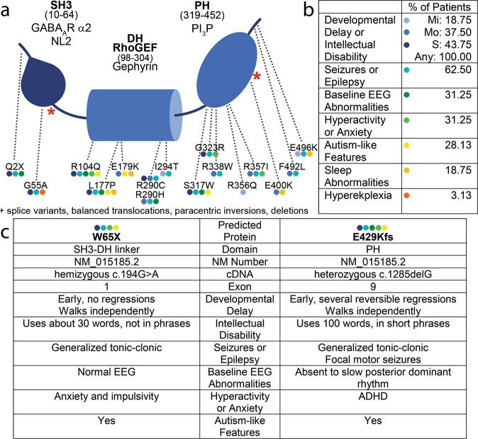
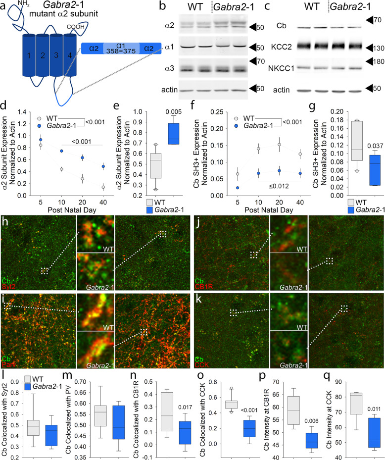
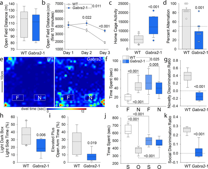
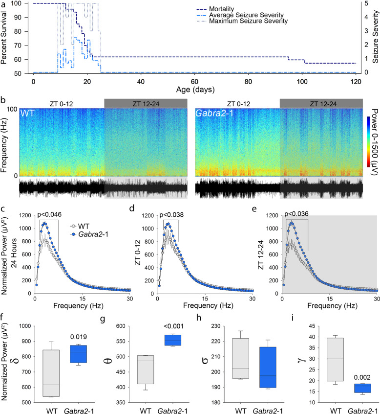
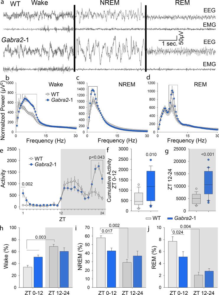
Intellectual disability (ID) is a common neurodevelopmental disorder that can arise from genetic mutations ranging from trisomy to single nucleotide polymorphism. Mutations in a growing number of single genes have been identified as causative in ID, including ARHGEF9. Evaluation of 41 ARHGEF9 patient reports shows ubiquitous inclusion of ID, along with other frequently reported symptoms of epilepsy, abnormal baseline EEG activity, behavioral symptoms, and sleep disturbances. ARHGEF9 codes for the Cdc42 Guanine Nucleotide Exchange Factor 9 collybistin (Cb), a known regulator of inhibitory synapse function via direct interaction with the adhesion molecule neuroligin-2 and the α2 subunit of GABAA receptors. We mutate the Cb binding motif within the large intracellular loop of α2 replacing it with the binding motif for gephyrin from the α1 subunit (Gabra2-1). The Gabra2-1 mutation causes a strong downregulation of Cb expression, particularly at cholecystokinin basket cell inhibitory synapses. Gabra2-1 mice have deficits in working and recognition memory, as well as hyperactivity, anxiety, and reduced social preference, recapitulating the frequently reported features of ARHGEF9 patients. Gabra2-1 mice also have spontaneous seizures during postnatal development which can lead to mortality, and baseline abnormalities in low-frequency wavelengths of the EEG. EEG abnormalities are vigilance state-specific and manifest as sleep disturbance including increased time in wake and a loss of free-running rhythmicity in the absence of light as zeitgeber. Gabra2-1 mice phenocopy multiple features of human ARHGEF9 mutation, and reveal α2 subunit-containing GABAA receptors as a druggable target for treatment of this complex ID syndrome.


Conflict of interest statement

The authors declare the following competing interests: SJM serves as a consultant for AstraZeneca, Bain Capital, and SAGE Therapeutics, relationships that are regulated by Tufts University. SJM is also a shareholder in SAGE Therapeutics.

Figures

Fig. 1
Fig. 1. ARHGEF9 mutations overlaid on collybistin protein structure, and relationship to the core phenotypes of the human ARHGEF9 mutation syndrome.
a Schematic diagram of Cb protein structure with reported point mutation sites leading to missense and nonsense mutations marked (new patients red asterisks). In addition to the 18 point mutations identified to date, the human ARHGEF9 ID syndrome has also been linked to splice variants, balanced translocations, paracentric inversions, and deletions. b Listing of the core reported phenotypes of the human ARHGEF9 mutation ID syndrome, and the proportion of patients reported to show each phenotype. c Clinical summary of the two patients characterized in the context of this study. Abbreviations: Src homology 3 (SH3) domain; Dbl homology (DH) domain; Rho Guanine Nucleotide Exchange Factor (RhoGEF); Plecstrin homology (PH) domain; neuroligin-2 (NL2); Phosphatidylinositol 3-phosphate (PI3P); mild (Mi); moderate (Mo); severe (S); electroencephalogram (EEG).
Fig. 2
Fig. 2. A mouse model with a mutation in the GABAA receptor α2 subunit large intracellular loop (Gabra2-1) strongly influences collybistin expression at specific subtypes of inhibitory synapses.
a Schematic diagram of the mutation generated in the large intracellular loop of the GABAA receptor α2 subunit large intracellular loop, at the site where Cb is characterized to interact. The Cb interaction motif has been replaced with a motif from the α1 subunit that preferentially interacts with gephyrin generating the Gabra2-1 mouse. b Western blotting of cortical lysate from PND 10 suggests that the Gabra2-1 mutation increases the total expression of the α2 subunit, but does not change expression of either α1 or α3 subunits. c Western blotting of cortical lysate from PND 40 suggests that the Gabra2-1 mutation increases the total expression of Cb. Expression of total KCC2 and NKCC1 appears unaltered by the Gabra2-1 mutation. d Quantification of changes in total α2 subunit expression across a time course of postnatal development in both WT and Gabra2-1 cortical lysates. e Quantification of α2 subunit expression from PND10 comparing WT and Gabra2-1. f Quantification of changes in total Cb expression across a time course of postnatal development in both WT and Gabra2-1 cortical lysates. g Quantification of Cb expression from PND40 comparing WT and Gabra2-1. h Immunostaining for Cb and Syt2 in WT and Gabra2-1 mice show closely opposed clusters on the soma of cortical cells. i Immunostaining for Cb and PV in WT and Gabra2-1 mice show colocalized clusters on the soma of cortical cells. j Immunostaining for Cb and CB1R in WT mice show closely opposed clusters on the soma of cortical cells. k Immunostaining for Cb and CCK in WT mice show closely opposed clusters on the soma of cortical cells. l, m. Quantification of colocalization (Pearson’s) of Cb with Syt2 (l) and PV (m) shows no change between WT and Gabra2-1. Colocalization (Pearson’s) of Cb with both CB1R (n) and CCK (o) is reduced on the soma of Gabra2-1 cortical cells. Quantification of Cb intensity at CB1R (p) and CCK (q) positive clusters on the soma of cortical cells reveals a significant reduction in Gabra2-1. Graphs d,f plot mean and standard error; graphs e, g, l-q plot median, first and third quartile, and range.
Fig. 3
Fig. 3. Gabra2-1 mice have behavioral impairments that model intellectual disability as well as other features of ARHGEF9 mutation.
a Gabra2-1 mice have normal levels of distance travelled in the open field, suggesting adequate motor performance. b Gabra2-1 mice maintain high levels of activity across repeated exposures to the open field, suggesting that they do not recall their prior experience in the open field. c Assessment of home cage activity comparing single housed WT and Gabra2-1 mice is suggestive of hyperactivity in Gabra2-1. d Gabra2-1 mice fail to spontaneously alternate arm choice in the T-maze, indicative of working memory impairment. e Heat maps of exploratory activity during the test session of the novel object recognition paradigm, showing representative exploration of the familiar (F) and novel (N) objects by WT and Gabra2-1 mice. f Analysis of time spent exploring the F and N objects during the novel object recognition test session. WT mice spend significantly more time with the N object, while Gabra2-1 mice do not. g Analysis of the discrimination ratio for the N object, which is positive for WT mice, but negative for Gabra2-1 mice. h Gabra2-1 mice spend significantly less time in the light side of the light-dark box. i In the elevated plus maze, Gabra2-1 mice spend less time in the open arms. j Analysis of time spent with novel social (S) or object (O) stimuli in the 3-chambered maze by WT and Gabra2-1 mice. WT mice spend significantly more time with the S stimulus, while Gabra2-1 do not. k Analysis of the discrimination ratio for the S stimulus, which is positive for WT mice, but neutral for Gabra2-1. Graphs b,d plot mean and standard error; graphs a, c, fk plot median, first and third quartile, and range.
Fig. 4
Fig. 4. Gabra2-1 mice are characterized by spontaneous epilepsy during development as well as background EEG abnormalities.
a Line plot superimposing the incidence and severity of spontaneous seizures observed in Gabra2-1 mice with early mortality. Mortality maps directly on to the period of time when the spontaneous seizures are observed. b Representative 24-hour spectrograms and traces from electroencephalogram (EEG) recordings of WT and Gabra2-1 mice. c Fast-Fourier transform analysis of complete 24-hour recordings of WT and Gabra2-1 mice. d, e Fast-Fourier transform analysis of light (ZT0-12) and dark (ZT12-24) phases of the 24 h cycle. f Analysis of δ waveform EEG power reveals an elevation in Gabra2-1 mice compared to WT controls. g θ waveform power is also significantly elevated in Gabra2-1 mice, while σ waveform power remains unchanged (h). i γ waveform power is significantly decreased in Gabra2-1 mice. Graphs c–e plot mean and standard error; graphs fi plot median, first and third quartile, and range.
Fig. 5
Fig. 5. Gabra2-1 mice show EEG abnormalities during all vigilance states, as well as a reduction in the time spent in NREM and REM.
a Representative EEG and EMG traces during Wake, NREM, and REM in WT and Gabra2-1 mice. b–d Fast-Fourier transform analysis of the EEG waveform during specific stages of sleep reveals an elevation in low-frequency waveform power in Gabra2-1 mice that is unique to each sleep stage. e Recording activity in the home cage reveals that Gabra2-1 mice are hyperactive, particularly around the transition from dark to light. f, g. Cumulative activity is increased in Gabra2-1 mice during both the light (ZT0-12) and dark (ZT12-24) phases of the cycle. h Epoch analysis of the EEG recordings reveals that Gabra2-1 mice spend significantly more time in wake during the light phase compared to WT controls, and that the time spent in wake does not differ between light and dark phases for Gabra2-1 mice. i, j Gabra2-1 mice spend significantly less time in NREM and REM during the light phase compared to WT controls. Graphs be, hj plot mean and standard error; graphs f,g plot median, first and third quartile, and range.

References

    1. Dierssen M. Down syndrome: the brain in trisomic mode. Nat Rev Neurosci. 2012;13:844–58. doi: 10.1038/nrn3314. - DOI - PubMed
    1. de Ligt J, Willemsen MH, van Bon BWM, Kleefstra T, Yntema HG, Kroes T, et al. Diagnostic exome sequencing in persons with severe intellectual disability. 101056/NEJMoa1206524. 2012. 10.1056/NEJMoa1206524. Accessed 21 April 2021. - PubMed
    1. Rauch A, Wieczorek D, Graf E, Wieland T, Endele S, Schwarzmayr T, et al. Range of genetic mutations associated with severe non-syndromic sporadic intellectual disability: an exome sequencing study. Lancet. 2012;380:1674–82. doi: 10.1016/S0140-6736(12)61480-9. - DOI - PubMed
    1. Alber M, Kalscheuer VM, Marco E, Sherr E, Lesca G, Till M, et al. ARHGEF9 disease. Neurol Genet. 2017;3:e148. doi: 10.1212/NXG.0000000000000148. - DOI - PMC - PubMed
    1. Shimojima K, Sugawara M, Shichiji M, Mukaida S, Takayama R, Imai K, et al. Loss-of-function mutation of collybistin is responsible for X-linked mental retardation associated with epilepsy. J Hum Genet. 2011;56:561–5. doi: 10.1038/jhg.2011.58. - DOI - PubMed

Publication types

Substances