Review
. 2022 Jul;20(7):385-400.
doi: 10.1038/s41579-022-00686-0. Epub 2022 Feb 15.

Skeletal infections: microbial pathogenesis, immunity and clinical management

Affiliations
Review

Skeletal infections: microbial pathogenesis, immunity and clinical management

Elysia A Masters et al. Nat Rev Microbiol. 2022 Jul.

Abstract

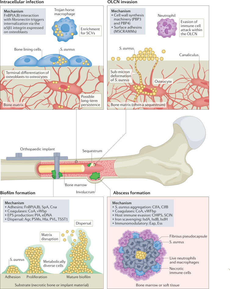
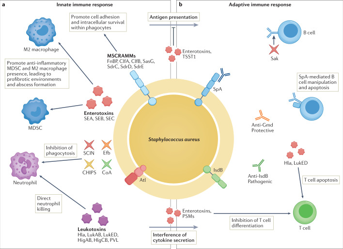
Osteomyelitis remains one of the greatest risks in orthopaedic surgery. Although many organisms are linked to skeletal infections, Staphylococcus aureus remains the most prevalent and devastating causative pathogen. Important discoveries have uncovered novel mechanisms of S. aureus pathogenesis and persistence within bone tissue, including implant-associated biofilms, abscesses and invasion of the osteocyte lacuno-canalicular network. However, little clinical progress has been made in the prevention and eradication of skeletal infection as treatment algorithms and outcomes have only incrementally changed over the past half century. In this Review, we discuss the mechanisms of persistence and immune evasion in S. aureus infection of the skeletal system as well as features of other osteomyelitis-causing pathogens in implant-associated and native bone infections. We also describe how the host fails to eradicate bacterial bone infections, and how this new information may lead to the development of novel interventions. Finally, we discuss the clinical management of skeletal infection, including osteomyelitis classification and strategies to treat skeletal infections with emerging technologies that could translate to the clinic in the future.


Conflict of interest statement

The authors declare no competing interests.

Figures

Fig. 1
Fig. 1. In vivo models of skeletal infection.
Animal studies have been instrumental in some of the most important discoveries that have advanced our understanding of musculoskeletal infection. In broad terms, these models may be classified into implant-related or non-implant-related models. Many of the implant-related models utilize simplified biomaterials to represent the orthopaedic device (grey) that may be placed in the subcutaneous space (for example, tissue cages or discs) or within the bone (for example, Kirschner wires or pins) and offer simplicity and a relatively low risk profile. Certain studies may require use of functional devices whereby the implant placed in the animal serves to fix a fracture or replace a joint (green) and these may be particularly useful where bone healing, implant biomechanics or regulatory approval is the goal. Finally, several models have been developed that do not fit within either category as they may model specific disease entities (purple), such as haematogenous inoculation, leading to osteomyelitis, tendon infection, vertebral osteomyelitis or the use of comorbid mice such as diabetic obese mice.
Fig. 2
Fig. 2. Staphylococcus aureus pathogenesis in osteomyelitis.
Staphylococcus aureus employs a variety of pathogenic mechanisms during skeletal infection. Intracellular infection of osteoblasts, osteoclasts and osteocytes has been investigated as a possible source of long-term S. aureus persistence during osteomyelitis and ‘Trojan horse’ macrophages have been shown to cause bacterial dissemination and multiorgan failure. S. aureus invasion of the osteocyte-lacuno canalicular network (OLCN), most commonly within a sequestrum, permits evasion of host immune cells during osteomyelitis and requires S. aureus deformation to invade canaliculi of bone. S. aureus biofilms formed on implant surfaces and necrotic bone confer resistance to immune cell attack and antibiotic therapy through diffusion limitations and metabolic diversity of S. aureus cells. Lastly, staphylococcal abscess communities can form within the medullary cavity of long bones and in associated soft tissue. Gram-positive S. aureus cells are found at the centre of an abscess surrounded by a fibrous pseudocapsule encasing bacterial cells, followed by layers of dead and live immune cells. Note the formation of a necrotic bone fragment (sequestrum) and new bone formation (involucrum) during prolonged infection. CHIPS, chemotaxis inhibitory protein of S. aureus; ClfA/B, clumping factor A/B; CoA, coagulase; Eap, extracellular adherence protein; eDNA, extracellular DNA; Ess, ESAT-6 secretion system; FnBPA/B, fibronectin-binding protein A/B; Hla, α-haemolysin; MSCRAMMs, microbial surface components recognizing adhesive matrix molecules; PBP3/4, penicillin-binding protein 3/4; PIA, polysaccharide intercellular adhesin; PSM, phenol-soluble modulins; PVL, Panton–Valentine leukocidin; SCIN, staphylococcal complement inhibitor; SCVs, small colony variants; TSST1, toxic shock syndrome toxin 1; vWbp, von Willebrand factor-binding protein.
Fig. 3
Fig. 3. Osteomyelitis pathogens beyond Staphylococcus spp. and Streptococcus spp.
Although Staphylococcus spp. are the most common pathogens across all skeletal infection types, other pathogens frequently cause infection within specific infection classes or patient populations. The figure highlights lesser common pathogens, beyond Staphylococcus spp. and Streptococcus spp., that are uniquely associated with skeletal infections across the human body. Salmonella spp. is most common in individuals with sickling haemoglobinopathies, Haemophilus influenza and Kingella kingae are most common in children, and Neisseria gonorrhoeae is found in sexually active patients.
Fig. 4
Fig. 4. Innate versus adaptive immune responses to Staphylococcus aureus infections.
Virulence proteins enable Staphylococcus aureus to successfully evade host immune responses. a | S. aureus utilizes several cell-associated microbial surface components recognizing adhesive matrix molecules (MSCRAMMs) and secretory proteins to thwart innate host defences. The bacterial cell surface protein SasG promotes cell adhesion, intracellular survival within neutrophils, macrophages and non-professional phagocytes. S. aureus impedes complement-mediated opsonization and phagocytosis by trapping complement proteins using extracellular fibronectin-binding protein (Efb), SpA and Sbi; inhibiting neutrophil recruitment using staphylococcal complement inhibitor (SCIN), chemotaxis inhibitory protein of S. aureus (CHIPS), SpA and coagulase (CoA); and secreting pore-forming toxins (α-haemolysin (Hla), β-haemolysin, γ-haemolysin (HlgAB, HlgCB), leukocidin AB (LukAB) and Panton–Valentine leukocidin (PVL)) to disrupt host membranes to kill innate cells. Superantigens (S. aureus enterotoxin B and C (SEB, SEC) and toxic shock syndrome toxin 1 (TSST1)) contribute to immune evasion by 1) skewing M2-macrophage polarization, 2) promoting myeloid-derived suppressor cell (MDSC) formation, leading to decreased phagocytosis, 3) promoting staphylococcal abscess community formation, and 4) contributing to dysregulation of antigen presentation and cytokine production that affects T and B cell activation. b | On the adaptive immunity side, superantigens can crosslink T cell receptors on tissue-resident T cells to cause antigen-independent stimulation, ultimately causing cell exhaustion. Hla, LukED and phenol soluble modulins (PSMs) can interfere with T cell differentiation and activation, and cause apoptosis. The multifunctional SpA protein can impede B cell function and staphylokinase (Sak) can cleave IgG to prevent antibody-mediated phagocytosis. Antibodies can either be protective or pathogenic during S. aureus infection, with anti-IsdB antibodies facilitating S. aureus survival within ‘Trojan horse’ macrophages causing sepsis, and anti-Gmd antibodies preventing biofilm formation and protecting against skeletal infections. Atl, autolysin; ClfA/B, clumping factor A/B; FnBP, fibronectin-binding protein.

References

    1. Kremers HM, et al. Trends in the epidemiology of osteomyelitis: a population-based study, 1969 to 2009. J. Bone Jt. Surg. Am. 2015;97:837. doi: 10.2106/JBJS.N.01350. - DOI - PMC - PubMed
    1. Tshefu K, Zimmerli W, Waldvogel FA. Short-term administration of rifampin in the prevention or eradication of infection due to foreign bodies. Rev. Infect. Dis. 1983;5(Suppl. 3):S474–S480. doi: 10.1093/clinids/5.Supplement_3.S474. - DOI - PubMed
    1. Li D, et al. Quantitative mouse model of implant-associated osteomyelitis and the kinetics of microbial growth, osteolysis, and humoral immunity. J. Orthop. Res. 2008;26:96–105. doi: 10.1002/jor.20452. - DOI - PMC - PubMed
    1. Arens D, et al. A rabbit humerus model of plating and nailing osteosynthesis with and without Staphylococcus aureus osteomyelitis. Eur. Cell Mater. 2015;30:148–161. doi: 10.22203/eCM.v030a11. - DOI - PubMed
    1. Shiels SM, Bedigrew KM, Wenke JC. Development of a hematogenous implant-related infection in a rat model. BMC Musculoskelet. Disord. 2015;16:255. doi: 10.1186/s12891-015-0699-7. - DOI - PMC - PubMed

Publication types