

Focal Cortical Dysplasia and Generalized Epileptiform Discharges: Case Report and Literature Review
- PMID: 35169375
- PMCID: PMC8802688
- DOI: 10.5455/medarh.2021.75.462-466
Focal Cortical Dysplasia and Generalized Epileptiform Discharges: Case Report and Literature Review
Abstract
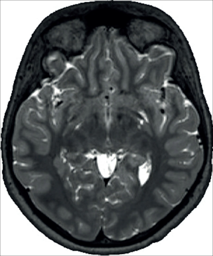
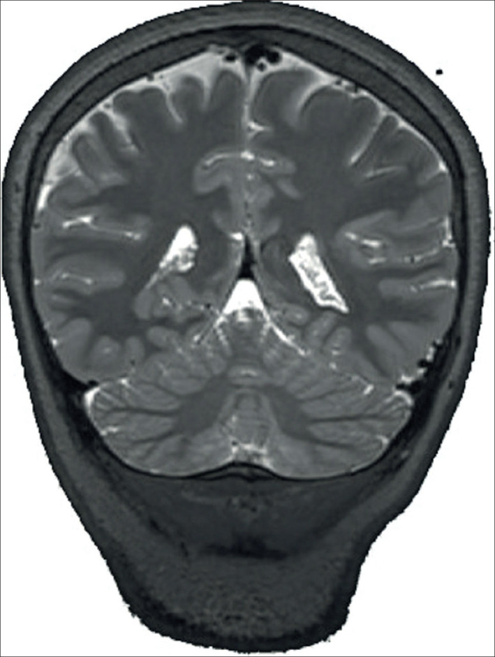
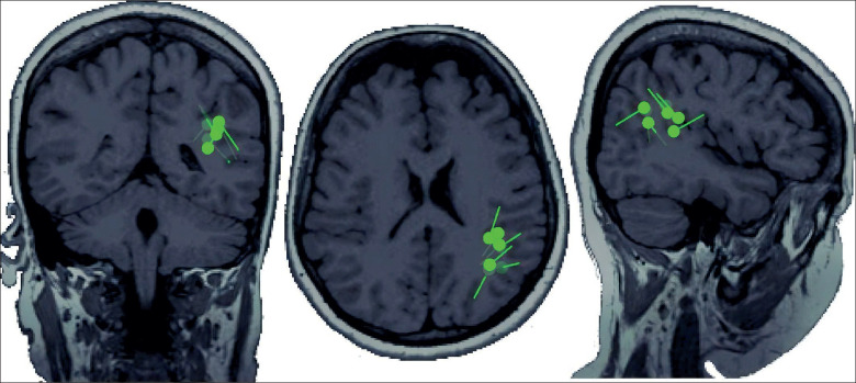
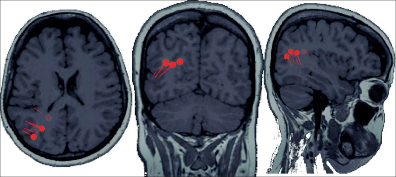
Background: Focal epilepsy can have a varied etiology, including malformations of cortical development (MCD), that can often be detected by Magnetic Resonance Imaging (MRI).Here we show a distinct characteristic of two forms of MCDs on MRI, with two tight dipole clusters in her MEG magnetoencephalography study, in a patient with electroencephalography (EEG) features of generalized epilepsy.
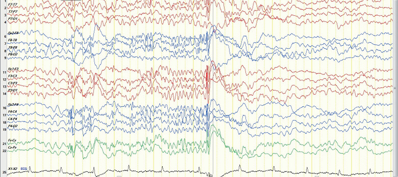
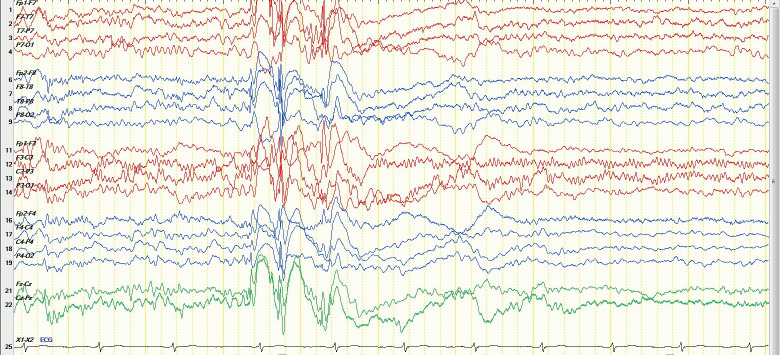
Case report: This is a case presentation of a 20 years old female with epilepsy, found to have upon EMU admission two pathologies (FCD, heterotropia) over the right side near the collateral sulcus, and two tight clusters of dipoles over the right parietal and left temporo-parietal region, with generalized inter ictal discharges in her EEG. FCD is a common etiology of medically intractable seizures and usually in EEG it will show either: pseudo-periodic spikes or rhythmic spikes, poly-spike or repetitive electrographic seizures or a brief discharge of fast rhythmic activity, atypical presentation with generalized epileptiform discharges were rarely reported.
Conclusion: The presence of MCD does not preclude a patient from having other types of epilepsy. Generalized epilepsy and focal related epilepsy have a distinct pathophysiology.
Keywords: EEG; Epilepsy; MEG; MRI; focal cortical dysplasia.
© 2021 Hanin Algethami, Vahe Poghosyan, Eman Baksh, Majed Alhameed.
Conflict of interest statement
There are no conflicts of interest.
Figures
References
Publication types
MeSH terms
LinkOut - more resources
Full Text Sources
Medical