

Case Reports
doi: 10.1002/ccr3.5441.
eCollection 2022 Feb.
Early identification of uterine scar defect by preconception magnetic resonance imaging to achieve successful pregnancy outcome after laparoscopic-assisted myomectomy: Two case reports
Affiliations
- PMID: 35169475
- PMCID: PMC8832169
- DOI: 10.1002/ccr3.5441
Item in Clipboard
Case Reports
Early identification of uterine scar defect by preconception magnetic resonance imaging to achieve successful pregnancy outcome after laparoscopic-assisted myomectomy: Two case reports
Clin Case Rep.
.
Abstract
Myomectomy improves the reproductive ability of women. However, the risk for uterine rupture and abnormal placentation remains a concern. In two cases with scar defects after laparoscopic-assisted myomectomy, one case developed amniocele, while other case showed abnormally invasive placenta. Obstetrical management measures with cesarean sections yielded uneventful postoperative courses.
Keywords: laparoscopic‐assisted myomectomy; magnetic resonance imaging; myomectomy scar defect; preconception management; pregnancy outcomes.
© 2022 The Authors. Clinical Case Reports published by John Wiley & Sons Ltd.
Conflict of interest statement
The authors report no conflict of interest.
Figures
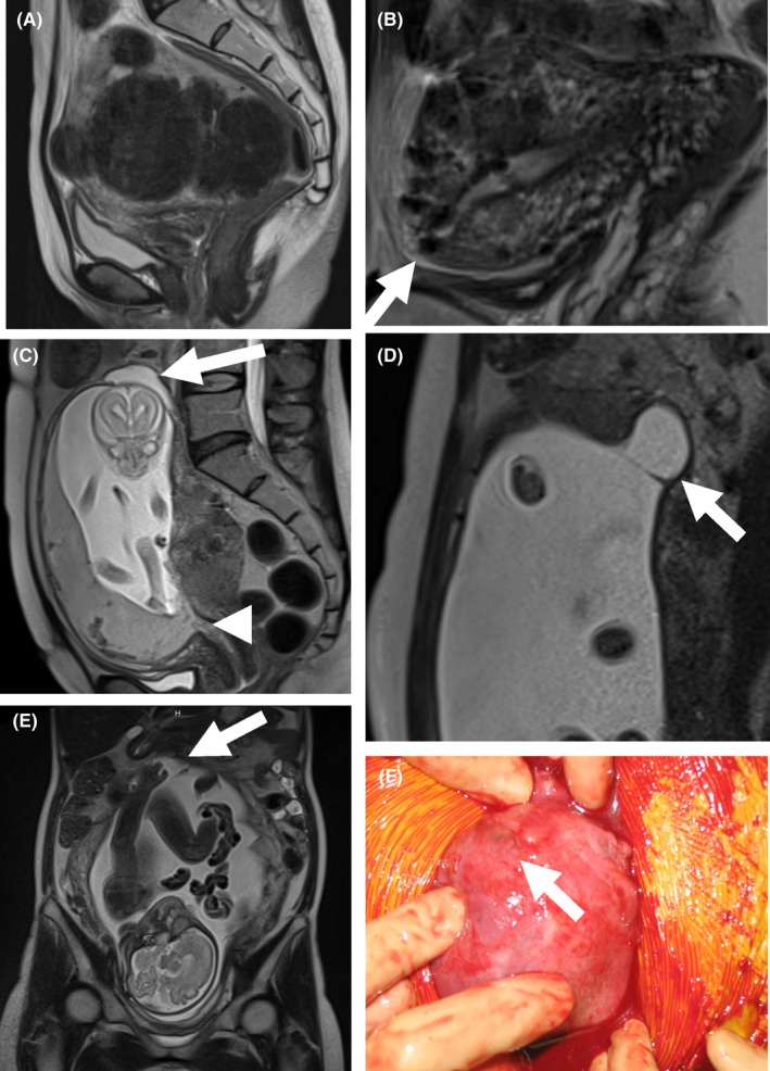
A case showing amniocele at the site of scar defect in pregnancy after laparoscopic‐assisted myomectomy. (A) Sagittal T2‐weighted magnetic resonance imaging (MRI) showed multiple intra‐myometrial and subserosal myomas at the initial examination. (B) Sagittal T2‐weighted MRI showed myomectomy scar thinning (arrow) at the fundal portion three months after LAM. (C) Sagittal T2‐weighted MRI showed extreme myomectomy scar thinning with partial dehiscence (arrow) and low‐lying placenta (arrowhead). (D) Sagittal T2‐weighted MRI showed the formation of an asymptomatic amniocele (arrow) with an intact uterine serosa at 24 weeks of gestation. (E) Coronal T2‐weighted MRI showed that the right hand of the fetus entered the enlarged amniocele at 31 weeks of gestation. (F) A thinned myomectomy scar (arrow) is shown at 34 weeks of gestation, elective cesarean section was performed
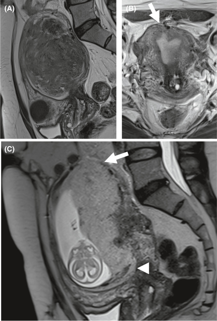
A case showing placenta increta at the site of scar defect in pregnancy after laparoscopic‐assisted myomectomy. (A) Sagittal T2‐weighted magnetic resonance imaging (MRI) showed multiple intra‐myometrial myomas at the initial examination. (B) Sagittal T2‐weighted MRI showing myomectomy scar thinning (arrow) at the fundal portion, three months after LAM. (C) Sagittal T2‐weighted MRI showing placenta increta (arrowhead) at the site of the thinned myomectomy scar and placenta previa (arrowhead) at 14 weeks of gestation
References
-
- Falcone T, Parker WH. Surgical management of leiomyomas for fertility or uterine preservation. Obstet Gynecol. 2013;121(4):856‐868. - PubMed
-
- Seracchioli R, Manuzzi L, Vianello F, et al. Obstetric and delivery outcome of pregnancies achieved after laparoscopic myomectomy. Fertil Steril. 2006;86(1):159‐165. - PubMed
-
- Koo YJ, Lee JK, Lee YK, et al. Pregnancy outcomes and risk factors for uterine rupture after laparoscopic myomectomy: a single‐center experience and literature review. J Minim Invasive Gynecol. 2015;22(6):1022‐1028. - PubMed
-
- Warshak CR, Eskander R, Hull AD, et al. Accuracy of ultrasonography and magnetic resonance imaging in the diagnosis of placenta accreta. Obstet Gynecol. 2006;108(3):573‐581. - PubMed
Publication types
LinkOut - more resources
Full Text Sources
