

An audit of CT brain findings in adults with new-onset seizures in a resource restricted setting in South Africa
- PMID: 35169503
- PMCID: PMC8831926
- DOI: 10.4102/sajr.v26i1.2294
An audit of CT brain findings in adults with new-onset seizures in a resource restricted setting in South Africa
Abstract
Background: Globally, adults presenting with seizures account for 1% - 2% of visits to emergency departments (EDs), of which 25% are new-onset seizures. Neuroimaging is essential as part of the initial workup. Multiple studies have demonstrated abnormal CT brain (CTB) findings in these patients.
Objectives: To review the CTB findings in adults presenting with new-onset seizures in a resource restricted setting.
Method: A retrospective review of 531 CTBs was conducted at a tertiary hospital in Gauteng on adults presenting to the ED with new-onset seizures.
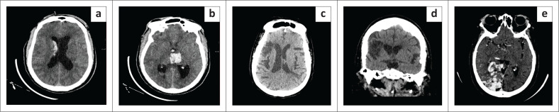
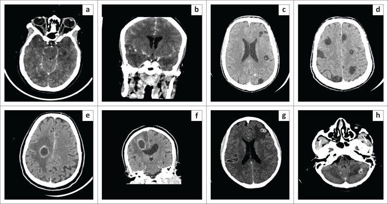
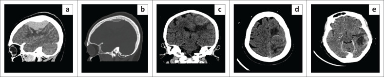
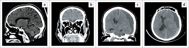
Results: The mean age of the patients was 45.6 ± 17.1 years, and the male to female ratio was 1.2:1. Generalised and focal seizure types were almost equally represented. Of the total 531 patients, 168 (31.6%) were HIV positive. The CTB findings were abnormal in 257 (48.4%) patients, albeit vascular pathology accounted for 21.9%. Infective pathology accounted for 14.1% with a statistically significant association with HIV (p = 0.003). Trauma related pathology was 2.4%, whilst neoplastic pathology was seen in 3.0%. Other causes included congenital pathology, calcifications, atrophy and gliosis. Clinical factors associated with abnormal CTB findings were age ≥ 40 years, HIV infection, hypertension, focal seizures, low Glasgow Coma Scale (GCS), raised cerebrospinal fluid (CSF) protein and presence of lymphocytes.
Conclusion: A high yield of abnormal CTB findings was noted in adult patients who presented with new-onset seizures, supporting the use of urgent CTB in patients with certain clinical risk factors. Patients without these risk factors can be scanned within 24-48 h in a resource restricted setting.
Keywords: CT findings; adult-onset; first-onset; new-onset; seizure.
© 2022. The Authors.
Conflict of interest statement
The authors declare that they have no financial or personal relationships that may have inappropriately influenced them in writing this article.
Figures
Similar articles
-
The role of brain computed tomography in evaluating children with new onset of seizures in the emergency department.Epilepsia. 2000 Aug;41(8):950-4. doi: 10.1111/j.1528-1157.2000.tb00277.x. Epilepsia. 2000. PMID: 10961619
-
Variation in CT use for paediatric head injuries across different types of emergency departments in Australia and New Zealand.Emerg Med J. 2020 Nov;37(11):686-689. doi: 10.1136/emermed-2020-209719. Epub 2020 Aug 17. Emerg Med J. 2020. PMID: 32816840
-
Predictors of abnormal findings of computed tomography of the head in pediatric patients presenting with seizures.Ann Emerg Med. 1997 Apr;29(4):518-23. doi: 10.1016/s0196-0644(97)70226-9. Ann Emerg Med. 1997. PMID: 9095014
-
Topiramate versus carbamazepine monotherapy for epilepsy: an individual participant data review.Cochrane Database Syst Rev. 2019 Jun 24;6(6):CD012065. doi: 10.1002/14651858.CD012065.pub3. Cochrane Database Syst Rev. 2019. PMID: 31233229 Free PMC article.
-
Carbamazepine versus phenytoin monotherapy for epilepsy: an individual participant data review.Cochrane Database Syst Rev. 2019 Jul 18;7(7):CD001911. doi: 10.1002/14651858.CD001911.pub4. Cochrane Database Syst Rev. 2019. PMID: 31318037 Free PMC article.
Cited by
-
Development of clinical prediction model to guide the use of CT head scans for non-traumatic Thai patient with seizure: A cross-sectional study.PLoS One. 2024 Jul 10;19(7):e0305484. doi: 10.1371/journal.pone.0305484. eCollection 2024. PLoS One. 2024. PMID: 38985708 Free PMC article.
-
The epidemiology of human Taenia solium infections: A systematic review of the distribution in Eastern and Southern Africa.PLoS Negl Trop Dis. 2023 Mar 31;17(3):e0011042. doi: 10.1371/journal.pntd.0011042. eCollection 2023 Mar. PLoS Negl Trop Dis. 2023. PMID: 37000841 Free PMC article.
-
Improving a Clinical Prediction Model for Computed Tomography Head Scan Use in Non-Traumatic Seizures: The SeizCT Optimized Model.J Clin Med Res. 2025 Jul 31;17(7):398-407. doi: 10.14740/jocmr6282. eCollection 2025 Jul. J Clin Med Res. 2025. PMID: 40809155 Free PMC article.
References
-
- Adams SM, Knowles PD. Evaluation of a first seizure. Am Fam Physician. 2007;75(9):1342–1347. - PubMed
LinkOut - more resources
Full Text Sources
Miscellaneous