

Case Reports
doi: 10.1016/j.xjtc.2021.09.057.
eCollection 2022 Feb.
Right pneumonectomy for invasive pulmonary mucormycosis
Affiliations
- PMID: 35169748
- PMCID: PMC8828794
- DOI: 10.1016/j.xjtc.2021.09.057
Item in Clipboard
Case Reports
Right pneumonectomy for invasive pulmonary mucormycosis
JTCVS Tech.
.
No abstract available
Figures

Right pneumonectomy space view through Eloesser flap, showing the Latissimus and omental pedicle flap closure of the bronchopleural fistula.
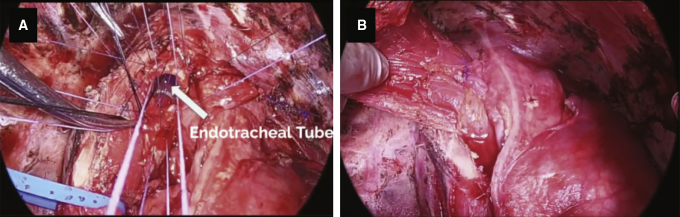
Carinal plasty (A), and reinforcement with latissimus dorsi flap (B).

Right pneumonectomy space view through the Eloesser flap, showing Latissimus flap (A), site of carinal dehiscence (B), omental pedicle flap (C), and incorporated omental flap (D).

Chest radiograph (A) and computed tomography scan (B-F) showing an inflammatory mass extending into the right hilum, encasing the right mainstem bronchus, and behind the right main pulmonary artery.

Sternotomy view of the right main pulmonary artery between the ascending aorta and superior vena cava (SVC).

Extensive inflammation in the posterior mediastinum. The carina is densely adhered to esophagus.

One-year follow up chest radiograph after Eloesser flap closure.

Pathology specimen. Hematoxylin and eosin stain (A), and Grocott methenamine silver stain (B). High magnification (200×) examination of necrotic areas of the lung reveals presence of wide-angle nonseptate fungal hyphae with ribbon-like appearance, characteristic of mucormycosis.
References
-
- Petrikkos G., Skiada A., Lortholary O., Roilides E., Walsh T.J., Kontoyiannis D.P. Epidemiology and clinical manifestations of mucormycosis. Clin Infect Dis. 2012;54(Suppl 1):S23–S34. - PubMed
-
- Connor B.A., Anderson R.J., Smith J.W. Mucor mediastinitis. Chest. 1979;75:524–526. - PubMed
-
- Majid A.A., Yii N.W. Granulomatous pulmonary zygomycosis in a patient without underlying illness: computed tomographic appearances and treatment by pneumonectomy. Chest. 1991;100:560–561. - PubMed
-
- Vercillo M.S., Liptay M.J., Seder C.W. Early pneumonectomy for pulmonary mucormycosis. Ann Thorac Surg. 2015;99:e67–e68. - PubMed
Publication types
LinkOut - more resources
Full Text Sources
