Meta-Analysis
. 2022 May;42(6):662-685.
doi: 10.1002/pd.6115. Epub 2022 May 7.

Diagnostic yield of exome sequencing for prenatal diagnosis of fetal structural anomalies: A systematic review and meta-analysis

Affiliations
Meta-Analysis

Diagnostic yield of exome sequencing for prenatal diagnosis of fetal structural anomalies: A systematic review and meta-analysis

Rhiannon Mellis et al. Prenat Diagn. 2022 May.

Abstract

Objectives: We conducted a systematic review and meta-analysis to determine the diagnostic yield of exome sequencing (ES) for prenatal diagnosis of fetal structural anomalies, where karyotype/chromosomal microarray (CMA) is normal.

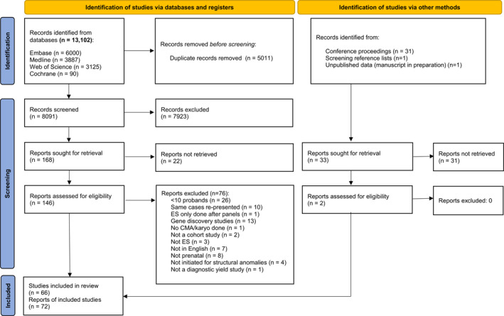
Methods: Following electronic searches of four databases, we included studies with ≥10 structurally abnormal fetuses undergoing ES or whole genome sequencing. The incremental diagnostic yield of ES over CMA/karyotype was calculated and pooled in a meta-analysis. Sub-group analyses investigated effects of case selection and fetal phenotype on diagnostic yield.

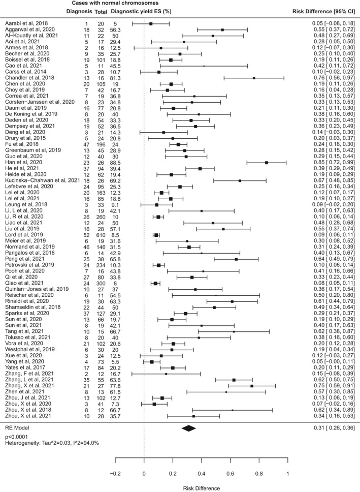
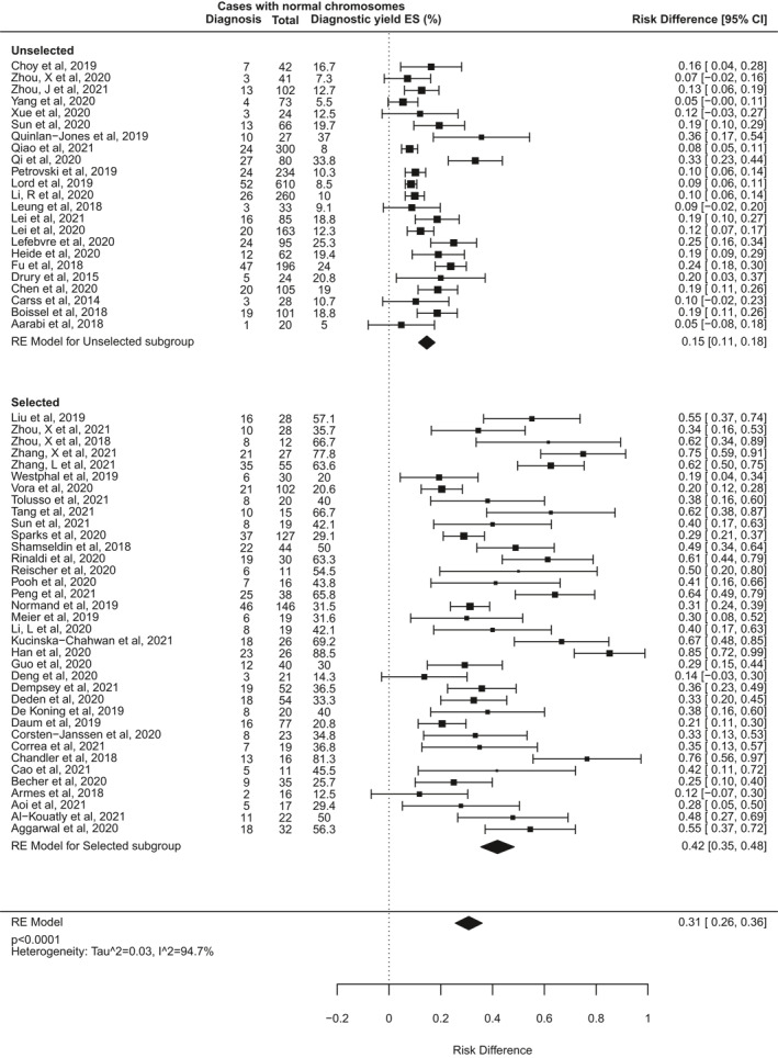
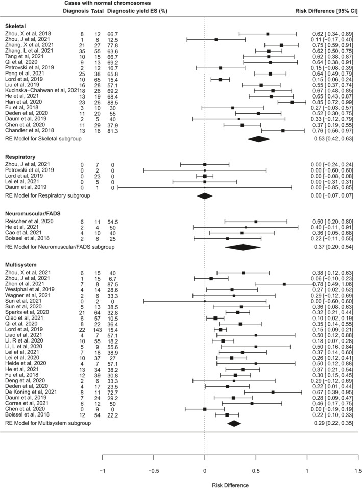
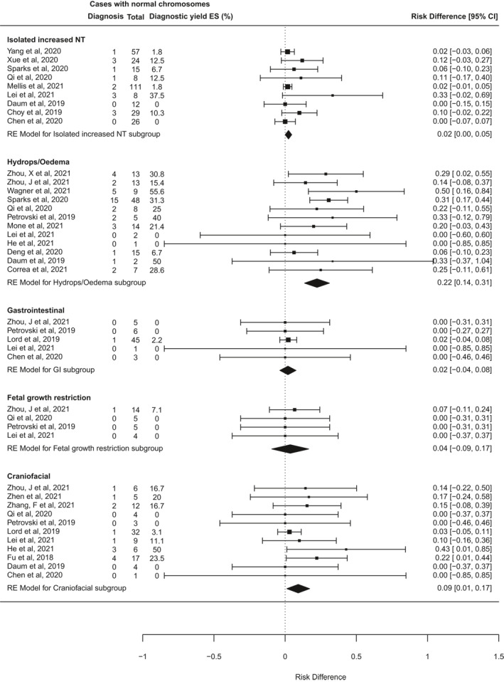
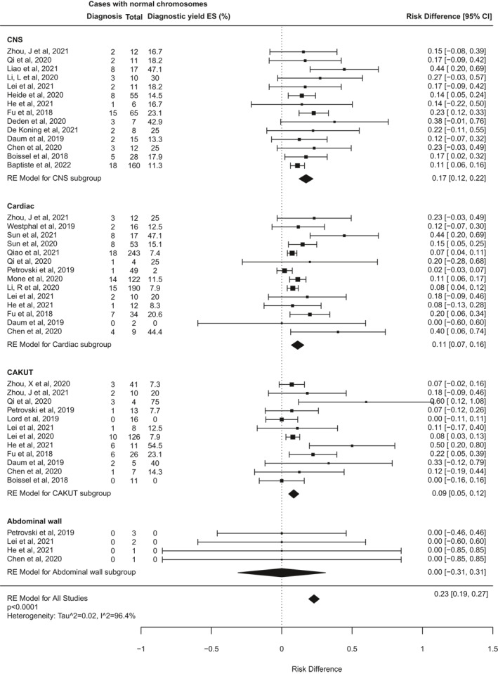
Results: We identified 72 reports from 66 studies, representing 4350 fetuses. The pooled incremental yield of ES was 31% (95% confidence interval (CI) 26%-36%, p < 0.0001). Diagnostic yield was significantly higher for cases pre-selected for likelihood of monogenic aetiology compared to unselected cases (42% vs. 15%, p < 0.0001). Diagnostic yield differed significantly between phenotypic sub-groups, ranging from 53% (95% CI 42%-63%, p < 0.0001) for isolated skeletal abnormalities, to 2% (95% CI 0%-5%, p = 0.04) for isolated increased nuchal translucency.

Conclusion: Prenatal ES provides a diagnosis in an additional 31% of structurally abnormal fetuses when CMA/karyotype is non-diagnostic. The expected diagnostic yield depends on the body system(s) affected and can be optimised by pre-selection of cases following multi-disciplinary review to determine that a monogenic cause is likely.


Conflict of interest statement

None of the authors have any conflict of interests to declare.

Figures

FIGURE 1
FIGURE 1
PRISMA flow diagram showing study screening and selection
FIGURE 2
FIGURE 2
Quality assessment of included study reports using modified Standards for Reporting of Diagnostic Accuracy (STARD) criteria. (n = 72, except or the item “structured summary of study”, where n = 71 because one report was a Letter to the Editor, which would not be expected to contain a structured abstract)
FIGURE 3
FIGURE 3
Forest plot showing individual and pooled incremental yield of prenatal exome sequencing (ES) over karyotype/chromosomal microarray from all 66 studies included in this review
FIGURE 4
FIGURE 4
Forest plot showing individual and pooled incremental yield of prenatal exome sequencing (ES) over karyotype/chromosomal microarray analysed by pre‐test case selection criteria: “unselected” cohorts underwent no prior review to determine clinical features indicating higher likelihood of monogenic aetiology; “selected” cohorts underwent pre‐test expert review to select fetuses with clinical features indicating higher likelihood of monogenic disorder or to rule out non‐genetic conditions
FIGURE 5
FIGURE 5
Forest plot showing individual and pooled incremental yield of prenatal exome sequencing (ES) over karyotype/chromosomal microarray analysed by phenotypic sub‐group in studies where fetuses with anomalies confined to a single body system were distinguishable from those with multi‐system anomalies
FIGURE 5
FIGURE 5
Forest plot showing individual and pooled incremental yield of prenatal exome sequencing (ES) over karyotype/chromosomal microarray analysed by phenotypic sub‐group in studies where fetuses with anomalies confined to a single body system were distinguishable from those with multi‐system anomalies

References

    1. Syngelaki A, Hammami A, Bower S, Zidere V, Akolekar R, Nicolaides KH. Diagnosis of fetal non‐chromosomal abnormalities on routine ultrasound examination at 11–13 weeks’ gestation. Ultrasound Obstet Gynecol. 2019;54(4):468‐476. - PubMed
    1. Wapner RJ, Martin CL, Levy B, et al. Chromosomal microarray versus karyotyping for prenatal diagnosis. N Engl J Med. 2012;367(23):2175‐2184. - PMC - PubMed
    1. Callaway JLA, Shaffer LG, Chitty LS, Rosenfeld JA, Crolla JA. The clinical utility of microarray technologies applied to prenatal cytogenetics in the presence of a normal conventional karyotype: a review of the literature. Prenat Diagn. 2013;33(12):1119‐1123. - PMC - PubMed
    1. Best S, Wou K, Vora N, Van der Veyver IB, Wapner R, Chitty LS. Promises, pitfalls and practicalities of prenatal whole exome sequencing. Prenat Diagn. 2017;38(1):10‐19. - PMC - PubMed
    1. Srivastava S, Love‐Nichols JA, Dies KA, et al. Meta‐analysis and multidisciplinary consensus statement: exome sequencing is a first‐tier clinical diagnostic test for individuals with neurodevelopmental disorders. Genet Med. 2019;21(11):2413‐2421. - PMC - PubMed