. 2022 Apr 1;322(4):L564-L580.
doi: 10.1152/ajplung.00408.2021. Epub 2022 Feb 16.

IRE1α drives lung epithelial progenitor dysfunction to establish a niche for pulmonary fibrosis

Affiliations

IRE1α drives lung epithelial progenitor dysfunction to establish a niche for pulmonary fibrosis

Vincent C Auyeung et al. Am J Physiol Lung Cell Mol Physiol. .

Abstract

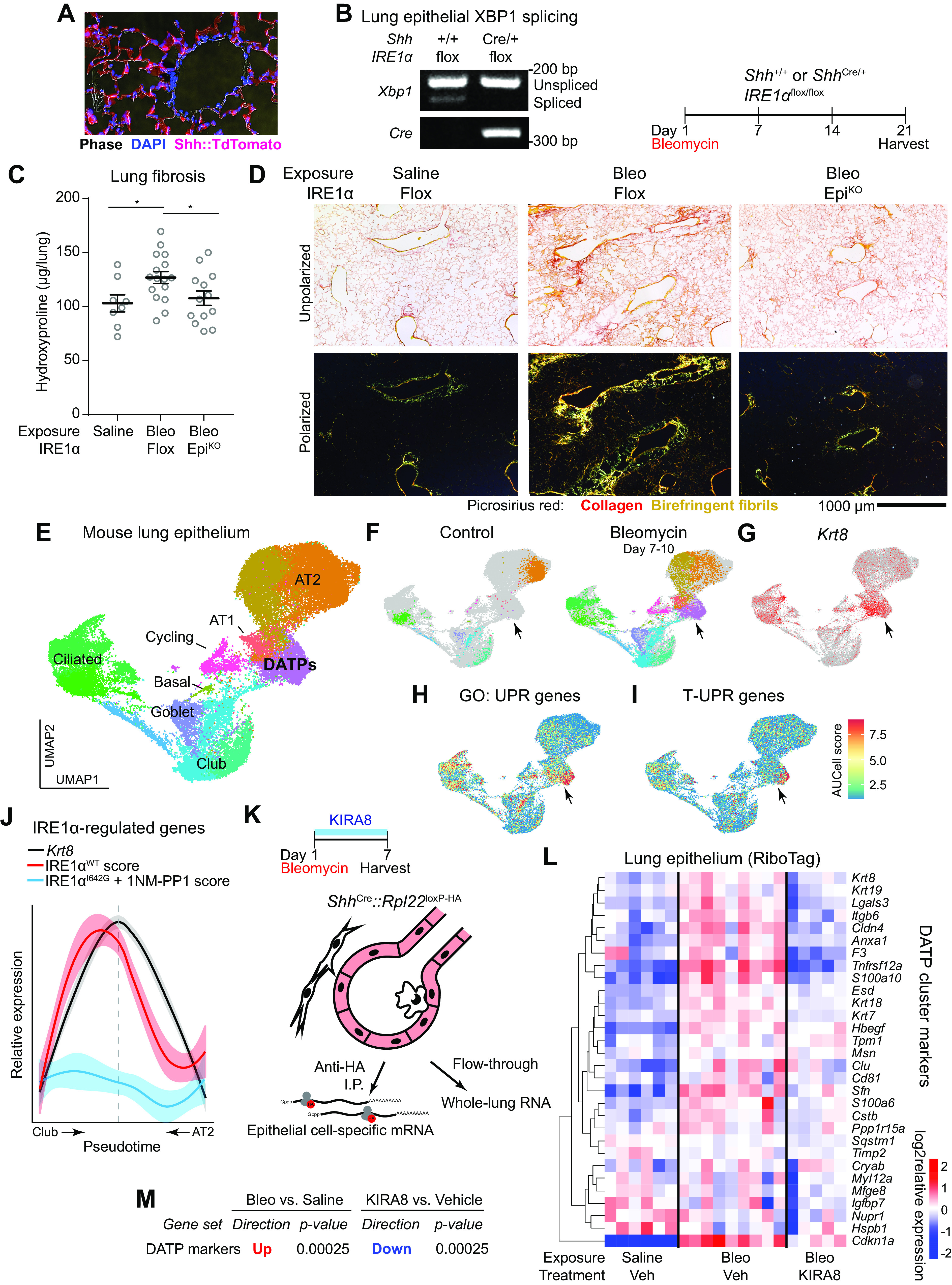
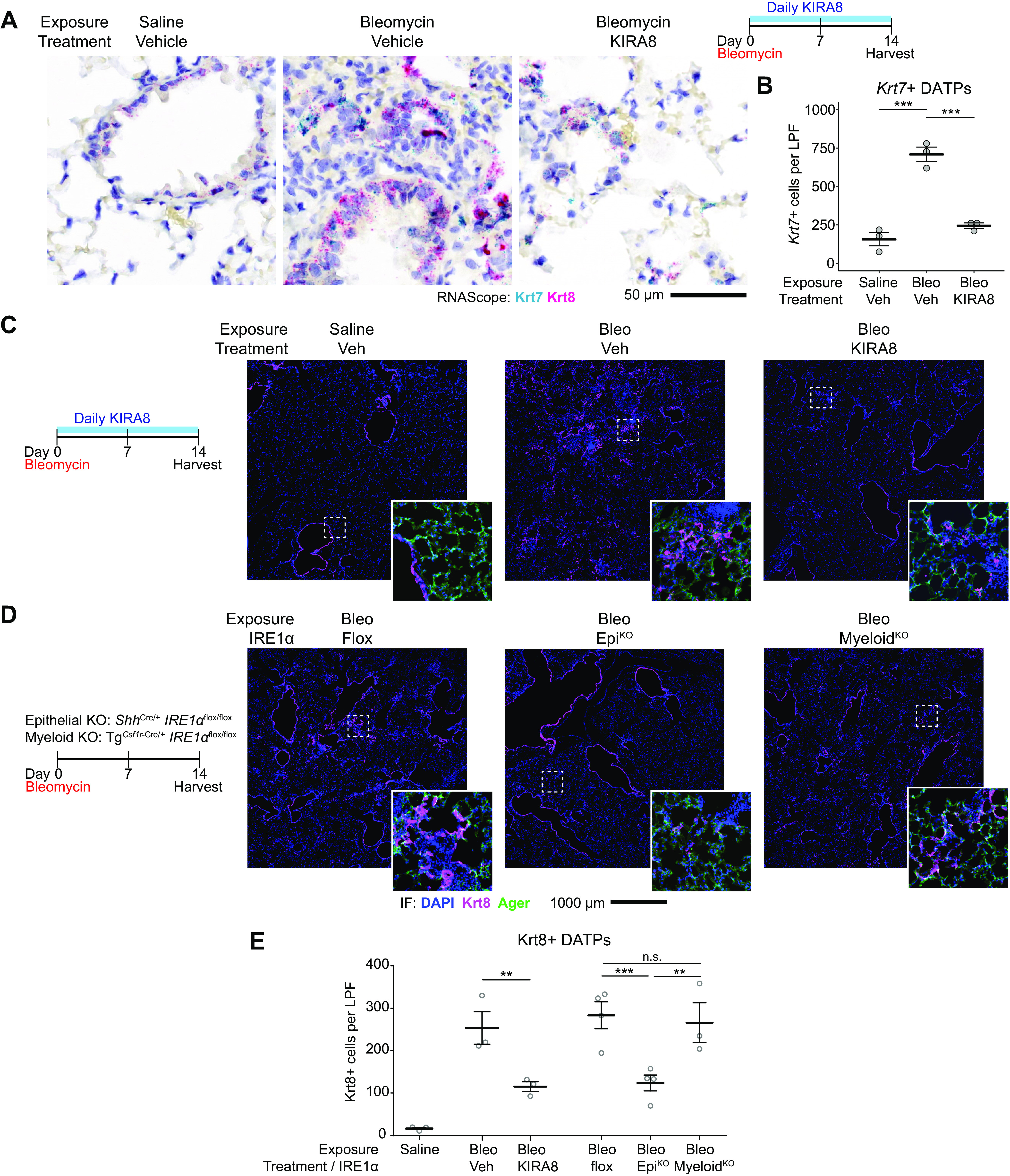
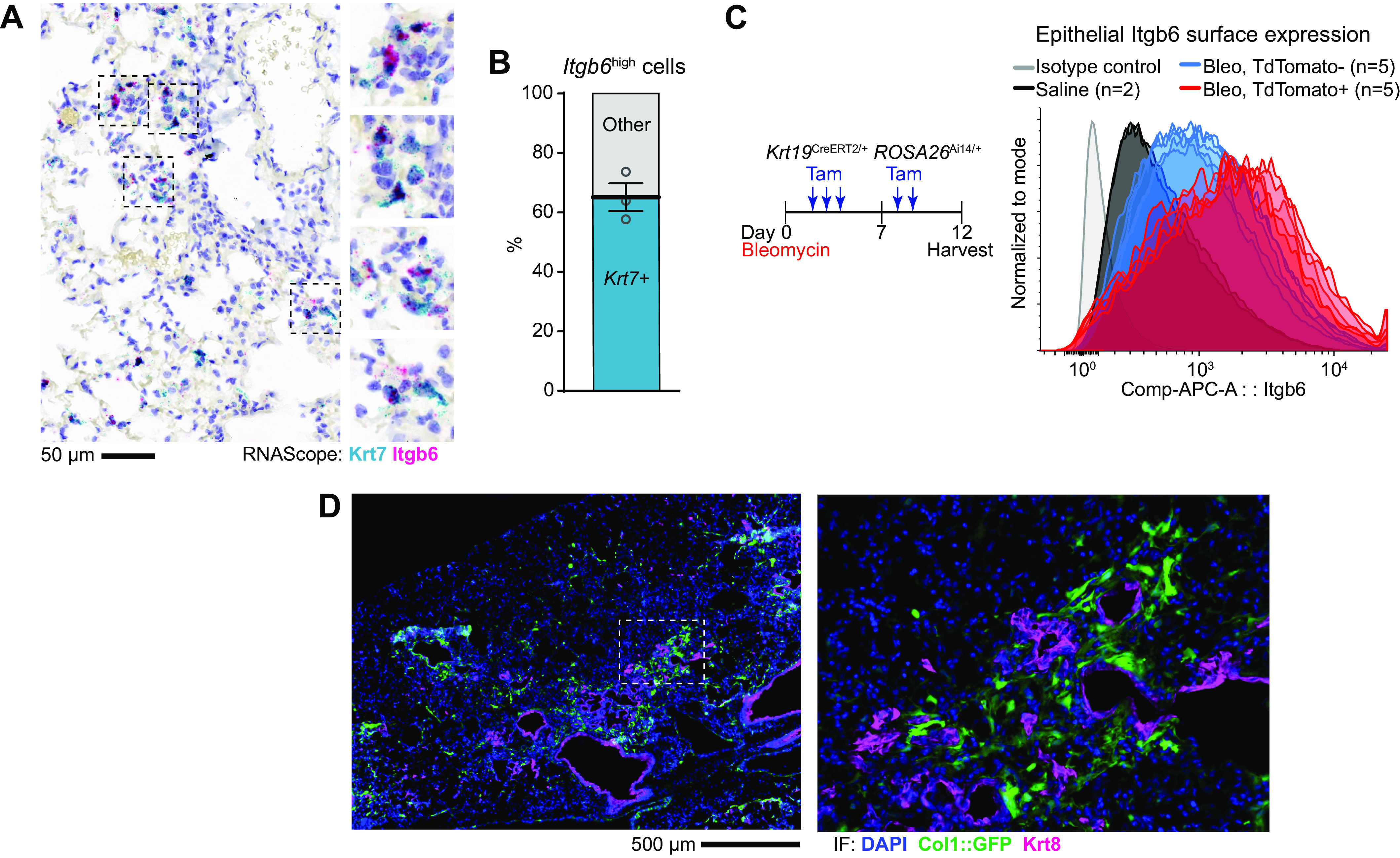
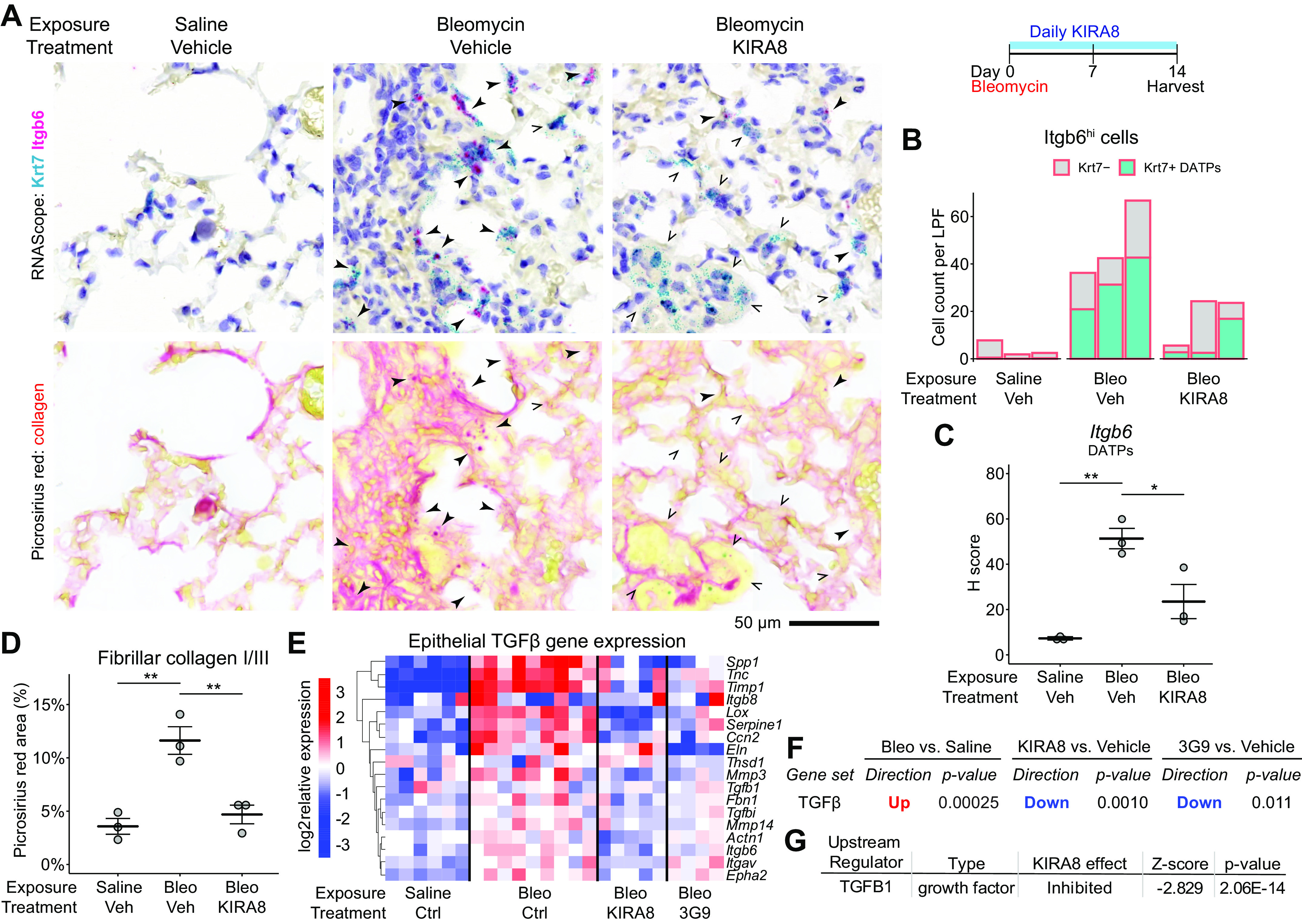
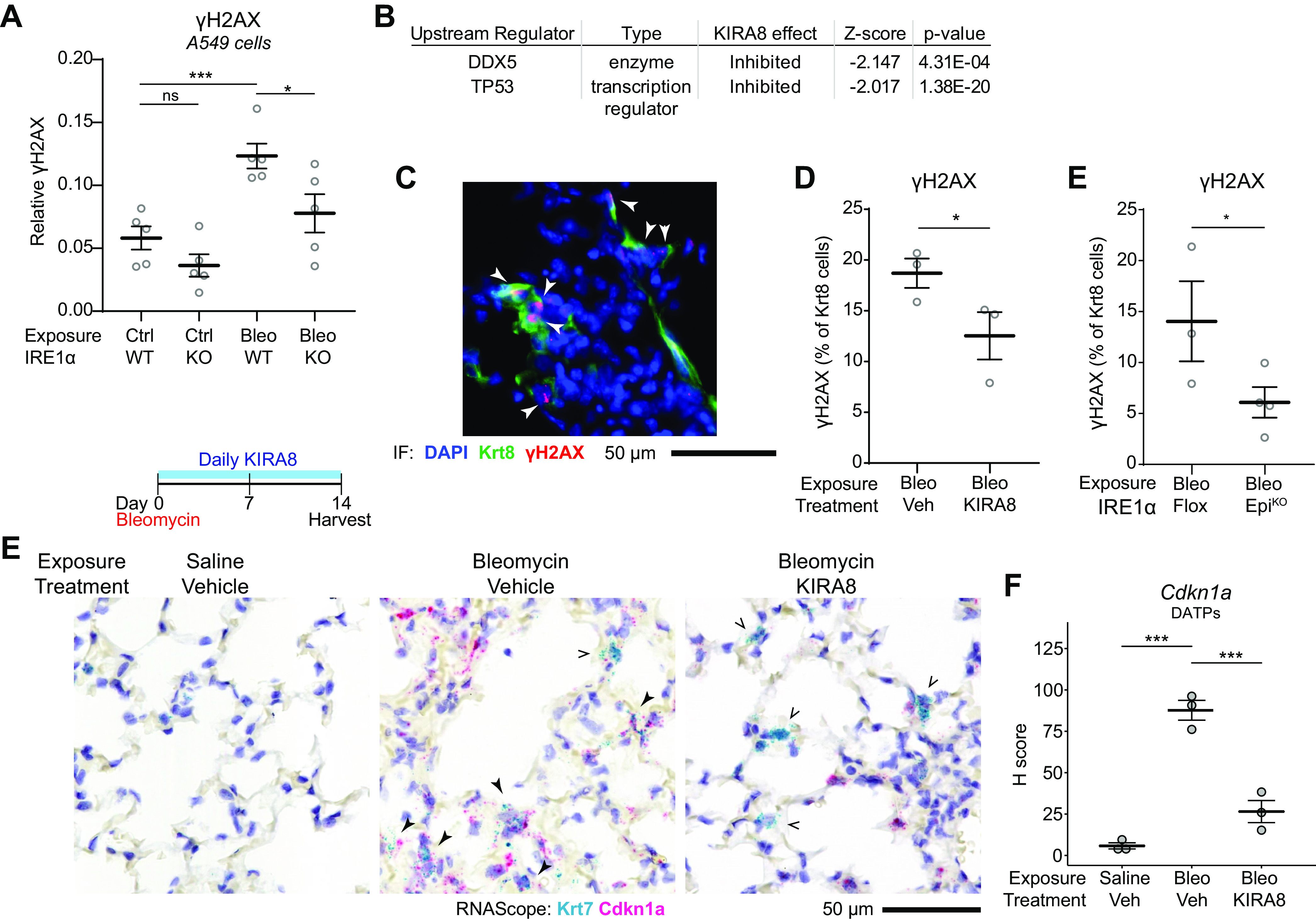
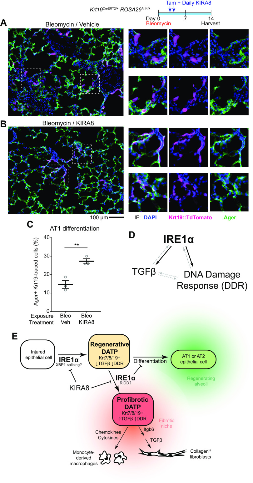
After lung injury, damage-associated transient progenitors (DATPs) emerge, representing a transitional state between injured epithelial cells and newly regenerated alveoli. DATPs express profibrotic genes, suggesting that they might promote idiopathic pulmonary fibrosis (IPF). However, the molecular pathways that induce and/or maintain DATPs are incompletely understood. Here we show that the bifunctional kinase/RNase-IRE1α-a central mediator of the unfolded protein response (UPR) to endoplasmic reticulum (ER) stress is a critical promoter of DATP abundance and function. Administration of a nanomolar-potent, monoselective kinase inhibitor of IRE1α (KIRA8)-or conditional epithelial IRE1α gene knockout-both reduce DATP cell number and fibrosis in the bleomycin model, indicating that IRE1α cell-autonomously promotes transition into the DATP state. IRE1α enhances the profibrotic phenotype of DATPs since KIRA8 decreases expression of integrin αvβ6, a key activator of transforming growth factor β (TGF-β) in pulmonary fibrosis, corresponding to decreased TGF-β-induced gene expression in the epithelium and decreased collagen accumulation around DATPs. Furthermore, IRE1α regulates DNA damage response (DDR) signaling, previously shown to promote the DATP phenotype, as IRE1α loss-of-function decreases H2AX phosphorylation, Cdkn1a (p21) expression, and DDR-associated secretory gene expression. Finally, KIRA8 treatment increases the differentiation of Krt19CreERT2-lineage-traced DATPs into type 1 alveolar epithelial cells after bleomycin injury, indicating that relief from IRE1α signaling enables DATPs to exit the transitional state. Thus, IRE1α coordinates a network of stress pathways that conspire to entrap injured cells in the DATP state. Pharmacological blockade of IRE1α signaling helps resolve the DATP state, thereby ameliorating fibrosis and promoting salutary lung regeneration.

Keywords: IRE1α; kinase inhibitor; lung regeneration; pulmonary fibrosis; unfolded protein response.


Conflict of interest statement

B.J.B. and F.R.P. are founders and equity holders of OptiKira, LLC (Cleveland, OH). No funding or chemical matter from OptiKira was used for the work described in this article. None of the other authors has any conflicts of interest, financial or otherwise, to disclose.

Figures

Figure 1.
Figure 1.
Epithelial IRE1α regulates fibrosis and DATPs exhibit maladaptive IRE1α activation. Confirmation of ShhCre epithelial expression using ROSA26Ai14 reporter mice (A) and IRE1α loss of function in the epithelium (B). Lung epithelial cells were isolated by dissociation and MACS enrichment for CD16/32 CD45 EPCAM+ cells, and IRE1α function evaluated by RT-PCR for XBP1 splice isoforms. Presence of the Cre allele was confirmed by PCR. C: bleomycin-induced fibrosis in IRE1α epithelial knockout mice. Mice were exposed to bleomycin, harvested at day 21, and hydroxyproline content measured. Each data point represents one mouse, with means ± SE indicated. Groups were compared by ANOVA with Fisher’s post hoc test. D: unpolarized and polarized images of lung sections from mice exposed as in C. To image birefringent fibrillar collagen, a circular polarizer was inserted in the illumination path and images were taken with a polarized analyzer in place. E: single-cell RNA sequencing of bleomycin-exposed lung epithelium. The UMAP projection and cluster identities were defined previously (5). F: UMAP projections of subsets of the dataset in for cells from control lungs (left) and lungs harvested at day 7 through day 10 after bleomycin exposure (right). Gray denotes cells in the data set that were collected at other time points; cluster colors and identities are as in E. G: overlay of Krt8 expression, marking some ciliated epithelial cells and the DATP population (arrowhead). Overlay of AUCell scores for Gene Ontology annotated UPR genes (H) and terminal UPR genes (I), concentrated in subset of DATPs (arrowhead). J: IRE1α-regulated gene expression over the course of distinct transdifferentiation trajectories from MHCIIhigh club cells or AT2s, converging on the DATP state marked by peak Krt8 expression (dashed line). Trajectories were defined previously (5). The relative expression level of Krt8 (black line), and AUCell scores for genes induced by wild-type IRE1α (red line) and IRE1αI642G with 1NM-PP1 administration (blue line) were mapped across the trajectory. Gene signatures were defined previously (16). K: RiboTag conditional ribosome tagging strategy. Lungs were harvested 7 days after bleomycin exposure and flash frozen. Epithelial ribosome-associated mRNA was purified by anti-HA immunoprecipitation and sequenced. L: heatmap of markers of the DATP cluster in the lung epithelium after bleomycin exposure and treatment with KIRA8. Markers were defined by the Seurat package using single-cell RNA sequencing data as described previously (5). M: unfiltered DATP marker gene expression changes and statistical testing using the ROAST function in the limma package. *P < 0.05. AUC, area under the curve; DATP, damage-associated transient progenitor; KIRA8, kinase inhibitor of IRE1α; AT2, alveolar type 2; UPR, unfolded protein response.
Figure 2.
Figure 2.
IRE1α regulates DATP number after bleomycin injury. A: RNA in situ hybridization (RNAscope) for Krt7 (teal) and Krt8 (magenta) at day 14 after bleomycin exposure and treatment with KIRA8. B: automated count of Krt7+ cells per 1,240-µm diameter low-power field (LPF). Each dot represents the average of 10 representative fields from one mouse with means ± SE indicated. Groups were compared by ANOVA with Fisher’s post hoc test. C: immunofluorescence staining for Krt8 (magenta) and Ager (green) at day 14 after bleomycin exposure and treatment with KIRA8. D: immunofluorescence staining as in (C) of mice with epithelial IRE1α conditional knockout (EpiKO) or myeloid conditional knockout (MyeloidKO). E: automated count of Krt8+ cells per low-power field. Each dot represents the average of eight representative fields from one mouse with means ± SE indicated. Groups were compared by ANOVA with Fisher’s post hoc test. **P < 0.01, ***P < 0.001. DATP, damage-associated transient progenitor; KIRA8, kinase inhibitor of IRE1α.
Figure 3.
Figure 3.
DATPs are the principal source of integrin αvβ6, a key activator of TGF-β in lung fibrosis. A: RNA in situ hybridization for Krt7 (teal) and Itgb6 (magenta) as in Fig. 2A. B: distribution of Itgb6high cells among Krt7+ or Krt7 cells. Each dot represents the average of 10 representative 1,240-µm diameter low-power fields from one mouse with means ± SE indicated. C: flow cytometric histogram of integrin αvβ6 expression on the surface of lung epithelial cells at day 12 after bleomycin. Krt19CreERT2/+ ROSA26Ai14/+ reporter mice were exposed to bleomycin and Krt19+ DATPs labeled by serial tamoxifen administration. Gated CD45 EPCAM+ epithelial cells are shown; in bleomycin-exposed mice, epithelial cells were subgated into TdTomato+ DATPs or the remaining TdTomato epithelial cells. Each overlaid curve represents data from a single mouse. D: immunofluorescence staining for Krt8+ DATPs and collagenhigh fibroblasts (green). Col1a1::GFP reporter mice were exposed to bleomycin and harvested on day 14. DATP, damage-associated transient progenitor; GFP, green fluorescent protein; TGF-β, transforming growth factor β.
Figure 4.
Figure 4.
IRE1α regulates DATP expression of Itgb6 and consequent TGF-β signaling and local collagen deposition. A: RNA in situ hybridization for Krt7 (teal) and Itgb6 (magenta) as in Fig. 2A followed by washing and restaining with picrosirius red for collagen. Closed arrowheads denote Krt7 and Itgb6 double-positive cells, whereas arrow outlines indicate Krt7+ cells without significant Itgb6 expression. B: automated count of Itgb6high cells, further divided into Krt7+ (teal) or Krt7 cells (gray). Each bar represents the average of 10 representative 1,240-µm diameter low-power fields from a single mouse. C: semiquantitative H-score of Itgb6 puncta in Krt7+ DATPs, averaged over 10 representative fields per dot as in B with means ± SE indicated. D: collagen deposition measured by picrosirius red area. Each dot represents the average of 10 representative fields as in B with means ± SE indicated. E: heatmaps of batch- and sex-adjusted log2 expression values for TGF-β-induced genes in epithelial RiboTag libraries. RiboTag mice were exposed to bleomycin and treated with daily KIRA8 or twice-weekly 3G9, an inhibitory antibody against integrin αvβ6, and epithelial transcriptomes obtained as in Fig. 1I. TGF-β-induced genes were defined previously (23). F: focused statistical testing on the unfiltered TGF-β gene signature in E using the ROAST function in the limma package. G: naïve upstream regulator analysis using the Ingenuity Pathway Analysis package on epithelial RiboTag data identifies TGF-β as an inhibited pathway after KIRA8 treatment. *P < 0.05, **P < 0.01. DATP, damage-associated transient progenitor; KIRA8, kinase inhibitor of IRE1α; TGF-β, transforming growth factor β.
Figure 5.
Figure 5.
IRE1α reinforces DNA damage response signaling in DATPs. A: quantification of γH2AX expression in A549 cells or CRISPR-knockout IRE1α A549 cells. Cells were exposed to 5 µg/mL bleomycin for 4 h. Each dot represents an independent well with means ± SE indicated. Groups were compared by ANOVA with Fisher’s post hoc test. B: naïve upstream regulator analysis using the Ingenuity Pathway Analysis package on epithelial RiboTag data from bleomycin-exposed mice as in Fig. 1I, identifies p53 and DDX5 as inhibited DNA damage response pathways after KIRA8 treatment. C: representative immunofluorescence staining for Krt8 (green) and γH2AX (red) in mice exposed to bleomycin for 14 days. Arrowheads denote Krt8+ γH2AX double-positive cells. Automated counting of γH2AX and Krt8+ cells and quantification of the percentage of Krt8+ cells that are also γH2AX positive in mice treated with KIRA8 (D) or mice with conditional epithelial IRE1α knockout (E). Each dot represents the average of eight low power fields from one mouse with means ± SE indicated. Groups were compared by one-sided Student’s t test. E: RNA in situ hybridization (RNAscope) for Krt7 (teal) and Cdkn1a (red) in mice exposed to bleomycin and treated with daily KIRA8 for 14 days. Solid arrowheads denote Krt7 and Cdkn1a double-positive cells while arrow outlines indicate Krt7+ cells without significant Cdkn1a expression. F: semiquantitative H-score of Cdkn1a puncta in Krt7+ DATPs, averaged over 10 representative fields per dot with means ± SE indicated. Groups were compared by ANOVA with Fisher’s post hoc test. *P < 0.05, ***P < 0.001. DATP, damage-associated transient progenitor; KIRA8, kinase inhibitor of IRE1α.
Figure 6.
Figure 6.
IRE1α impairs DATP differentiation into mature alveolar epithelial cells. Lineage tracing of DATPs after bleomycin exposure. Krt19CreERT2/+ ROSA26Ai14/+ mice were exposed to bleomycin and treated daily with vehicle (A) or KIRA8 (B). Krt19+ DATPs were pulse-labeled by tamoxifen administration on day 3 and day 4 after bleomycin injury. Lungs were harvested at day 14 and evaluated for TdTomato lineage tracing (magenta) and stained for Ager (green). C: automated quantification of Ager staining in TdTomato lineage-traced DATPs. Each dot represents the average of five representative low power fields from a single mouse. Groups were compared by two-sided Student’s t test. D: model of IRE1α at the center of a mutually reinforcing gene expression network that induces and maintains the DATP phenotype and attendant profibrotic gene expression. Solid arrows represent relationships directly interrogated in this study. Dashed arrows represent relationships described in the literature. E: model of the role of IRE1α in the induction of DATPs and their functional phenotypes. IRE1α activity contributes to the induction of the DATP state, but hyperactivation or abnormal persistence of IRE1α signaling enhances TGF-β and DDR signaling and consequent establishment of a fibrotic niche. Relief of IRE1α signaling by kinase inhibitors decreases DATP number, but also dampens TGF-β and DDR signaling in DATPs, promoting their differentiation into mature alveolar epithelial cells. **P < 0.01. DATP, damage-associated transient progenitor; DDR, DNA damage response; KIRA8, kinase inhibitor of IRE1α; TGF-β, transforming growth factor β.

References

    1. Matthay MA, Zemans RL, Zimmerman GA, Arabi YM, Beitler JR, Mercat A, Herridge M, Randolph AG, Calfee CS. Acute respiratory distress syndrome. Nat Rev Dis Primers 5: 18, 2019. doi:10.1038/s41572-019-0069-0. - DOI - PMC - PubMed
    1. Wolters PJ, Blackwell TS, Eickelberg O, Loyd JE, Kaminski N, Jenkins G, Maher TM, Molina-Molina M, Noble PW, Raghu G, Richeldi L, Schwarz MI, Selman M, Wuyts WA, Schwartz DA. Time for a change: is idiopathic pulmonary fibrosis still idiopathic and only fibrotic? Lancet Respir Med 6: 154–160, 2018. doi:10.1016/S2213-2600(18)30007-9. - DOI - PMC - PubMed
    1. Riemondy KA, Jansing NL, Jiang P, Redente EF, Gillen AE, Fu R, Miller AJ, Spence JR, Gerber AN, Hesselberth JR, Zemans RL. Single cell RNA sequencing identifies TGFβ as a key regenerative cue following LPS-induced lung injury. JCI Insight 5: e123637, 2019. doi:10.1172/jci.insight.123637. - DOI - PMC - PubMed
    1. Joshi N, Watanabe S, Verma R, Jablonski RP, Chen CI, Cheresh P, Markov NS, Reyfman PA, McQuattie-Pimentel AC, Sichizya L, Lu Z, Piseaux-Aillon R, Kirchenbuechler D, Flozak AS, Gottardi CJ, Cuda CM, Perlman H, Jain M, Kamp DW, Scott Budinger GR, Misharin AV. A spatially restricted fibrotic niche in pulmonary fibrosis is sustained by M-CSF/M-CSFR signalling in monocyte-derived alveolar macrophages. Eur Respir J 55: 1900646, 2020. doi:10.1183/13993003.00646-2019. - DOI - PMC - PubMed
    1. Strunz M, Simon LM, Ansari M, Kathiriya JJ, Angelidis I, Mayr CH, Tsidiridis G, Lange M, Mattner LF, Yee M, Ogar P, Sengupta A, Kukhtevich I, Schneider R, Zhao Z, Voss C, Stoeger T, Neumann JHL, Hilgendorff A, Behr J, O’Reilly M, Lehmann M, Burgstaller G, Königshoff M, Chapman HA, Theis FJ, Schiller HB. Alveolar regeneration through a Krt8+ transitional stem cell state that persists in human lung fibrosis. Nat Commun 11: 3559, 2020. doi:10.1038/s41467-020-17358-3. - DOI - PMC - PubMed

MeSH terms

Substances