. 2022 Mar 2;144(8):3696-3705.
doi: 10.1021/jacs.1c12697. Epub 2022 Feb 16.

Rapid Inhibitor Discovery by Exploiting Synthetic Lethality

Affiliations

Rapid Inhibitor Discovery by Exploiting Synthetic Lethality

Jacob D Muscato et al. J Am Chem Soc. .

Abstract

Synthetic lethality occurs when inactivation of two genes is lethal but inactivation of either single gene is not. This phenomenon provides an opportunity for efficient compound discovery. Using differential growth screens, one can identify biologically active compounds that selectively inhibit proteins within the synthetic lethal network of any inactivated gene. Here, based purely on synthetic lethalities, we identified two compounds as the only possible inhibitors of Staphylococcus aureus lipoteichoic acid (LTA) biosynthesis from a screen of ∼230,000 compounds. Both compounds proved to inhibit the glycosyltransferase UgtP, which assembles the LTA glycolipid anchor. UgtP is required for β-lactam resistance in methicillin-resistant S. aureus (MRSA), and the inhibitors restored sensitivity to oxacillin in a highly resistant S. aureus strain. As no other compounds were pursued as possible LTA glycolipid assembly inhibitors, this work demonstrates the extraordinary efficiency of screens that exploit synthetic lethality to discover compounds that target specified pathways. The general approach should be applicable not only to other bacteria but also to eukaryotic cells.


Conflict of interest statement

The authors declare no competing financial interest.

Figures

Figure 1
Figure 1
LTA assembled on Glc2DAG plays a critical role in S. aureus cell envelope integrity. (A) Schematic showing assembly of Glc2DAG-LTA having d-alanine modifications. A simplified pathway for WTA synthesis is also shown and highlights the role of TarO in catalyzing the first step. (B) Chemical structures of Glc2DAG-LTA and DAG-LTA showing that the polymers have different numbers of phosphoglycerol repeats. (C) Selected synthetic lethal relationships between LTA proteins (red), the WTA pathway (blue), and the d-alanylation pathway (yellow) that were exploited for compound discovery in this work.
Figure 2
Figure 2
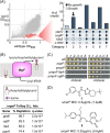
Two potential LTA pathway inhibitors were identified from a ∼230,000 compound screen using only synthetic lethal growth assays. (A) A differential growth screen of wildtype and ΔtarOS. aureus (left panel) identified 68 hits that were classified into categories based on growth profiles against a four-strain mutant panel (right panel). Category 1 (light gray) contained d-alanylation inhibitors. Category 2 (arrow), the only category containing compounds that do not inhibit growth of mutants with disruptions in the LTA pathway, contained the possible LTA pathway inhibitors. (B) MprF synthesizes lysyl-phosphatidyglycerol intracellularly and translocates it to the extracellular surface of the membrane. A TnSeq experiment in a ΔmprF background showed depletion of transposon insertion reads in LTA pathway genes. (C) Spot dilution assays confirm that strains lacking LTA pathway genes ugtP or ltaA grow only if MprF is expressed. (D) Structures of the two compounds from category 2 that prevent growth of ΔmprF, with minimum inhibitory concentrations (MICs) against this strain.
Figure 3
Figure 3
Compound 1 blocks Glc2DAG synthesis, and overexpression analysis nominated the Glc2DAG synthase, UgtP, as the target of 1. (A) Representative western blot showing dose-dependent length changes in LTA upon treatment of wildtype S. aureus with 1. See Figure S5A for data on 2. (B) Bar graph showing normalized Glc2DAG levels from compound-treated wildtype S. aureus and LTA pathway mutants (n = 3 with individual data points shown; error bars = mean + SD). Inset shows a representative TLC image with the lanes from left to right in the same order as that of the bar graph (ctrl indicates the spike-in control used for quantification; see the Supporting Information Materials and Methods section). See Figure S5B for data on 2. (C) Spot dilution assays testing if overexpression of the genes involved in Glc2DAG synthesis from a multi-copy plasmid permits growth of ΔmprF on 1. Only ugtP overexpression rescued growth. See Figure S5C for data on 2.
Figure 4
Figure 4
UgtP is the target of both 1 and 2. (A) Bar graph showing normalized Glc2DAG levels in the presence of 1 when S. aureusugtP (red bars) or B. subtilisugtP (pink bars) is expressed from a plasmid in a ΔugtP background (n = 3 with individual data points shown; error bars = mean + SD, representative inset). See Figure S5D for data on 2. (B) Schematic (left panel) depicting cells tested in spot dilution assays (right panel). Chromosomal expression of B. subtilisugtP, but not S. aureusugtP, from an ectopic locus permits growth of ΔmprF cells on 1 or 2. (C) Schematic depicting selection and sorting of target mutants resistant to 1 or 2. Mutants were selected by plating ΔtarO on 1 or 2. ∼50 mutants resistant to each compound were then grown on either 1 or 2 and compound 3, which inhibits growth of ΔugtP but not ΔtarO strains. Targeted sequencing of the surviving mutants showed that all contained mutations in or upstream of ugtP (see Figure S9). (D) Spot dilution assays show that point mutations in ugtP that change a single amino acid permit growth of ΔmprF on 1 or 2. (E) Left panel: Cartoon representation of the AlphaFold model for S. aureus UgtP (gray), showing UDP-Glc (yellow spheres) in the active site. The locations of the residues that confer resistance when altered (F75 and P113) are shown in red and blue. Right panel: Close-up view of surface representation of a hydrophobic tunnel that leads to the active site cleft. F75 and P113 flank this tunnel.
Figure 5
Figure 5
UgtP inhibitors restore β-lactam sensitivity in MRSA. (A) Structures and ΔmprF and ΔtarO MICs of a small survey of commercially available analogues of 1. The survey identified 4 and 5 as more potent analogues. (B) Checkerboard assay showing decreasing oxacillin MIC for the MRSA strain COL in the presence of increasing concentrations of 4 or 2. Individual checkers are colored based on percentage growth relative to that of the untreated; values are a mean of three replicates. 4 and 2 are not lethal to wildtype S. aureus up to at least 16 μg/mL.

References

    1. Silhavy T. J.; Kahne D.; Walker S. The bacterial cell envelope. Cold Spring Harbor Perspect. Biol. 2010, 2, a000414. 10.1101/cshperspect.a000414. - DOI - PMC - PubMed
    1. Rajagopal M.; Walker S. Envelope Structures of Gram-Positive Bacteria. Curr. Top. Microbiol. Immunol. 2017, 404, 1–44. 10.1007/82_2015_5021. - DOI - PMC - PubMed
    1. Brown S.; Maria J. P.; Walker S. Wall Teichoic Acids of Gram-Positive Bacteria. Annu. Rev. Microbiol. 2013, 67, 313. 10.1146/annurev-micro-092412-155620. - DOI - PMC - PubMed
    1. Percy M. G.; Gründling A. Lipoteichoic acid synthesis and function in gram-positive bacteria. Annu. Rev. Microbiol. 2014, 68, 81–100. 10.1146/annurev-micro-091213-112949. - DOI - PubMed
    1. Brown S.; Xia G.; Luhachack L. G.; Campbell J.; Meredith T. C.; Chen C.; Winstel V.; Gekeler C.; Irazoqui J. E.; Peschel A.; Walker S. Methicillin resistance in Staphylococcus aureus requires glycosylated wall teichoic acids. Proc. Natl. Acad. Sci. U.S.A. 2012, 109, 18909–18914. 10.1073/pnas.1209126109. - DOI - PMC - PubMed

Publication types

MeSH terms