. 2022 Feb 16;14(632):eabh1478.
doi: 10.1126/scitranslmed.abh1478. Epub 2022 Feb 16.

Antimicrobial production by perifollicular dermal preadipocytes is essential to the pathophysiology of acne

Affiliations

Antimicrobial production by perifollicular dermal preadipocytes is essential to the pathophysiology of acne

Alan M O'Neill et al. Sci Transl Med. .

Abstract

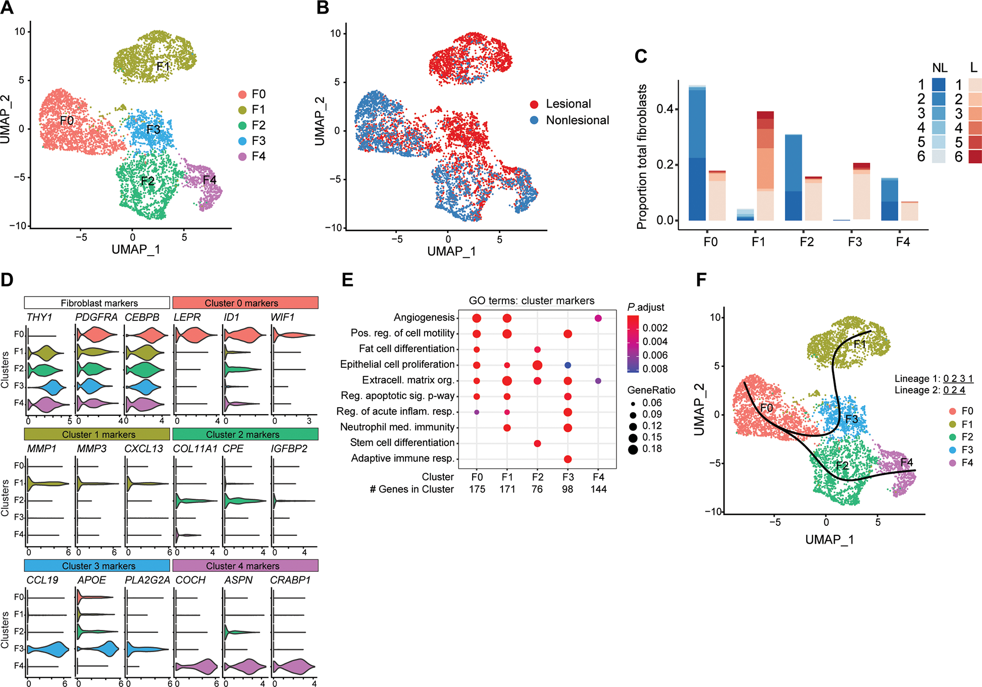
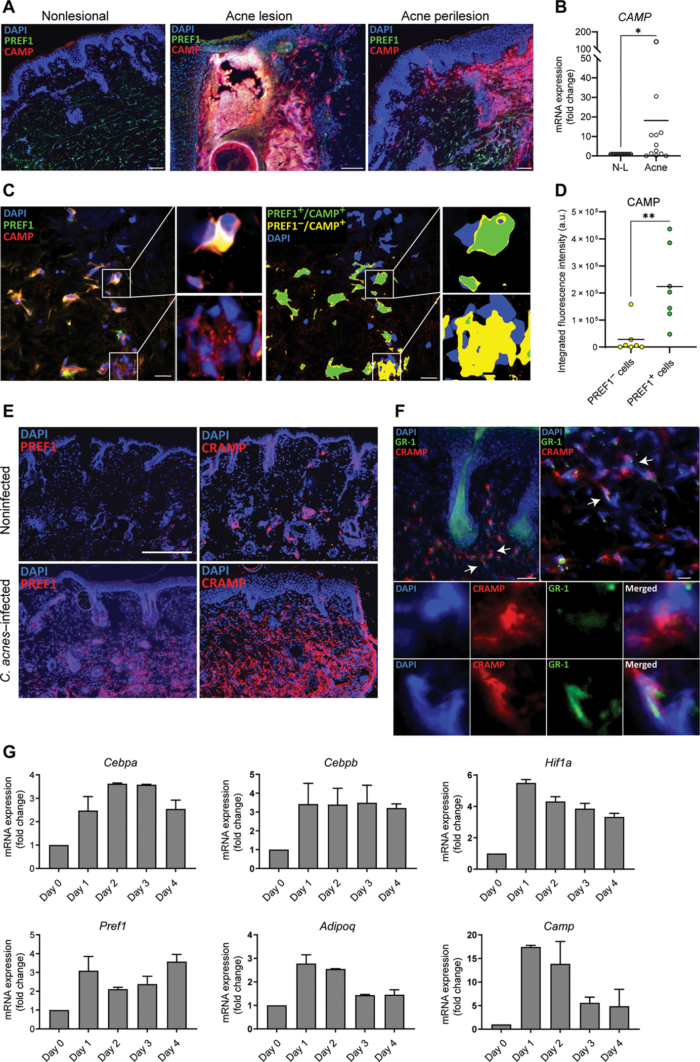
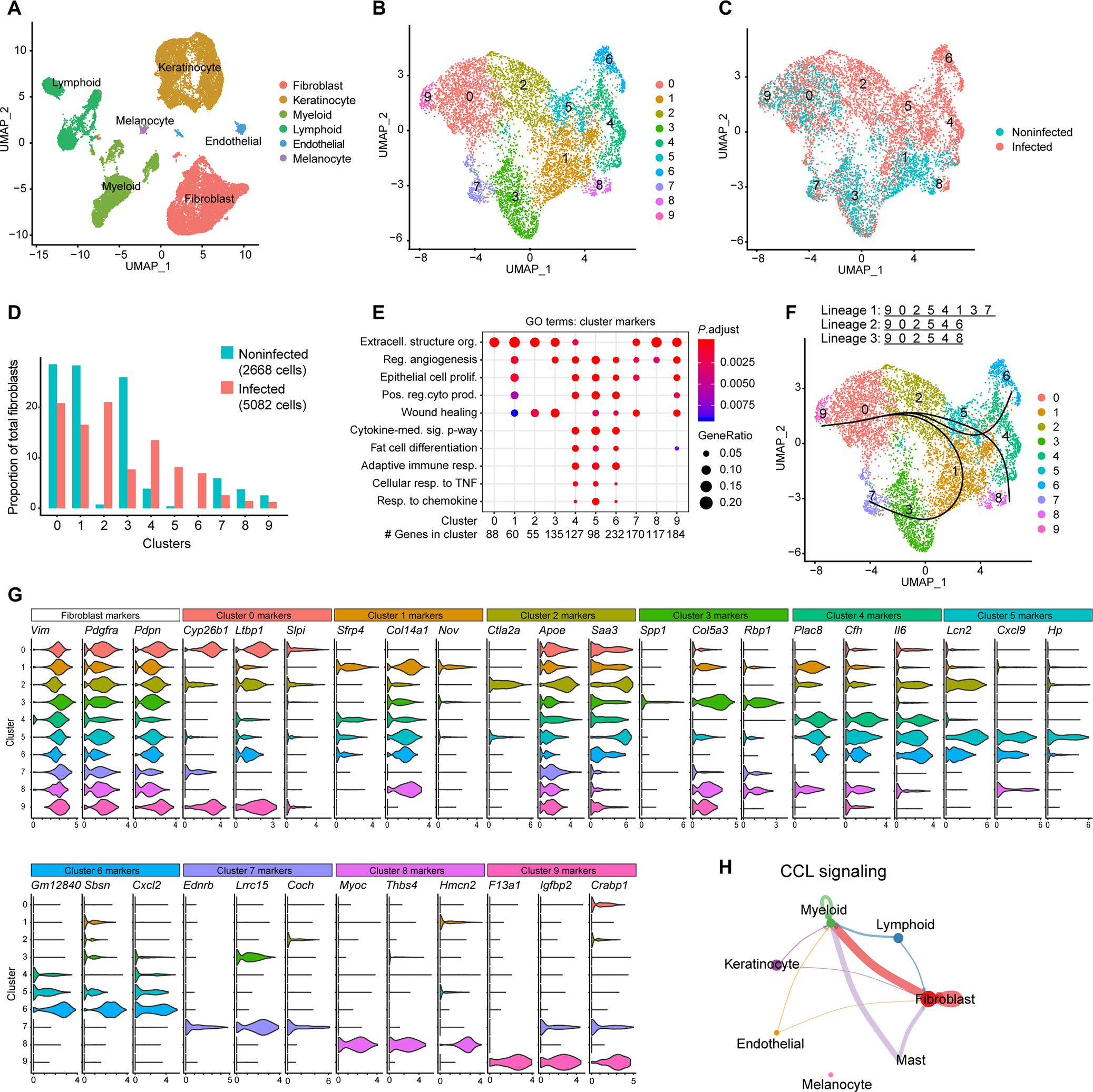
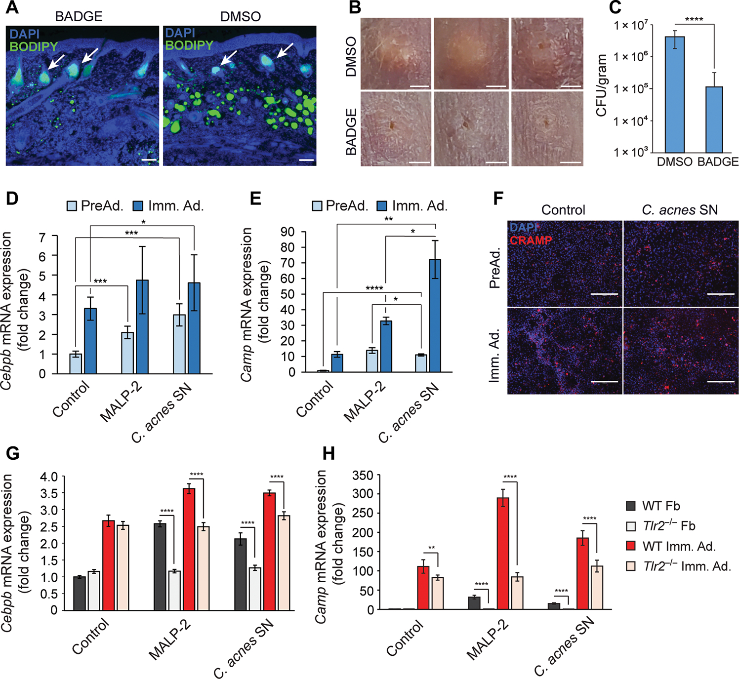
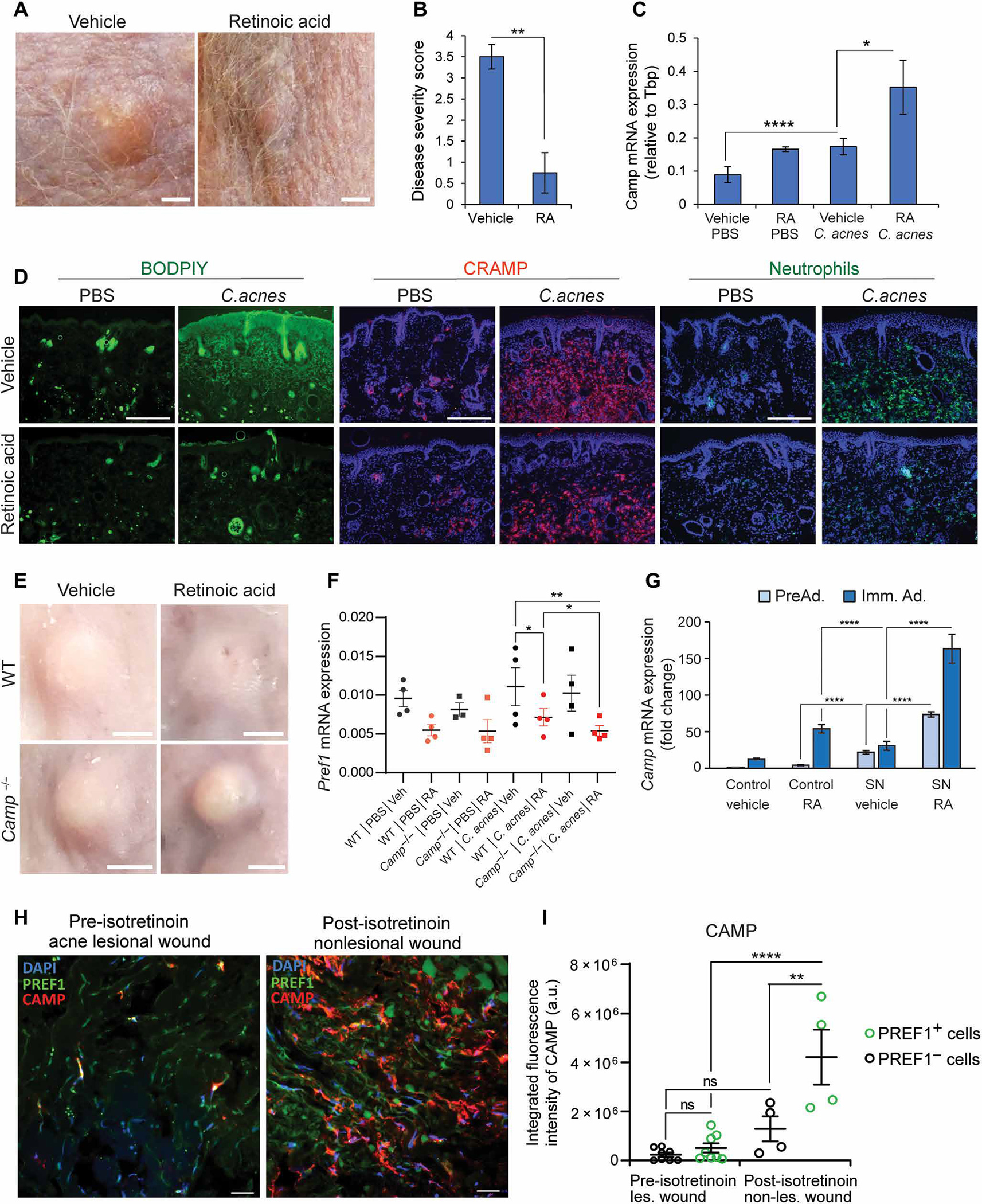
Innate immune defense against deep tissue infection by Staphylococcus aureus is orchestrated by fibroblasts that become antimicrobial when triggered to differentiate into adipocytes. However, the role of this process in noninfectious human diseases is unknown. To investigate the potential role of adipogenesis by dermal fibroblasts in acne, a disorder triggered by Cutibacterium acnes, single-cell RNA sequencing was performed on human acne lesions and mouse skin challenged by C. acnes. A transcriptome consistent with adipogenesis was observed within specific fibroblast subsets from human acne and mouse skin lesions infected with C. acnes. Perifollicular dermal preadipocytes in human acne and mouse skin lesions showed colocalization of PREF1, an early marker of adipogenesis, and cathelicidin (Camp), an antimicrobial peptide. This capacity of C. acnes to specifically trigger production of cathelicidin in preadipocytes was dependent on TLR2. Treatment of wild-type mice with retinoic acid (RA) suppressed the capacity of C. acnes to form acne-like lesions, inhibited adipogenesis, and enhanced cathelicidin expression in preadipocytes, but lesions were unresponsive in Camp-/- mice, despite the anti-adipogenic action of RA. Analysis of inflamed skin of acne patients after retinoid treatment also showed enhanced induction of cathelicidin, a previously unknown beneficial effect of retinoids in difficult-to-treat acne. Overall, these data provide evidence that adipogenic fibroblasts are a critical component of the pathogenesis of acne and represent a potential target for therapy.


Conflict of interest statement

Competing interests: R.L.G. is a cofounder, scientific advisor, and consultant and has equity in MatriSys Bioscience and is a consultant, receives income, and has equity in Sente Inc.

Figures

Fig. 1.
Fig. 1.. scRNA-seq analysis of human acne lesions.
(A) UMAP plot of PDGFRA+ fibroblasts showing five distinct subtypes colored by cluster or (B) by disease association. (C) Stacked bar plot showing the donor and lesion type composition for each of the fibroblast cell types. Lesional cells (L) are shown in red, and nonlesional cells (NL) are shown in blue. (D) Violin plots of conserved pan-fibroblast marker gene expression and expression of major cluster markers for all five fibroblast subsets from the top three differentially expressed genes. (E) Dot plot of selected GO terms across all fibroblast subsets. Circle size corresponds to the proportion of markers annotated to a given term, while the fill color indicates the adjusted P value for the enrichment score. (F) Pseudotime analysis projected onto UMAP plot from (A). Curves represent inferred trajectories.
Fig. 2.
Fig. 2.. Reactive adipogenesis occurs in the perifollicular stroma of acne.
(A) Representative immunofluorescence images from biopsy sections of human acne lesional and nonlesional skin stained for LL-37 (red), PREF1 (green), and 4′,6-diamidino-2-phenylindole (DAPI) (blue). Scale bars, 100 μm for nonlesional and perilesion and 150 μm for lesional skin. (B) RNA was extracted from tissue sections of 12 biopsies of eight acne patients, and CAMP mRNA expression from acne was measured by qPCR and normalized to nonlesional (N-L) skin. (C) Representative confocal microscopy image of the perifollicular dermis of an acne lesion stained for cathelicidin protein (CAMP), PREF1, and DAPI, with higher magnification images provided from the boxed areas. Right panel shows the same representative image after thresholding, with green indicating areas where CAMP and PREF1 overlap and yellow color indicating areas where CAMP does not overlap with PREF1. (D) Quantification of the integrated fluorescence intensity of CAMP in overlapping and nonoverlapping PREF1-positive cells. Each spot represents a 212 μm × 212 μm perifollicular image from the dermis of acne lesions sampled from two patients. a.u., arbitrary units. (E) Representative immunofluorescence images of CAMP and PREF1 staining from skin biopsies of SKH-1 mice that were intradermally infected with C. acnes or noninfected after 7 days. (F) Representative immunofluorescence images of CRAMP, GR-1, and DAPI in the perifollicular area of SKH-1 mouse skin infected with C. acnes. White arrows indicate examples of CAMP-expressing cells that are positive or negative for GR-1 expression, with corresponding higher magnification images provided in the lower panel. Scale bar, 100 μm. (G) C. acnes–infected and noninfected SKH-1 mouse skin biopsies were collected on indicated days, and mRNA expression of the indicated genes was analyzed by qPCR with values shown relative to Gapdh control and normalized to noninfected (day 0) skin. n = 3 mice per condition. Data are means ± SEM. *P < 0.05 and **P < 0.01, two-tailed paired Student’s t test.
Fig. 3.
Fig. 3.. scRNA-seq analysis of C. acnes–infected mouse skin reveals enrichment of adipogenic fibroblast subpopulations.
scRNA-seq was conducted on skin lesions 3 days after C. acnes infection or mock-infected control skin of SKH-1 mice. (A) UMAP plot of all cells passing initial quality control, showing cell type assignment based on established lineage markers. (B) UMAP plot after subclustering of Pdgfra+ fibroblasts, showing 10 distinct subtypes (0 to 9) colored by cluster. (C) Subclustered fibroblasts colored by disease association. (D) Bar chart of the percentage of cells in each fibroblast cluster for C. acnes– and mock-infected skin. (E) Dot plot of selected GO terms across fibroblast clusters. Circle size corresponds to the proportion of markers annotated to a given term, while the fill color indicates the adjusted P value for the enrichment score. (F) Pseudotime analysis projected onto UMAP plot from (A). Curves represent inferred trajectories. (G) Violin plots of conserved pan-fibroblast marker gene expression and expression of major cluster markers for all 10 fibroblast subsets from the top 3 differentially expressed genes. (H) CellChat circle plot of the CCL signaling pathway network. Edge color indicates sender. Edge weight is proportional to strength of communication signal. Circle sizes are proportional to the number of cells per cluster.
Fig. 4.
Fig. 4.. C. acnes triggers adipocyte differentiation and Camp expression in dermal fibroblasts.
(A to C) SKH-1 mice were intraperitoneally injected daily with BADGE or vehicle (DMSO) as a control starting 1 day before intradermal infection with C. acnes. On day 6, skin was collected for analysis (n = 5). (A) Skin was stained with DAPI to stain nuclei or BODIPY to stain for the presence of lipid droplets. White arrows indicate sebaceous glands. Scale bar, 100 μm. (B) Representative images of the PBS control or C. acnes injection sites on mouse back after BADGE or DMSO vehicle treatment. Scale bar, 2 mm. (C) Enumeration of bacterial CFU from infected and mock-infected skin lesions of the infected area. (D and E) 3T3-L1 preadipocytes were cultured as nondifferentiated preadipocytes (PreAd.) or differentiated into immature adipocytes (Imm. Ad.) and stimulated for 24 hours with MALP-2 (100 ng/ml), 2.5% sterile conditioned C. acnes supernatant (SN), or bacterial culture medium (RCM) as control, and the relative mRNA expression of Cebpb (D) or Camp (E) was assessed by real-time qPCR. (F) Undifferentiated 3T3-L1 preadipocytes were stimulated with C. acnes SN for 48 hours, immunostained for CRAMP (red), and counterstained with DAPI (blue). Scale bar, 300 μm. (G and H) Primary dermal fibroblasts (Fb) isolated from wild-type (WT) or TLR2 knockout (Tlr2−/−) mice were stimulated with MALP-2 (100 ng/ml), 2.5% C. acnes SN, or RCM control for 24 hours in the presence or absence of differentiation medium, and the relative mRNA expression of Cebpb (G) or Camp (H) was assessed by real-time qPCR. All data are means ± SD. *P < 0.05, **P < 0.01, ***P < 0.001, and ****P < 0.0001 using Student’s paired t test.
Fig. 5.
Fig. 5.. RA inhibits adipogenesis and enhances cathelicidin in response to C. acnes infection.
SKH-1 mice were intradermally infected with C. acnes on day 0. Starting on day 3, mice were injected subcutaneously at a distal site with retinoic acid (RA) or olive oil (control) for 3 days (n = 4 per group). On day 6, skin was collected for analysis. (A) Representative day 6 images of acne-like lesions in mice treated with vehicle (left) or RA (right). Scale bar, 2 mm. (B) Disease severity score of C. acnes–infected lesions (n = 4) by investigator assessment of lesion severity ranging from 0 (no lesion detected) to 4 (most severe). (C) Camp mRNA expression in C. acnes–infected or mock PBS–infected mouse skin treated with RA or vehicle control (n = 4 mice). (D) Fluorescent microscopy images of C. acnes–infected and mock PBS–infected mouse skin (n = 4), showing representative staining of total skin lipids (BODIPY dye, green), or immunolabeling of CRAMP (red) or neutrophils (anti–GR-1, green) for each treatment group (RA versus vehicle, n = 4 sections). All sections were counterstained with DAPI. Scale bar, 300 μm. (E and F) WT or Camp−/− C57BL/6 mice were intradermally infected with C. acnes and treated identically to SKH-1 mice listed above, and findings were assayed on day 6. (E) Representative day 6 images of acne-like lesions from WT and Camp−/− mice treated with vehicle (oil) or RA after injection with PBS control or infected with C. acnes (representative of 8 lesions, n = 4 mice per group, n = 2 lesions per mouse). Scale bar, 2 mm. (F) C. acnes lesions and control skin were collected on day 6, and mRNA was measured by qPCR for expression of Pref1. (G) 3T3-L1 preadipocytes were cultured as nondifferentiated preadipocytes (PreAd.) or differentiated into immature adipocytes (Imm. Ad.) and stimulated with 2.5% C. acnes SN or RCM control with or without 1 μM RA or vehicle (ethanol) controls for 24 hours, and the relative mRNA expression of Camp was assessed by qPCR. (H) Representative confocal images of dermal preadipocytes in 24-hour wounds from acne patients before and after isotretinoin therapy. Sections were stained for PREF1, CAMP, and DAPI, and thresholding was conducted on regions of CAMP staining with overlap of PREF1 and regions of CAMP staining without overlap of PREF1. Scale bar, 20 μm. (I) Quantification of the integrated fluorescence intensity of CAMP in PREF1-positive cells and PREF1-negative cells before and after isotretinoin treatment. Each spot represents a 212 μm × 212 μm perifollicular dermal image from acne sections from two acne patients. Data are means ± SEM. *P < 0.05, **P < 0.01, ***P < 0.001, and ****P < 0.0001 using Student’s paired t test. Data in (I) represent means ± SEM with ordinary one-way ANOVA with Tukey’s multiple comparisons test.

References

    1. Sanford JA, O’Neill AM, Zouboulis CC, Gallo RL, Short-chain fatty acids from Cutibacterium acnesActivate both a canonical and epigenetic inflammatory response in human sebocytes. J. Immunol. 202, 1767–1776 (2019). - PMC - PubMed
    1. O’Neill AM, Gallo RL, Host-microbiome interactions and recent progress into understanding the biology of acne vulgaris. Microbiome 6, 177 (2018). - PMC - PubMed
    1. Chen B, Li P, Li J, Chen J, Putative genes and pathways involved in the acne treatment of isotretinoin via microarray data analyses. Biomed. Res. Int. 2020, 1–14 (2020). - PMC - PubMed
    1. Laughlin JM, Watterson S, Layton AM, Bjourson AJ, Barnard E, Dowell AM, Propionibacterium acnes and acne vulgaris: New insights from the integration of population genetic, multi-omic, biochemical and host-microbe studies. Microorganisms 7, 128 (2019). - PMC - PubMed
    1. Kovács D, Lovászi M, Póliska S, Oláh A, Bíró T, Veres I, Zouboulis CC, Ståhle M, Rühl R, Remenyik É, Törőcsik D, Sebocytes differentially express and secrete adipokines. Exp. Dermatol. 25, 194–199 (2016). - PubMed

Publication types