. 2022 Feb 18;8(7):eabk3139.
doi: 10.1126/sciadv.abk3139. Epub 2022 Feb 16.

2.4-Å structure of the double-ring Gemmatimonas phototrophica photosystem

Affiliations

2.4-Å structure of the double-ring Gemmatimonas phototrophica photosystem

Pu Qian et al. Sci Adv. .

Abstract

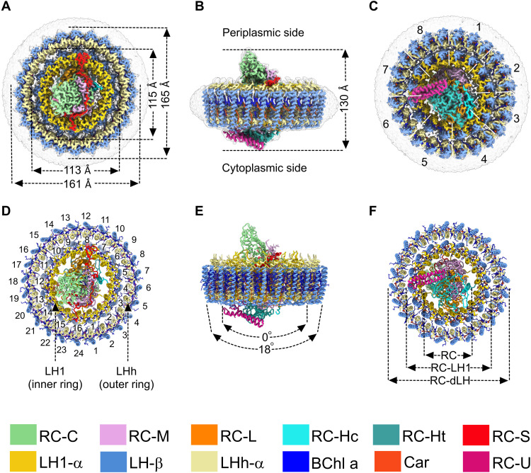
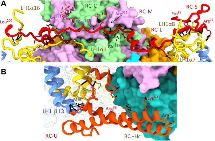
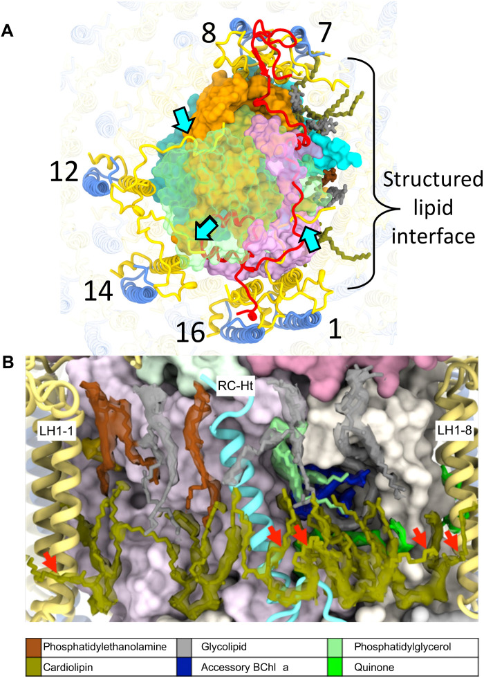
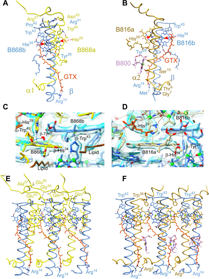
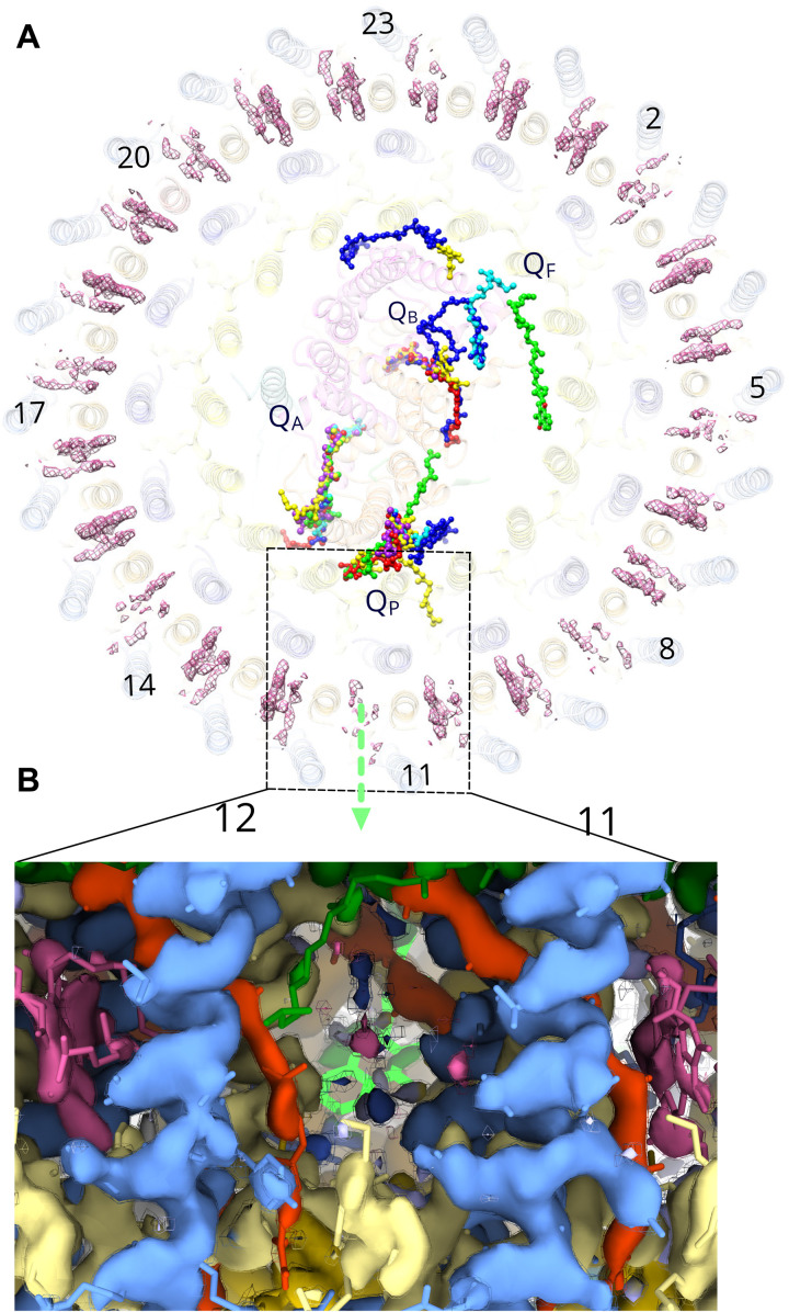
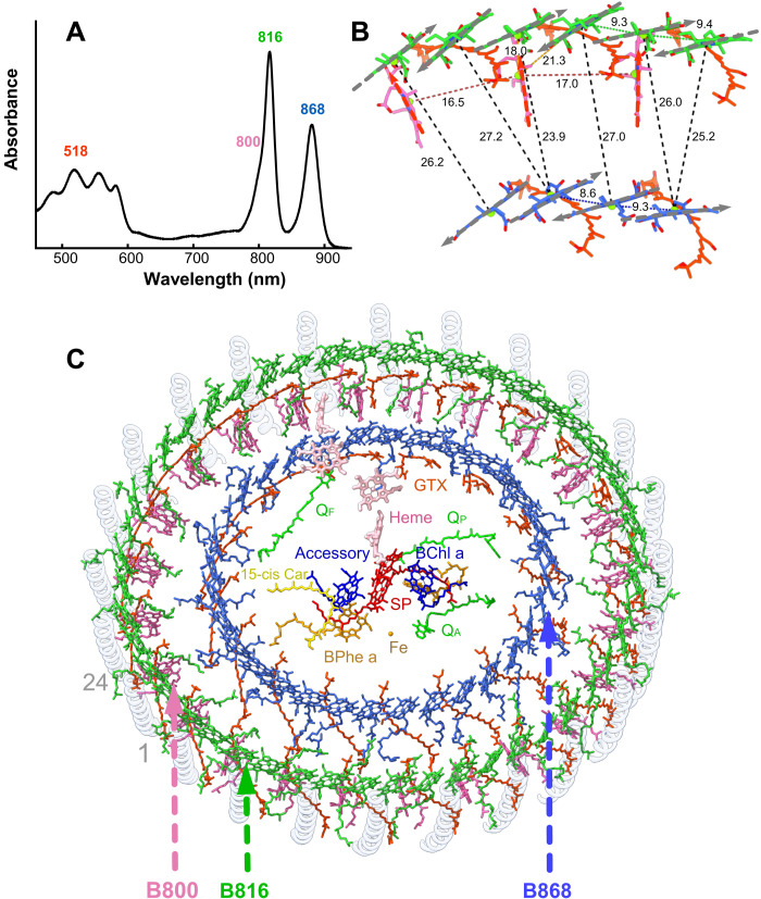
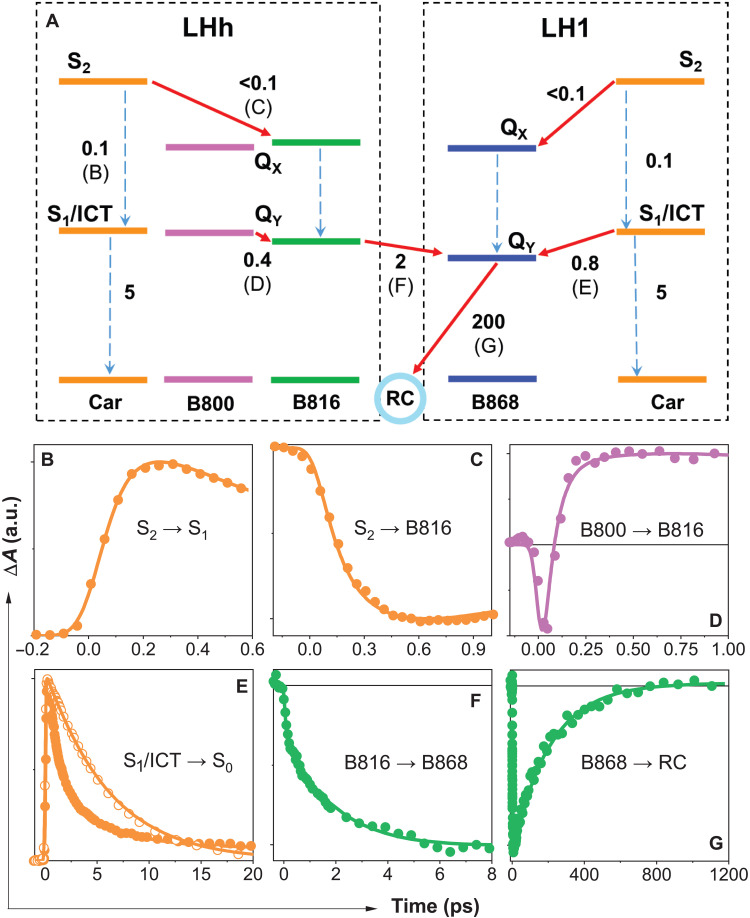
Phototrophic Gemmatimonadetes evolved the ability to use solar energy following horizontal transfer of photosynthesis-related genes from an ancient phototrophic proteobacterium. The electron cryo-microscopy structure of the Gemmatimonas phototrophica photosystem at 2.4 Å reveals a unique, double-ring complex. Two unique membrane-extrinsic polypeptides, RC-S and RC-U, hold the central type 2 reaction center (RC) within an inner 16-subunit light-harvesting 1 (LH1) ring, which is encircled by an outer 24-subunit antenna ring (LHh) that adds light-gathering capacity. Femtosecond kinetics reveal the flow of energy within the RC-dLH complex, from the outer LHh ring to LH1 and then to the RC. This structural and functional study shows that G. phototrophica has independently evolved its own compact, robust, and highly effective architecture for harvesting and trapping solar energy.


Figures

Fig. 1.
Fig. 1.. Cryo-EM structure of the RC-dLH complex from G. phototrophica.
(A to C) Views of the color-coded RC-dLH density map. Detergent and other disordered molecules are in gray. (A) Perpendicular view from the periplasmic side of the membrane, with the diameters of the two LH rings indicated. (B) View within the plane of the membrane showing the overall height of the complex. (C) Perpendicular view from the cytoplasmic side. White dashed lines indicate pseudo-C8 symmetry of the double LHh-LH1 ring. (D to F) Ribbon models corresponding to (A) to (C). In (D), the LH1 and LHh subunits are numbered in black. (E) The two arrows indicate the degree of curvature of the outer and inner LH rings. (F) Concentric nature of the complex proceeding outward from the central RC. The color code is shown below the panels. Images were produced using ChimeraX (15).
Fig. 2.
Fig. 2.. Protein-protein interactions between the LH1 and RC complexes.
(A) The RC-S polypeptide occupies a deep groove spanning the periplasmic surface of the RC complex and involves residues from RC-C, RC-M, and RC-L. Its N- and C-terminal ends engage the C termini of LH1 α7 and α16 chains, respectively. Intermolecule H-bonds involving RC-S are shown in black. (B) RC-U forms a short coiled-coil spanning between the cytoplasmic surface of RC-Hc and LH1 subunit 13, with a largely hydrophobic N-terminal protrusion buried deep in the transmembrane space.
Fig. 3.
Fig. 3.. Stabilization of RC-LH1 complex.
(A) View of the periplasmic face of the RC-dLH complex, showing interactions (cyan arrows) between the C termini of LH1-α1, 12, and 14 (yellow) and the RC-M and RC-C subunits (RC-C subunit is shown in translucent green). The region surrounding RC-Ht (covering the QA and spirilloxanthin sites) is dominated by a tightly packed lipid interface. On the cytoplasmic side, six cardiolipins (olive) interdigitate between RC and LH1 chains. The periplasmic interface consists of three uncharged glyceroglycolipids (gray), two phosphatidylethanolamines (brown), and a single phosphatidylglycerol (pale green). (B) View of the structured lipid interface in (A) with a 6-σ density map around the lipids. Outward-projecting cardiolipin tails are marked with red arrows.
Fig. 4.
Fig. 4.. Protein-protein and protein-pigment interactions within the two rings of light-harvesting antenna complexes.
(A) Key interactions involved in stabilizing a single α/β subunit of the 16-member LH1 ring. Histidine residues that are involved in coordination with central Mg atom of the BChls are indicated by red arrows. (B) Single α/β subunit of the 24-member LHh ring. (C) Interaction between BChl a molecules and protein in LH1 subunit. The density map is shown in blue. (D) Interaction between BChl a molecules and protein in the LHh subunit. (E) Three consecutive subunits of the LH1 ring. Interfaces between subunits are primarily mediated by the bound pigments, with few specific protein-protein interactions. (F) Three consecutive subunits of the outer LHh ring, showing H-bonds between the C-terminal domains in three LHh α/β units. GTX, gemmatoxanthin. Colors as in Fig. 1 with the exception of B816 and B868 BChls, which are colored by chain, and B800 is colored pink.
Fig. 5.
Fig. 5.. The QP quinol export site in the RC-dLH complex.
(A) The RC-dLH complex viewed from the cytoplasmic side of the membrane, with all details partly obscured, and showing the densities for B800 BChls (pink) in the outer LHh ring. Structurally resolved quinones for the G. phototrophica complex are shown (green), with those for the RC-LH1 complexes from Rhodopseudomonas palustris W-minus (16) (blue), R. palustris W-plus (16) (cyan), T. tepidum (11) (yellow), Thiorhodovibrio stain 970 (17) (purple), and Blastochloris viridis (9) (red). The green arrow indicates a likely path for quinol export, at a position of weak B800 density. The positions of LHh subunit are numbered. (B) Two LHh α/β units 11 and 12 viewed in the plane of the membrane showing the proposed position for quinol export that corresponds to a low or zero occupancy B800 site; LHh-α (pale yellow), LHh-β (cornflower blue), gemmatoxanthin (orange red), B800 (pink), B816 (dark green), and QP (bright green). The cryo-EM map is shown at two contour levels (opaque surface = 7.0σ; wireframe = 7.5σ).
Fig. 6.
Fig. 6.. Structural and kinetic basis for the ET network in RC-dLH.
(A) The 77K absorption spectrum of the RC-dLH complex, colored according to the approximate absorption ranges of carotenoids (orange) and the B800 (pink), B816 (green), and B868 (blue) BChls. (B) Relative positions of BChl a molecules in the inner and outer rings for a unit of pseudo-C8 symmetry. Gray arrows indicate QY dipole moments of the BChls. The proteins have been removed for clarity. (C) Outer B800-B816 and inner B868 rings of BChls, and gemmatoxanthin (GTX, red orange), enclosing the central RC pigments. Other colors as in Figs. 1 and 2.
Fig. 7.
Fig. 7.. ET kinetics in the RC-dLH1 complex.
(A) The two boxes (black dashed lines) map the ET events in the outer LHh and inner LH1 antenna rings. Solid red arrows represent ET channels; dashed arrows correspond to internal conversion processes. A time constant (in picoseconds) is associated with each process. The letters B to G associated with ET/internal conversion processes in the scheme correspond to the kinetics shown in (B) to (G), which monitor the dynamics of these processes. Color of each kinetic corresponds to excitation of carotenoids (orange), B800 (purple), and B816 (green). The probing wavelengths are within the bands corresponding to the gemmatoxanthin S1/ICT state (620 nm; B and E), B816 bleaching (820 nm; C), B816 excited-state absorption (805 nm; D), and B868 bleaching (880 nm; F and G). The kinetics shown as open symbols in (E) corresponds to the S1/ICT lifetime of gemmatoxanthin in methanol. Full transient absorption spectra showing the spectral bands and a detailed description are shown in fig. S13. a.u., arbitrary units.

References

    1. R. E. Blankenship, Molecular Mechanisms of Photosynthesis (John Wiley & Sons, ed. 2, 2014), pp. 312.
    1. Hohmann-Marriott M. F., Blankenship R. E., Evolution of photosynthesis. Annu. Rev. Plant Biol. 62, 515–548 (2011). - PubMed
    1. Martin W. F., Bryant D. A., Beatty J. T., A physiological perspective on the origin and evolution of photosynthesis. FEMS Microbiol. Rev. 42, 205–232 (2018). - PMC - PubMed
    1. Sener M., Strumpfer J., Abhishek S., Hunter C. N., Schulten K., Overall energy conversion efficiency of a photosynthetic vesicle. eLife 5, e09541 (2016). - PMC - PubMed
    1. Bryant D. A., Costas A. M., Maresca J. A., Chew A. G., Klatt C. G., Bateson M. M., Tallon L. J., Hostetler J., Nelson W. C., Heidelberg J. F., Ward D. M., Candidatus Chloracidobacterium thermophilum: An aerobic phototrophic Acidobacterium. Science 317, 523–526 (2007). - PubMed