. 2022 Feb 18;8(7):eabe4375.
doi: 10.1126/sciadv.abe4375. Epub 2022 Feb 16.

A balanced Oct4 interactome is crucial for maintaining pluripotency

Affiliations

A balanced Oct4 interactome is crucial for maintaining pluripotency

Dong Han et al. Sci Adv. .

Abstract

Oct4 collaborates primarily with other transcriptional factors or coregulators to maintain pluripotency. However, how Oct4 exerts its function is still unclear. Here, we show that the Oct4 linker interface mediates competing yet balanced Oct4 protein interactions that are crucial for maintaining pluripotency. Oct4 linker mutant embryonic stem cells (ESCs) show decreased expression of self-renewal genes and increased expression of differentiation genes, resulting in impaired ESC self-renewal and early embryonic development. The linker mutation interrupts the balanced Oct4 interactome. In mutant ESCs, the interaction between Oct4 and Klf5 is decreased. In contrast, interactions between Oct4 and Cbx1, Ctr9, and Cdc73 are increased, disrupting the epigenetic state of ESCs. Control of the expression level of Klf5, Cbx1, or Cdc73 rebalances the Oct4 interactome and rescues the pluripotency of linker mutant ESCs, indicating that such factors interact with Oct4 competitively. Thus, we provide previously unidentified molecular insights into how Oct4 maintains pluripotency.


Figures
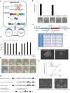
Fig. 1.
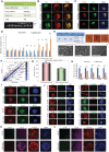
Fig. 1.. Oct4 linker is crucial for maintaining pluripotency in vitro.
(A) Schematic of complementation assay system. In ZHBTc4 ESCs, both Oct4 alleles have been disrupted, and a Dox-suppressible transgene Oct4 maintains ESC pluripotency. The WT or linker mutant Oct4 was transfected into ZHBTc4 ESCs by a lentivirus overexpression system. (B) Rescue index of WT Oct4 and chimeric Oct4 with different species linker orthologs in the presence of Dox and AP staining of ZHBTc4 ESC colonies rescued by different Oct4 proteins. (C) Representation of mouse Oct4 protein (WT) and different alanine mutations on the linker segment. N-terminal transactivation domain (TAD), POU-specific domain, linker, POU homeodomain, and C-terminal TAD are shown in different colors. Amino acids that are replaced are colored in red. (D) Rescue index of WT Oct4 and mutant Oct4 with different alanine mutations on the linker segment. (E) AP staining of ZHBTc4 ESCs colonies rescued by different Oct4 mutants. (F) Morphology of WT and L80A Oct4–rescued ZHBTc4 ESC colony. (G) Proliferation rate of WT, L80A Oct4–rescued ZHBTc4 ESCs, and ZHBTc4 ESCs cultured without or with Dox. *** represents P value between WT and L80A Oct4–rescued ZHBTc4 ESCs. (H) Strategy of WT and L80A Oct4–KI ZHBTc4 ESCs expressing FLPe. Black boxes represent Oct4 exons. Star represents the position of L80 amino acid in the linker region. FLPe, FLP recombinase; SA, splice acceptor; BSD, blasticidin; Hyg, hygromycin; Puro, puromycin. (I) Morphology of L80A and WT Oct4–KI ZHBTc4 cells cultured with Dox.
Fig. 2.
Fig. 2.. Oct4 linker is crucial for maintaining pluripotency in preimplantation embryos.
(A) Number of pups with different genotypes from intercross matings of WT/L80A Oct4 mice and representative genotyping gel picture. (B) Immunocytochemistry on ~E3.5 blastocysts using anti-Oct4 antibody and confocal microscopy. Scale bar, 30 μm. (C) Immunocytochemistry on ~E4.5 embryos using anti-Oct4 and anti-Gata6 antibodies and confocal microscopy. Scale bar, 30 μm. (D) RT-qPCR gene expression analysis of E4.5 embryos. All data are calibrated to the WT/WT embryos, whose expression is considered to be 1 for all genes. (E) Derivation of ESC line with different genotypes from E4.5 embryos. Upright pictures show outgrowths from embryos. Scale bar, 50 μm. Landscape pictures below show established ESCs lines with different genotypes. (F) Pairwise scatterplot analysis of the global gene expression profiles of L80A/L80A and WT/WT Oct4 ESCs obtained from the microarray analysis. (G) Bar plot of the number of differentially expressed genes between WT/WT and L80A/L80A Oct4 ESCs. (H) Relative mRNA expression of self-renewal–related genes in WT/WT and L80A/L80A Oct4 ESC lines. (I and J) Immunofluorescence microscopy images of WT/WT and L80A/L80A Oct4 ESCs using antibodies against Oct4, Sox2, and Nanog (I) and Oct4, Cdx2, Eomes, Gata3, Gata6, Nestin, and Sox17 (J). Scale bars, 50 μm. (K and L) Immunofluorescence microscopy images of WT/WT and L80A/L80A Oct4 ESCs expressing Cdx2-ERT2-RFP cultured under the TSC culture condition without (K) or with (L) tamoxifen for 3 weeks using antibodies against Oct4, Cdx2, and Troma1. Scale bars, 50 μm. DAPI, 4′,6-diamidino-2-phenylindole.
Fig. 3.
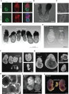
Fig. 3.. Effect of L80A Oct4 on postimplantation embryo development.
(A) Immunocytochemistry on ~E5.5 embryos using anti-Oct4 and anti-Cdx2 antibodies and confocal microscopy. Scale bars, 50 μm. (B) Morphology of embryos from intercross matings of WT/L80A Oct4 mice on ~E6.5. (C) Histological sections of ~E6.5 embryos from intercross matings of WT/L80A Oct4 mice showed growth defects in L80A/L80A Oct4 embryos. (D and E) Morphology of embryos from intercross matings of WT/L80A Oct4 mice on ~E7.75. (F and G) Morphology of embryos from intercross matings of WT/L80A Oct4 mice on ~E9.5 (F) and ~E9.75 (G). (H and I) Morphology of embryos with placentas from intercross matings of WT/L80A Oct4 mice on ~E12.5. (J) Morphology of embryos from intercross matings of WT/L80A Oct4 mice on ~E13.5.
Fig. 4.
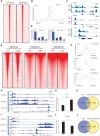
Fig. 4.. Genomic binding and transactivation ability of L80A Oct4 are similar to those of WT Oct4 in ESCs, but L80A mutation interrupts the balanced Oct4 interactome.
(A) Heatmaps of Oct4 ChIP-seq signals in WT and L80A Oct4 ESCs. (B) Representative Oct4 ChIP-seq tracks showing similar occupancy profiles of WT and L80A Oct4. (C) Oct4 ChIP-qPCR assay of WT and L80A Oct4 ESCs. Occupancy relative to a negative control region is shown. Error bars represent SD of biological triplicates. (D) The sox-oct motif is significantly enriched at the top 1000 WT and L80A Oct4 target sites. (E) The relative transcriptional activity levels of WT and L80A Oct4 were measured by luciferase reporter assay in WT/WT and L80A/L80A Oct4 ESCs. (F) Comparison of our Oct4 interactome with the three published Oct4 network studies. Overlapping proteins in different published datasets and novel proteins found in this study are listed in different color boxes. (G) Co-IP and Western blot (WB) showed decreased Oct4-Klf5 interaction and increased Oct4-Cbx1, Oct4-Ctr9, and Oct4-Cdc73 interactions in L80A Oct4 ESCs.
Fig. 5.
Fig. 5.. Chromatin accessibility and H3K4me3 level are not affected, but H3K36me and H3K9me3 levels are increased in L80A/L80A Oct4 ESCs.
(A) Heatmaps of ATAC-seq signals in WT and L80A Oct4 ESCs. (B) Average signal intensity of ATAC-seq in WT and L80A Oct4 ESCs. (C) Representative ATAC-seq tracks showing similar peak profile in WT and L80A Oct4 ESCs. (D) Genomic distribution of ATAC-seq signal in WT and L80A Oct4 ESCs. (E) Heatmaps of H3K4me3, H3K36me3, and H3K9me3 ChIP-seq profiles in WT and L80A Oct4 ESCs. (F) Average signal intensity of H3K4me3, H3K36me3, and H3K9me3 ChIP-seq in WT and L80A Oct4 ESCs. (G) H3K4me3, H3K36me3, and H3K9me3 ChIP-seq profiles in Tead4 locus. Higher H3K36me3 peak signal in L80A Oct4 ESCs is shown in red box. (H) Relative expression level of Tead4 in WT and L80A Oct4 ESCs from microarray data. Error bars represent SD of biological duplicates. (I) H3K4me3, H3K36me3, and H3K9me3 ChIP-seq profiles in Epha2 locus. Higher H3K9me3 peak signal in L80A ESCs is shown in red box. (J) Relative expression level of Epha2 in WT and L80A Oct4 ESCs from microarray data. Error bars represent SD of biological duplicates. (K and L) Venn diagram of Oct4 target genes overlapping with those genes that have higher H3K36me3 level (K) or H3K9me3 level (L) in L80A Oct4 ESCs.
Fig. 6.
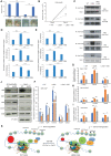
Fig. 6.. Overexpression of Klf5 rescues the phenotype of L80A Oct4 ZHBTc4 ESCs by rebalancing the Oct4 interactome.
(A) Overexpression of Klf5 increased the L80A Oct4 rescue efficiency in ZHBTc4 ESCs. Bottom: AP-positive colonies rescued by WT, L80A Oct4, and L80A Oct4 + Klf5. (B) Proliferation rate of WT, L80A Oct4–, and L80A Oct4 + Klf5–rescued ZHBTc4 ESCs. *** represents P values between L80A Oct4 and L80A Oct4 + Klf5–rescued ZHBTc4 ESCs. (C) Overexpression of Klf5 rebalanced L80A Oct4 interactions with Klf5, Cbx1, Ctr9, and Cdc73, as shown by Western blot after Co-IP with FLAG antibody. (D and E) ChIP-qPCR analysis of H3K36me3 (D) and H3K9me3 (E) in WT, L80A Oct4–, and L80A Oct4 + Klf5–rescued ZHBTc4 ESCs. (F to I) Klf5 (F), Cbx1 (G), Cdc73 (H), and Ctr9 (I) ChIP-qPCR assay of WT, L80A Oct4–, and L80A Oct4 + Klf5–rescued ZHBTc4 ESCs. Occupancy relative to a negative control region is shown. (J) Knockdown of Klf5 changed the interactions between WT Oct4 and Klf5, Cbx1, Ctr9, and Cdc73, as shown by Western blot after Co-IP with FLAG antibody. (K) Schematic diagram of the Oct4 linker interface, which mediates competing yet balanced Oct4-protein interactions. L80A Oct4 recruits less Klf5 but more Cbx1, Ctr9, and Cdc73 to Oct4 downstream genomic regions. The decreased Klf5 DNA binding and increased DNA binding of Cbx1, Ctr9, and Cdc73 result in dysregulated gene expression and mediate abnormal epigenetic changes at those specific loci.

References

    1. Li M., Belmonte J. C., Ground rules of the pluripotency gene regulatory network. Nat. Rev. Genet. 18, 180–191 (2017). - PubMed
    1. Schöler H. R., Balling R., Hatzopoulos A. K., Suzuki N., Gruss P., Octamer binding proteins confer transcriptional activity in early mouse embryogenesis. EMBO J. 8, 2551–2557 (1989). - PMC - PubMed
    1. Rosner M. H., Vigano M. A., Ozato K., Timmons P. M., Poirier F., Rigby P. W., Staudt L. M., A POU-domain transcription factor in early stem cells and germ cells of the mammalian embryo. Nature 345, 686–692 (1990). - PubMed
    1. Nichols J., Zevnik B., Anastassiadis K., Niwa H., Klewe-Nebenius D., Chambers I., Schöler H., Smith A., Formation of pluripotent stem cells in the mammalian embryo depends on the POU transcription factor Oct4. Cell 95, 379–391 (1998). - PubMed
    1. Niwa H., Miyazaki J., Smith A. G., Quantitative expression of Oct-3/4 defines differentiation, dedifferentiation or self-renewal of ES cells. Nat. Genet. 24, 372–376 (2000). - PubMed