. 2022 Mar 1;38(9):110428.
doi: 10.1016/j.celrep.2022.110428. Epub 2022 Feb 7.

Cryo-EM structure of the SARS-CoV-2 Omicron spike

Affiliations

Cryo-EM structure of the SARS-CoV-2 Omicron spike

Gabriele Cerutti et al. Cell Rep. .

Abstract

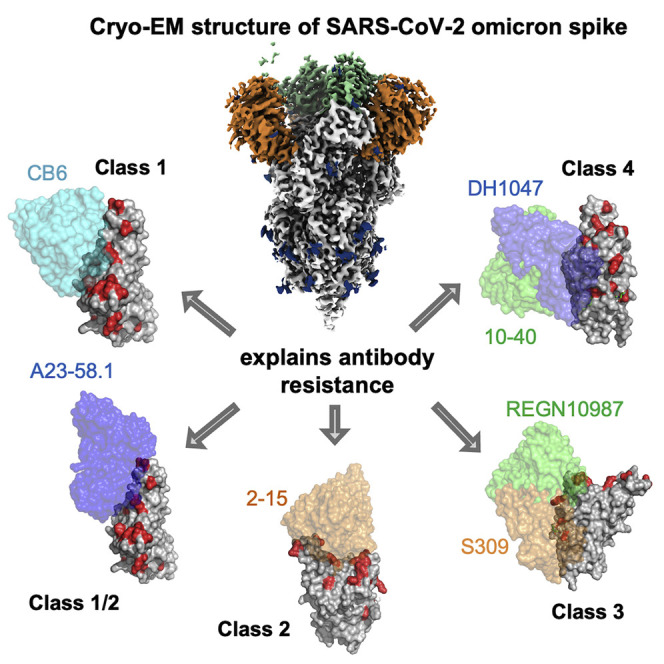
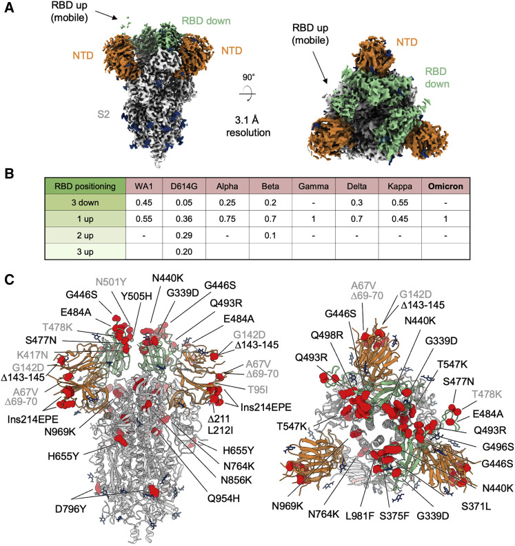
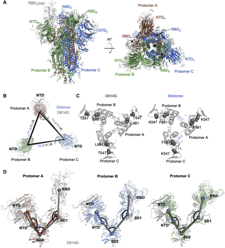
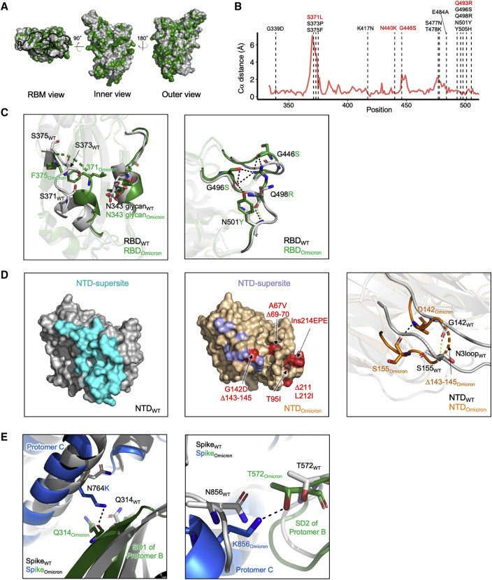
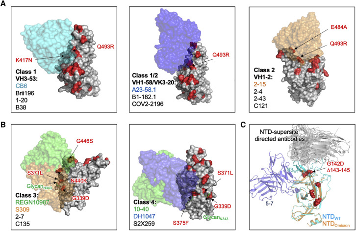
The recently reported B.1.1.529 Omicron variant of severe acute respiratory syndrome coronavirus-2 (SARS-CoV-2) includes 34 mutations in the spike protein relative to the Wuhan strain, including 15 mutations in the receptor-binding domain (RBD). Functional studies have shown Omicron to substantially escape the activity of many SARS-CoV-2-neutralizing antibodies. Here, we report a 3.1 Å-resolution cryoelectron microscopy (cryo-EM) structure of the Omicron spike protein ectodomain. The structure depicts a spike that is exclusively in the 1-RBD-up conformation with high mobility of RBD. Many mutations cause steric clashes and/or altered interactions at antibody-binding surfaces, whereas others mediate changes of the spike structure in local regions to interfere with antibody recognition. Overall, the structure of the Omicron spike reveals how mutations alter its conformation and explains its extraordinary ability to evade neutralizing antibodies.

Keywords: B.1.1.529; COVID-19; NTD; Omicron; RBD; SARS-CoV-2; cryo-EM; neutralizing antibody; spike; variant of concern.


Conflict of interest statement

Declaration of interests The authors declare no competing interests.

Figures

None
Graphical abstract
Figure 1
Figure 1
Cryo-EM structure of prefusion SARS-CoV-2 Omicron (B.1.1.529) spike (A) Cryo-EM map of SARS-CoV-2 Omicron S2P spike in the prefusion state shown in two orthogonal views. The density for the single RBD up is barely visible at the optimal contour level due to high mobility of the domain. NTD is colored in orange, RBD in green, glycans in blue, the rest of the trimer in gray. (B) Relative population of RBD states observed in cryo-EM structures of SARS-CoV-2 spike for different variants. (C) Structure of SARS-CoV-2 Omicron spike in the 1 RBD-up state with mutations highlighted in red. Mutations observed in previous variants are labeled in gray, new Omicron mutations are labeled in black. See also Figures S1, S2, and S3; Tables S1 and S2.
Figure 2
Figure 2
Structural comparison of SARS-CoV-2 Omicron spike with D614G WT (A) Superposition of Omicron spike with D614G spike. The S2 subunit is used for superimposition. (B) Distance between NTDs of Omicron spike and D614G spike. (C) The inter-protomer distance between S2 helices in Omicron is shorter than that observed in D614G spike. (D) Measured angles between NTD, NTD′, SD2, SD1, and RBD showed that protomer A with an up-RBD has altered angles between NTD′, SD2, and SD1. The two protomers with RBDs down show similar domain orientation. Thus, only angles of the Omicron spike are shown. See also Table S3.
Figure 3
Figure 3
Omicron spike mutations alter local conformation and polar interaction pattern (A) Superposition of Omicron RBD with WT RBD. The Omicron RBD is colored in green and WT RBD in gray. The dark line shows the footprint of the RBM. (B) Per-residue Ca distance between Omicron and WT RBD. RBD mutations in Omicron are labeled. The Omicron-specific mutations labeled in red resulted in dramatic and broad antibody neutralization resistance. (C) Details of conformation changes in Omicron RBDs. Left: 367–375 conformation change. Right: 444–448 loop conformation change. Black and yellow dashed lines show hydrogen bonds in Omicron and WT RBDs respectively. (D) Comparison of Omicron and WT NTD. Left: structure for WT NTD with the NTD supersite colored cyan. Middle: structure for Omicron NTD; the blue residues show the NTD supersite on Omicron, the red residues labeled the Omicron mutations. Right: details of N3 loop change in Omicron compared with WT. (E) S2 mutations N764K and N856K in Omicron form new polar interactions with SD2 and SD1 from adjacent protomers respectively. See also Figure S4.
Figure 4
Figure 4
Structural basis of neutralizing antibody escape by Omicron (A) Surface diagram of class 1 and 2 antibodies bound to RBD. Left: VH3-53-derived antibodies with CB6 as an example (PDB: 7C01). Middle: VH1-58/VK3-20-derived antibodies with A23-58.1 (PDB: 7LRS)-binding mode shown. Right: VH1-2-derived antibody with 2-15 (PDB: 7L57)-binding mode shown. Mutations in Omicron RBD are colored in red. (B) Surface diagram of the class 3 and 4 antibodies bound to Omicron RBD, antibody REGN10987 (PDB: 6XDG), S309 (PDB: 6WPT), 10–40 (PDB: 7SD5), and DH1047 (PDB: 7LD1) are shown. (C) Cartoon diagrams of the Omicron and WT NTDs in complex with antigenic supersite-directed antibodies and 5-7 (PDB: 7RW2). Omicron mutations are shown as red spheres. See also Figure S5.

References

    1. Adams P.D., Afonine P.V., Bunkoczi G., Chen V.B., Davis I.W., Echols N., Headd J.J., Hung L.W., Kapral G.J., Grosse-Kunstleve R.W., et al. PHENIX: a comprehensive Python-based system for macromolecular structure solution. Acta Crystallogr. D Biol. Crystallogr. 2010;66:213–221. - PMC - PubMed
    1. Amanat F., Thapa M., Lei T., Ahmed S.M.S., Adelsberg D.C., Carreno J.M., Strohmeier S., Schmitz A.J., Zafar S., Zhou J.Q., et al. SARS-CoV-2 mRNA vaccination induces functionally diverse antibodies to NTD, RBD, and S2. Cell. 2021;184:3936–3948 e3910. - PMC - PubMed
    1. Andreano E., Paciello I., Piccini G., Manganaro N., Pileri P., Hyseni I., Leonardi M., Pantano E., Abbiento V., Benincasa L., et al. Hybrid immunity improves B cells and antibodies against SARS-CoV-2 variants. Nature. 2021;600:530–535. - PMC - PubMed
    1. Barad B.A., Echols N., Wang R.Y., Cheng Y., DiMaio F., Adams P.D., Fraser J.S. EMRinger: side chain-directed model and map validation for 3D cryo-electron microscopy. Nat. Methods. 2015;12:943–946. - PMC - PubMed
    1. Barnes C.O., Jette C.A., Abernathy M.E., Dam K.A., Esswein S.R., Gristick H.B., Malyutin A.G., Sharaf N.G., Huey-Tubman K.E., Lee Y.E., et al. SARS-CoV-2 neutralizing antibody structures inform therapeutic strategies. Nature. 2020;588:682–687. - PMC - PubMed

Publication types

Substances

Supplementary concepts