. 2022 Feb 17;22(1):61.
doi: 10.1186/s12872-022-02508-w.

Exosomes derived from human placental mesenchymal stem cells ameliorate myocardial infarction via anti-inflammation and restoring gut dysbiosis

Affiliations

Exosomes derived from human placental mesenchymal stem cells ameliorate myocardial infarction via anti-inflammation and restoring gut dysbiosis

Libo Yang et al. BMC Cardiovasc Disord. .

Abstract

Background: Myocardial infarction (MI) represents a severe cardiovascular disease with limited therapeutic agents. This study was aimed to elucidate the role of the exosomes derived from human placental mesenchymal stem cells (PMSCs-Exos) in MI.

Methods: PMSCs were isolated and cultured in vitro, with identification by both transmission electron microscopy (TEM) and nanoparticle tracking analysis (NTA). To further investigate the effects of PMSC-Exos on MI, C57BL/6 mice were randomly divided into Sham group, MI group, and PMSC-Exos group. After 4 weeks of the intervention, cardiac function was assessed by cardiac echocardiography, electrocardiogram and masson trichrome staining; lipid indicators were determined by automatic biochemical instrument; inflammatory cytokines were measured by cytometric bead array (CBA); gut microbiota, microbial metabolites short chain fatty acids (SCFAs) as well as lipopolysaccharide (LPS) were separately investigated by 16S rRNA high throughput sequencing, gas chromatography mass spectrometry (GC-MS) and tachypleus amebocyte lysate kit; transcriptome analysis was used to test the transcriptional components (mRNA\miRNA\cirRNA\lncRNA) of PMSC-Exos.

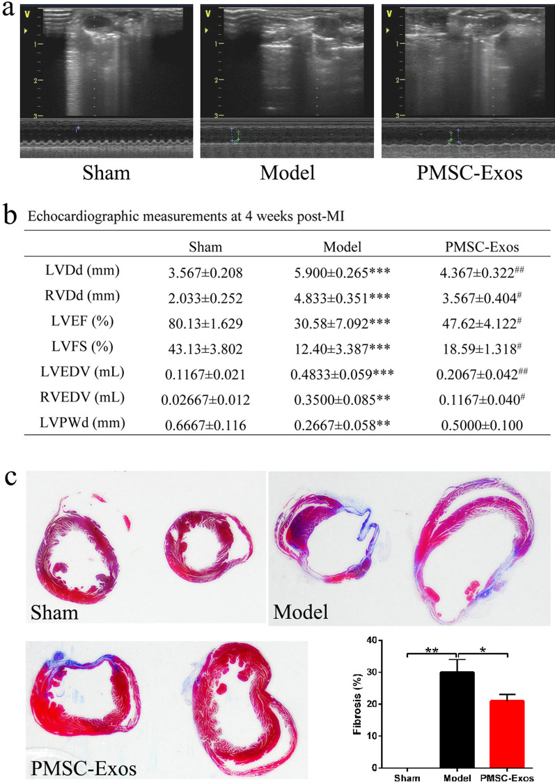
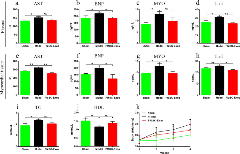
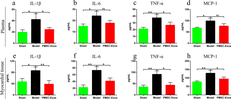
Results: We found that human PMSC-Exos were obtained and identified with high purity and uniformity. MI model was successfully established. Compared to MI group, PMSC-Exos treatment ameliorated myocardial fibrosis and left ventricular (LV) remodeling (P < 0.05). Moreover, PMSC-Exos treatment obviously decreased MI molecular markers (AST/BNP/MYO/Tn-I/TC), pro-inflammatory indicators (IL-1β, IL-6, TNF-α, MCP-1), as well as increased HDL in comparison with MI group (all P < 0.05). Intriguingly, PMSC-Exos intervention notably modulated gut microbial community via increasing the relative abundances of Bacteroidetes, Proteobacteria, Verrucomicrobia, Actinobacteria, Akkermansia, Bacteroides, Bifidobacterium, Thauera and Ruminiclostridium, as well as decreasing Firmicutes (all P < 0.05), compared with MI group. Furthermore, PMSC-Exos supplementation increased gut microbiota metabolites SCFAs (butyric acid, isobutyric acid and valeric acid) and decreased LPS in comparison with MI group (all P < 0.05). Correlation analysis indicated close correlations among gut microbiota, microbial SCFAs and inflammation in MI.

Conclusions: Our study highlighted that PMSC-Exos intervention alleviated MI via modulating gut microbiota and suppressing inflammation.

Keywords: Anti-inflammation; Gut microbiota; LPS; MI; PMSC-Exos; SCFAs.


Conflict of interest statement

All the authors declare that there are no conflicts of interest.

Figures

Fig.1
Fig.1
Morphological characteristics and identification of PMSCs and exosomes. a Morphological characteristics of PMSCs. b Positive rates of cell surface antigens CD73, CD90, CD105, CD11b, CD19, CD34, CD45 and HLA-DR in PMSCs were detected by flow cytometry. c Observation of exosomes by transmission electron microscopy (TEM). d Observation of exosomes by nanoparticle tracking analysis (NTA)
Fig. 2
Fig. 2
Identification of myocardial infarction in mice. a Electrocardiogram of mice left anterior descending coronary artery (LAD) before ligation. b Electrocardiogram of mice LAD after ligation
Fig. 3
Fig. 3
PMSCs-derived exosomes improved cardiac function and myocardial infarction (MI) area of mice with MI. a Representative images of echocardiograms in diverse groups. b Determination of LVDd, RVDd, LVEF, LVFS, LVEDV, RVEDV and LVPWd of mice in each group by echocardiography. Data were expressed as mean ± SD. **P < 0.01, ***P < 0.001 versus sham. #P < 0.05, ##P < 0.01 versus model. c Exosomes secreted by PMSCs reduced myocardial infarction area of mice with MI. Myocardial tissue was stained in red and collagen fibers were stained in blue by Masson staining. Data were expressed as mean ± SD. *P < 0.05, **P < 0.01
Fig. 4
Fig. 4
PMSCs-derived exosomes reduced plasma and myocardial biochemical indicators. a Plasma aspartate transaminase (AST) levels. b Plasma brain natriuretic peptide (BNP) levels. c Plasma myoglobin (MYO) levels. d Plasma troponin- I (Tn-I) levels. e Myocardial AST levels. f Myocardial BNP levels. g Myocardial MYO levels. h Myocardial Tn-I levels. i Plasma total cholesterol (TC) levels. j Plasma high density lipoprotein cholesterol (HDL-C) levels. k BWs growth curve. Data were expressed as mean ± SD. *P < 0.05, **P < 0.01, NS: P > 0.05
Fig. 5
Fig. 5
PMSCs-derived exosomes ameliorated plasma and myocardial inflammation factors. a Plasma interleukin 1β (IL-1β) levels. b Plasma IL-6 levels. c Plasma tumor necrosis factor α (TNF-α) levels. d Plasma monocyte chemotactic protein 1 (MCP-1) levels. e Myocardial IL-1β levels. f Myocardial tissue IL-6 levels. g Myocardial TNF-α levels. h Myocardial MCP-1 levels. Data were expressed as mean ± SD. *P < 0.05, **P < 0.01, NS: P > 0.05
Fig. 6
Fig. 6
Alpha-diversity and β diversity of gut microbial in the feces of mice. a Shannon index on OTU level. b Rarefaction curves (Alpha-diversity analysis). c OUT analysis. d PCA analysis on OTU level. e PCoA analysis on OTU level. f NMDS analysis on OTU level
Fig. 7
Fig. 7
Relative abundances of gut microbial species at the phylum and genus levels in the feces of mice. a, c The relative abundances of gut microbial species in phylum level. b, d The relative abundances of microbial species in genus level. Data were expressed as mean ± SD. *P < 0.05
Fig. 8
Fig. 8
PMSC-Exos reduced plasma LPS levels. Data were expressed as mean ± SD. *P < 0.05, **P < 0.01
Fig. 9
Fig. 9
Effects of exosomes secreted by PMSCs on short-chain fatty acids (SCFAs) of diverse groups. a Sample chromatogram of rats stool. b Acetic acid. c Isobutyric acid. d Butyric acid. e Isovaleric acid. f Valeric acid. Data were expressed as mean ± SD. *P < 0.05, **P < 0.01, NS: P > 0.05
Fig. 10
Fig. 10
Correlation analyses between relative abundance (%) of microbiota and inflammation indicators. *P < 0.05, **P < 0.01
Fig. 11
Fig. 11
GO analysis. a mRNA. b sRNA. c cirRNA. d TvsN
Fig. 12
Fig. 12
KEGG functional enrichment analysis. a mRNA. b sRNA. c cirRNA. d TvsN. KEGG pathway were analyzed using the KEGG database of the Kanehisa laboratory– (www.kegg.jp/kegg/kegg1.html)

References

    1. Jiang W, Wang M. New insights into the immunomodulatory role of exosomes in cardiovascular disease. Rev Cardiovasc Med. 2019;20(3):153–160. - PubMed
    1. Reichert K, Colantuono B, McCormack I, Rodrigues F, Pavlov V, Abid MR. Murine left anterior descending (LAD) coronary artery ligation: an improved and simplified model for myocardial infarction. J Vis Exp. 2017 doi: 10.3791/55353. - DOI - PMC - PubMed
    1. Mangion K, Gao H, Husmeier D, Luo X, Berry C. Advances in computational modelling for personalised medicine after myocardial infarction. Heart. 2018;104(7):550–557. - PubMed
    1. Cung TT, Morel O, Cayla G, Rioufol G, Garcia-Dorado D, Angoulvant D, Bonnefoy-Cudraz E, Guerin P, Elbaz M, Delarche N, et al. Cyclosporine before PCI in patients with acute myocardial infarction. N Engl J Med. 2015;373(11):1021–1031. - PubMed
    1. Wang X, Guo Z, Ding Z, Mehta JL. Inflammation, autophagy, and apoptosis after myocardial infarction. J Am Heart Assoc. 2018 doi: 10.1161/JAHA.117.008024. - DOI - PMC - PubMed

Publication types

MeSH terms