. 2022 Feb 16;22(1):57.
doi: 10.1186/s12872-022-02505-z.

Mapping and ablation of left atrial roof-dependent tachycardias using an ultra-high resolution mapping system

Affiliations

Mapping and ablation of left atrial roof-dependent tachycardias using an ultra-high resolution mapping system

Shinsuke Miyazaki et al. BMC Cardiovasc Disord. .

Abstract

Background: Left atrial roof-dependent tachycardias (LARTs) are common macroreentrant atrial tachycardias (ATs). We sought to characterize clinical LARTs using an ultra-high resolution mapping system.

Methods: This study included 22 consecutive LARTs in 21 patients who underwent AT mapping/ablation using Rhythmia systems.

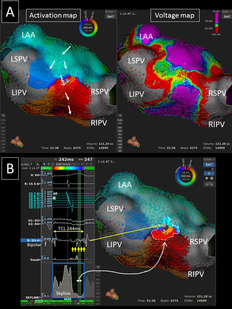
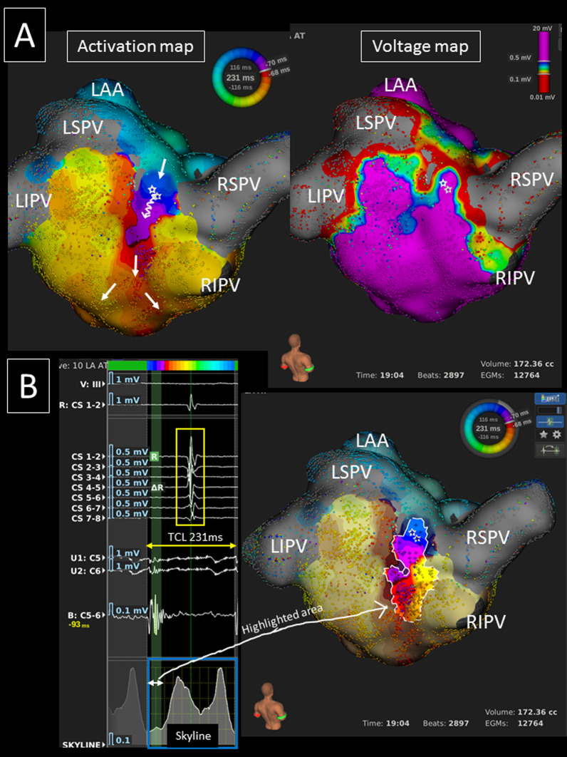
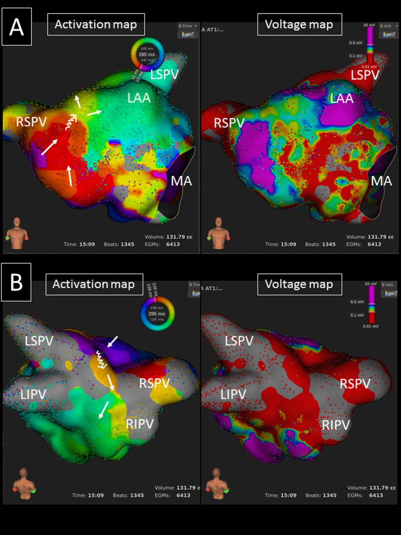
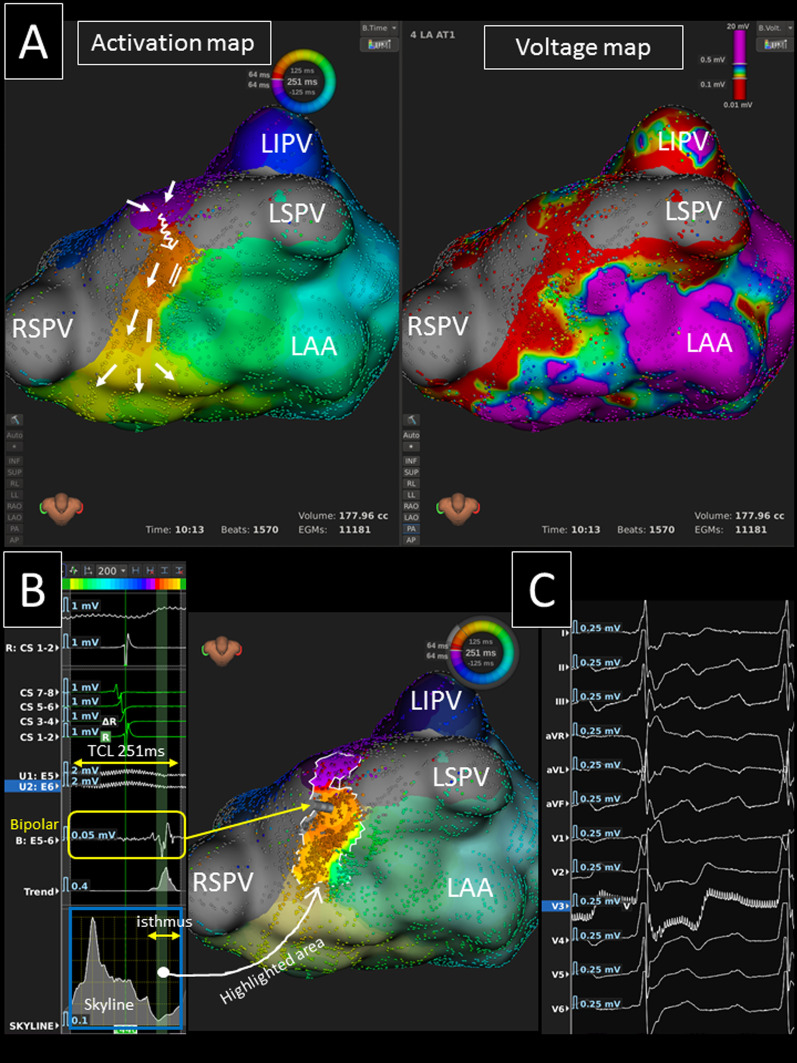
Results: Three, 13, 4, and 2 LART patients were cardiac intervention naïve (Group-A), post-roof line ablation (Group-B), post-atrial fibrillation ablation without linear ablation (Group-C), and post-cardiac surgery (Group-D), respectively. The mean AT cycle length was 244 ± 43 ms. Coronary sinus activation was proximal-to-distal or distal-to-proximal in 16 (72.7%) ATs. The activation map revealed 13 (59.1%) clockwise and 9 (40.9%) counter-clockwise LARTs. A 12-lead synchronous isoelectric interval was observed in 10/19 (52.6%) LARTs. The slow conduction area was identified on the LA roof, anterior/septal wall, and posterior wall in 18, 6, and 2 ATs, respectively. Twenty concomitant ATs among 13 procedures were also eliminated, and peri-mitral AT coexisted in 7 of 9 non-group-B patients. In group-B, the conduction gap was predominantly located on the mid-roof. Sustained LARTs were terminated by a single application and linear ablation in 6 (27.3%) and 9 (40.9%), while converting to other ATs in 7 (31.8%) LARTs. Complete linear block was created without any complications in all, however, ablation at the mid-posterior wall was required to achieve block in 4 (18.2%) procedures. During 14.0 (6.5-28.5) months of follow-up, 17 (81.0%) and 19 (90.5%) patients were free from any atrial tachyarrhythmias after single and last procedures.

Conclusions: The LART mechanisms were distinct in individual patients, and elimination of all concomitant ATs was required for the management.

Keywords: Atrial tachycardia; Catheter ablation; Roof line; Ultra-high resolution mapping.


Conflict of interest statement

Dr. Miyazaki belongs to the endowed departments of Medtronic, Boston, Abbott, and Japan Lifeline.

Figures

Fig. 1
Fig. 1
A An ultra-high resolution activation map revealed a clockwise roof-dependent AT with slow conduction on the LA roof (TCL = 244 ms) (left panel). The white arrows show the propagation of the activation. A voltage map shows an LVA on the LA roof (right panel). B The valley (green curtain, white arrow) on the global activation histogram (skyline, blue square) corresponds to the isthmus of the AT (highlighted area). Please note that low-voltage fractionated signals (yellow arrows) during the time period of the valley of the skyline were identified in the critical isthmus. LAA left atrial appendage, LI(S)PV left inferior (superior) pulmonary vein, RI(S)PV right inferior (superior) pulmonary vein, TCL tachycardia cycle length
Fig. 2
Fig. 2
A An ultra-high resolution activation map revealed a clockwise roof-dependent AT with slow conduction on the LA posterior roof (TCL = 231 ms) (left panel). The white arrows show the propagation of the activation. A voltage map shows scar on the LA posterior roof (right panel). B The valley (green curtain, white arrow) on the global activation histogram (skyline, blue square) corresponds to the isthmus of the AT (highlighted area). Please note the straight CS activation pattern (yellow square). LAA left atrial appendage, LI(S)PV left inferior (superior) pulmonary vein, RI(S)PV right inferior (superior) pulmonary vein, TCL tachycardia cycle length
Fig. 3
Fig. 3
An ultra-high resolution activation map revealed a clockwise roof-dependent AT with slow conduction on both the LA anterior wall (A) and roof (B) (TCL = 295 ms) (left panels). The white arrows show the propagation of the activation. A voltage map reveals extensive scar and an LVA on the LA anterior (A) and posterior walls (B) (right panels). LAA left atrial appendage, LI(S)PV left inferior (superior) pulmonary vein, MA mitral annulus, RI(S)PV right inferior (superior) pulmonary vein
Fig. 4
Fig. 4
A An ultra-high resolution activation map revealed a counter-clockwise roof-dependent AT with a conduction path and slow conduction on the LA roof (TCL = 251 ms) (left panel). The white arrows show the propagation of the activation. A voltage map reveals scar on the LA roof (right panel). B The valley (green curtain, white arrow) on the global activation histogram (skyline, blue square) corresponds to the isthmus of the AT (highlighted area). Please note that fractionated signals (yellow square) during the time period of the valley of the skyline were identified in the critical isthmus. C During the AT, continuous electrical activity was observed on the 12-lead ECG. LAA left atrial appendage, LI(S)PV left inferior (superior) pulmonary vein, RSPV right superior pulmonary vein, TCL tachycardia cycle length
Fig. 5
Fig. 5
A An ultra-high resolution activation map revealed a counter-clockwise roof-dependent AT (TCL = 212 ms) (left panel). The white arrows show the propagation of the activation. A voltage map reveals an LVA on the LA posterior roof (right panel). Please note that double potentials are observed at point-A and point-C, but a low-amplitude fractionated signal is identified during the time period of the valley of the skyline at point-B (yellow square). B A 12-lead ECG during the AT exhibits a synchronous isoelectric interval. C The valley (green curtain, white arrow) on the global activation histogram (skyline) corresponds to the isthmus of the AT (highlighted area). LAA left atrial appendage, LI(S)PV left inferior (superior) pulmonary vein, RI(S)PV right inferior (superior) pulmonary vein, TCL tachycardia cycle length
Fig. 6
Fig. 6
An ultra-high resolution activation map revealed a clockwise roof-dependent AT (TCL = 309 ms) (left panel) in a patient with a history of cardiac surgery. The white arrows show the propagation of the activation. A voltage map reveals an extensive LVA on the LA anterior wall (right panel). LAA left atrial appendage, LI(S)PV left inferior (superior) pulmonary vein, MA mitral annulus, RI(S)PV right inferior (superior) pulmonary vein

References

    1. Shah AJ, Jadidi A, Liu X, Miyazaki S, Forclaz A, Nault I, Rivard L, Linton N, Xhaet O, Derval N, Sacher F, Bordachar P, Ritter P, Hocini M, Jais P, Haissaguerre M. Atrial tachycardias arising from ablation of atrial fibrillation: a proarrhythmic bump or an antiarrhythmic turn? Cardiol Res Pract. 2010;2010:950763. doi: 10.4061/2010/950763. - DOI - PMC - PubMed
    1. Hocini M, Jaïs P, Sanders P, Takahashi Y, Rotter M, Rostock T, Hsu LF, Sacher F, Reuter S, Clémenty J, Haïssaguerre M. Techniques, evaluation, and consequences of linear block at the left atrial roof in paroxysmal atrial fibrillation: a prospective randomized study. Circulation. 2005;112:3688–3696. doi: 10.1161/CIRCULATIONAHA.105.541052. - DOI - PubMed
    1. Jaïs P, Matsuo S, Knecht S, Weerasooriya R, Hocini M, Sacher F, Wright M, Nault I, Lellouche N, Klein G, Clémenty J, Haïssaguerre M. A deductive mapping strategy for atrial tachycardia following atrial fibrillation ablation: importance of localized reentry. J Cardiovasc Electrophysiol. 2009;20:480–491. doi: 10.1111/j.1540-8167.2008.01373.x. - DOI - PubMed
    1. Mantziari L, Butcher C, Kontogeorgis A, Panikker S, Roy K, Markides V, Wong T. Utility of a novel rapid high-resolution mapping system in the catheter ablation of arrhythmias: an initial human experience of mapping the atria and the left ventricle. JACC Clin Electrophysiol. 2015;1:411–420. doi: 10.1016/j.jacep.2015.06.002. - DOI - PubMed
    1. Laţcu DG, Bun SS, Viera F, Delassi T, El Jamili M, Al Amoura A, Saoudi N. Selection of critical isthmus in scar-related atrial tachycardia using a new automated ultrahigh resolution mapping system. Circ Arrhythm Electrophysiol. 2017;10:e004510. doi: 10.1161/CIRCEP.116.004510. - DOI - PubMed

Publication types

LinkOut - more resources