. 2022 Feb 16;13(1):7.
doi: 10.1186/s13227-022-00191-8.

Heterochrony and repurposing in the evolution of gymnosperm seed dispersal units

Affiliations

Heterochrony and repurposing in the evolution of gymnosperm seed dispersal units

Juca A B San Martin et al. Evodevo. .

Abstract

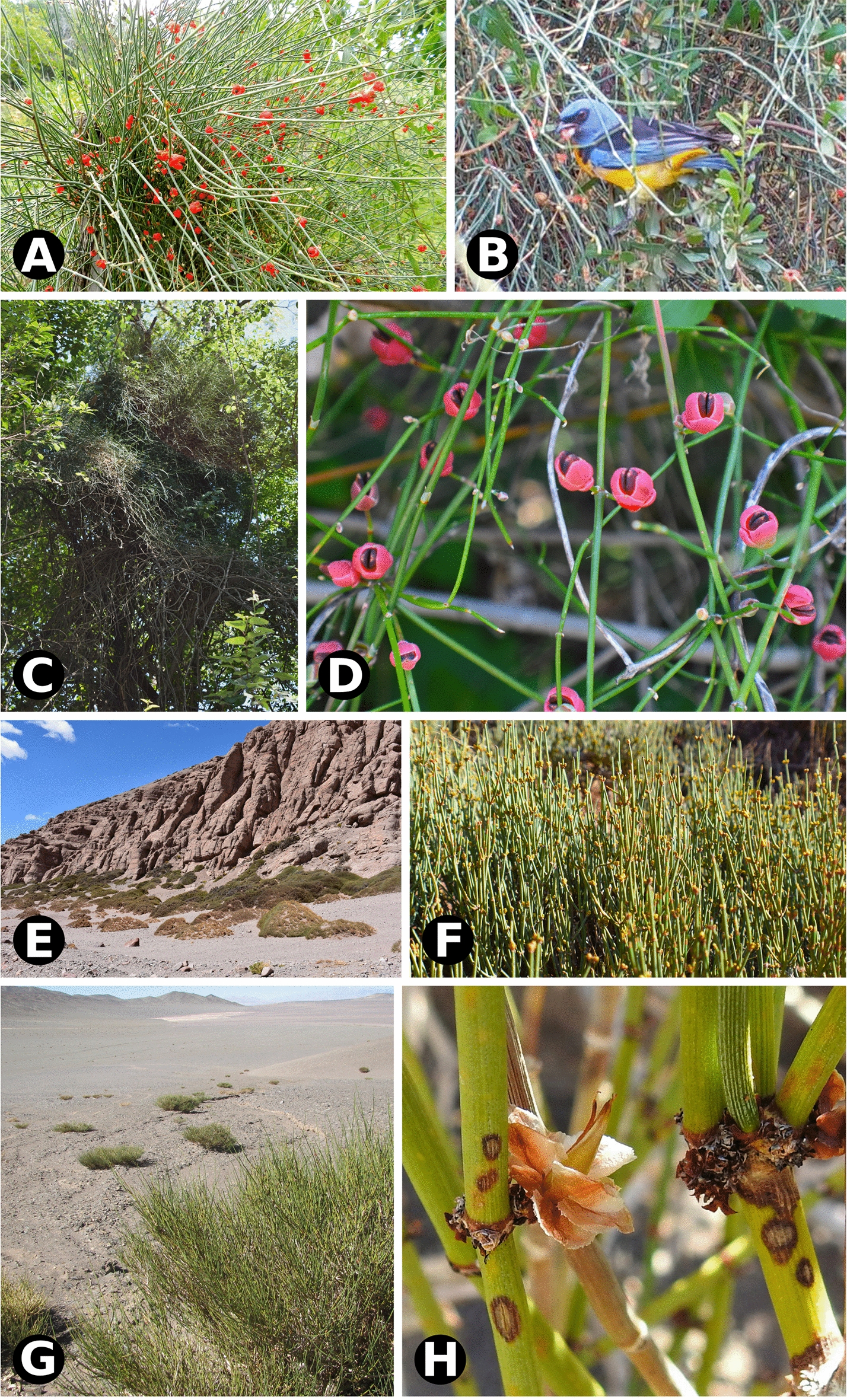
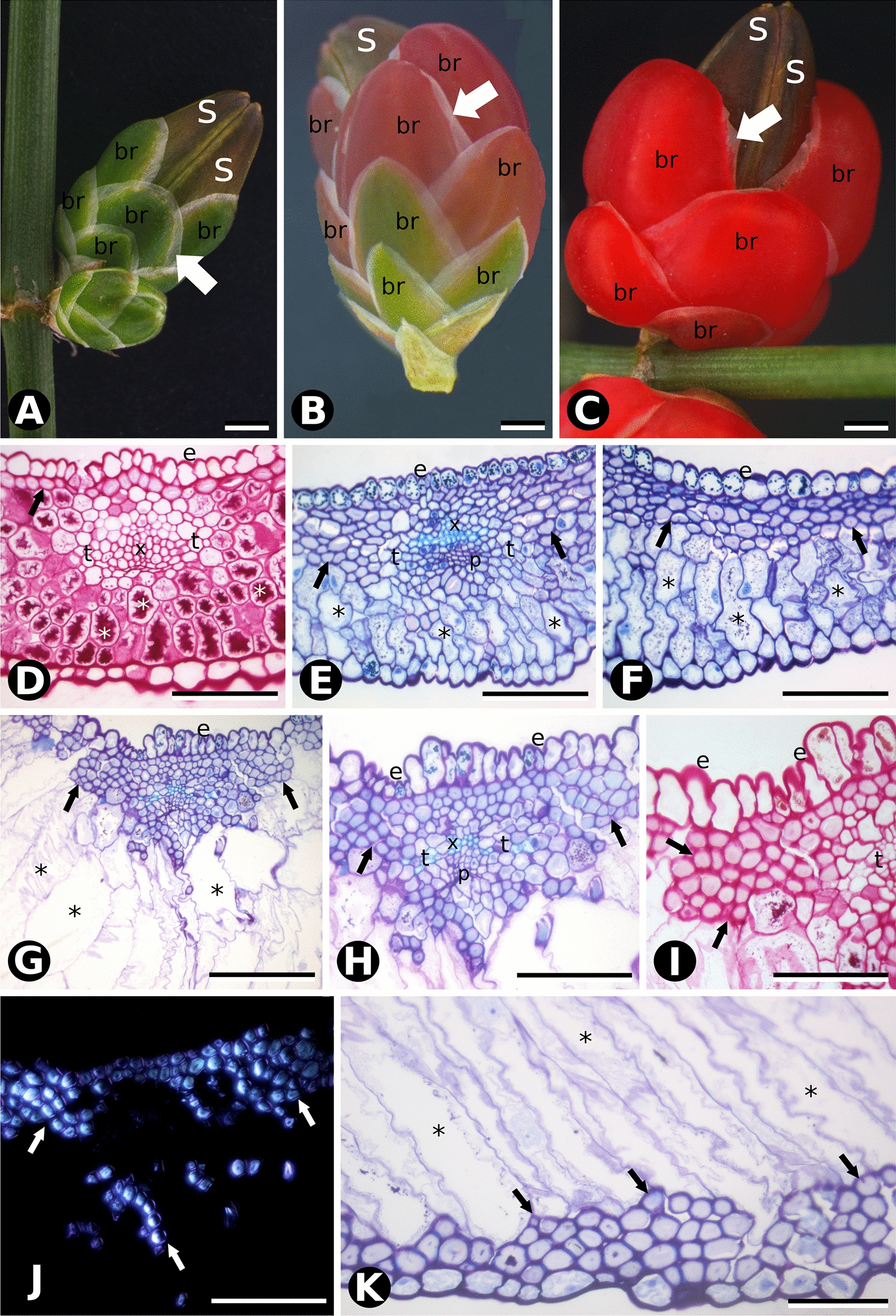
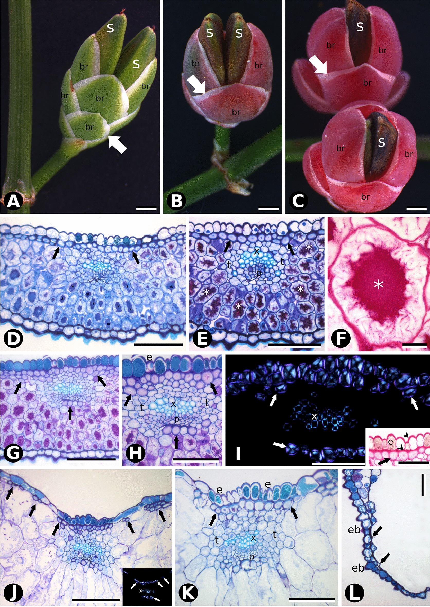
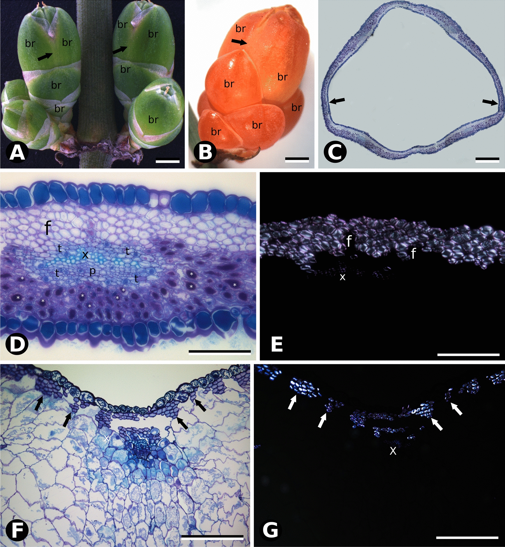
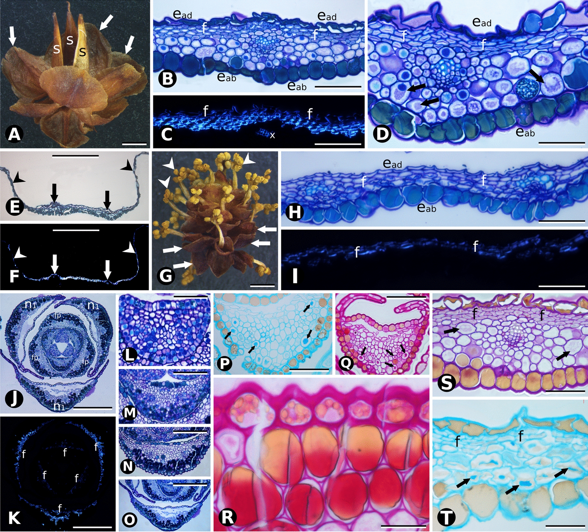
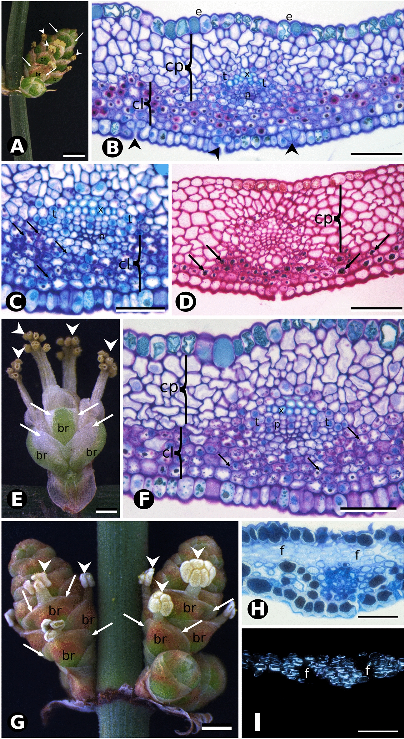
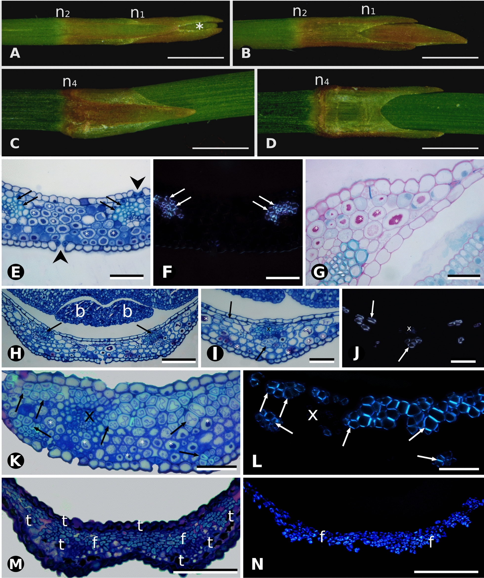
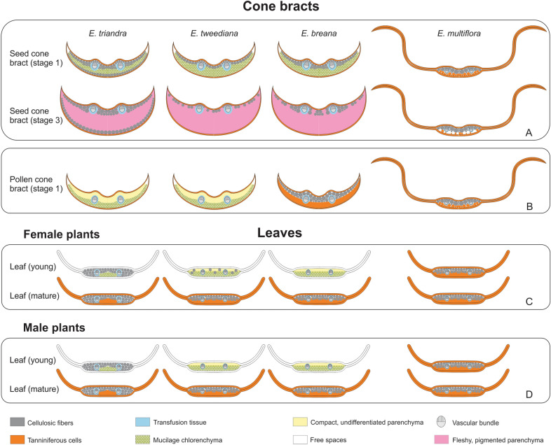
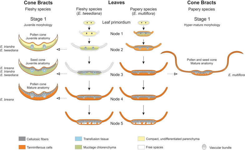
Background: Plant dispersal units, or diaspores, allow the colonization of new environments expanding geographic range and promoting gene flow. Two broad categories of diaspores found in seed plants are dry and fleshy, associated with abiotic and biotic dispersal agents, respectively. Anatomy and developmental genetics of fleshy angiosperm fruits is advanced in contrast to the knowledge gap for analogous fleshy structures in gymnosperm diaspores. Improved understanding of the structural basis of modified accessory organs that aid in seed dispersal will enable future work on the underlying genetics, contributing to hypotheses on the origin of angiosperm fruits. To generate a structural framework for the development and evolution of gymnosperm fleshy diaspores, we studied the anatomy and histochemistry of Ephedra (Gnetales) seed cone bracts, the modified leaves surrounding the reproductive organs. We took an ontogenetic approach, comparing and contrasting the anatomy and histology of fleshy and papery-winged seed cone bracts, and their respective pollen cone bracts and leaves in four species from the South American clade.

Results: Seed bract fleshiness in Ephedra derives from mucilage accumulated in chlorenchyma cells, also found in the reduced young leaves before they reach their mature, dry stage. Cellulosic fibers, an infrequent cell type in gymnosperms, were found in Ephedra, where they presumably function as a source of supplementary apoplastic water in fleshy seed cone bracts. Papery-winged bract development more closely resembles that of leaves, with chlorenchyma mucilage cells turning into tanniniferous cells early on, and hyaline margins further extending into "wings".

Conclusions: We propose an evolutionary developmental model whereby fleshy and papery-winged bracts develop from an early-stage anatomy shared with leaves that differs at the pollination stage. The ancestral fleshy bract state may represent a novel differentiation program built upon young leaf anatomy, while the derived dry, papery-winged state is likely built upon an existing differentiation pattern found in mature vegetative leaves. This model for the evolution of cone bract morphology in South American Ephedra hence involves a novel differentiation program repurposed from leaves combined with changes in the timing of leaf differentiation, or heterochrony, that can further be tested in other gymnosperms with fleshy diaspores.

Keywords: Bracts; Cellulosic fibers; Fruit-like; Gnetales; Gymnosperm; Heterochrony; Histochemistry; Mucilage; Ontogeny; Seed dispersal.


Conflict of interest statement

The authors declare that they have no competing interests.

Figures

Fig. 1
Fig. 1
Natural environment, habit and dispersal biology of Ephedra species studied. A E. triandra with seed cones; B Bird disperser Rauenia bonariensis, “naranjero” feeding on the fleshy cones (Anillaco, la Rioja); photos in A, B by Adriana Aranda-Rickert. C E. tweediana climbing on a tree; D detail of fleshy cones (El Rodeo, Catamarca). E E. breana in high elevation semi-arid environment; F plant detail (Laguna Brava, La Rioja). G Sparse populations of E. multiflora in high elevation semi-dessert environment; photo by Javier Torréns; H detail of plant habit (Laguna Brava, La Rioja); photo by Adriana Aranda-Rickert
Fig. 2
Fig. 2
Comparative morphology and bract anatomy of Ephedra triandra seed cones at three developmental stages: A green (Stage 1), B red (Stage 2), and C fleshy (Stage 3). The white arrow shows a papyraceous lateral region that decreases as bracts (br) and seeds (s) mature. D Cross section of a green bract stained with PAS showing a continuous single layer of cellulosic fibers beneath the epidermis (black arrow), and a mesophyll of cells with substantial amounts of insoluble carbohydrates (*). Adaxial epidermis (e), xylem (x) and transfusion tissue (t). E, F Cross sections of red bracts stained with PAS and toluidine blue O, showing the proliferation of cellulosic fibers (black arrows) beneath the adaxial epidermis. Mesophyll cells (*) no longer contain insoluble carbohydrates. G Cross section of red and fleshy bracts showing an adaxial epidermis with columnar cells and cellulosic fibers organized in several subepidermal layers (arrows). Mesophyll cells (*) look enlarged, with thin primary walls and enlarged vacuoles. H Detail of the vascular bundle in G, adaxial epidermis, xylem and transfusion tissue. I Detail of cellulosic fibers (arrows) showing intense PAS staining on their primary walls (black arrows) and lighter staining on their secondary walls. Adaxial epidermis and transfusion tissue. J Polarized light microscopy showing the intense birefringence of cellulosic fibers in cross section (arrows). K Detail of the abaxial face showing the distribution of cellulosic fibers (arrows) in subepidermal layers. Mesophyll cells (*) are enlarged, with thin, sinuous walls and enlarged vacuoles in their cytoplasm. Scale bars: 1 mm (AC); 100 µm (D); 50 µm (E, F, IK); 100 µm (G, H)
Fig. 3
Fig. 3
Comparative morphology and bract anatomy of Ephedra tweediana seed cones at three developmental stages: A green (Stage 1); B red (Stage 2), and C fleshy (Stage 3). White arrows show the papyraceous lateral region of the bracts (br); two seeds (s) are present per cone. D Cross section of a green bract stained with toluidine blue O and PAS showing a homogeneous mesophyll consisting of cells with substantial amounts of insoluble carbohydrates (*) and cellulosic fibers (black arrows) distributed in a single adaxial subepidermal layer. E Detail of vascular bundle in D, with mesophyll cells with substantial amounts of insoluble carbohydrates, phloem (p), xylem (x) and transfusion tissue (t). F Detail of mesophyll cell stained with PAS showing its vacuole filled with insoluble carbohydrates. G Overview of a red bract in cross section showing an increase in the number of cellulosic fiber layers (black arrows). H Detail of the vascular bundle shown in G, with adaxial epidermal cells (e) filled with phenolic compounds (tannins), and cellulosic fibers organized into one or two subepidermal layers (black arrows). I Polarized light microscopy showing the intense birefringence of cellulosic fibers in cross section (white arrows). Inset, detail of cellulosic fibers showing intense PAS staining on primary walls (arrowheads) and weak staining on secondary walls (black arrow). Adaxial epidermis (e) with thickening of the external and internal periclinal walls (arrowheads). JL Cross section of fleshy bracts (Stage 3). J Cellulosic fibers (black arrows) are organized in separate bundles of one or two layers beneath the adaxial epidermis. Mesophyll cells are large with thin, sinuous walls and devoid of cytoplasmic content. Inset: polarized light microscopy showing the intense birefringence of cellulosic fibers (white arrows) and xylem. K Detail of the vascular bundle area beneath the adaxial epidermis with cellulosic fibers (black arrows) above, phloem, xylem and transfusion tissue. L Detail of papery lateral region of the bract, consisting solely of epidermis, with adaxial cells collapsed and reduced to the periclinal, juxtaposed walls (black arrows) and abaxial epidermis (eb) with tanniniferous cells. Scale bars: 1 mm (AC); 100 µm (D, E, G, JL); 50 µm (I); 20 µm (F)
Fig. 4
Fig. 4
Comparative morphology and bract anatomy of E. breana seed cones. A Seed cone in Stage 1, with green bracts (br). Black arrows indicate the fused region of the distal bracts. B Seed cone at Stage 3, with fleshy red bracts. Black arrows indicate the fused region of the distal bracts. Note the highly developed distal bracts that completely enclose the seeds, which are not visible as in the other species studied. C Overview of anatomical section in the region of seed cone bract fusion (black arrows). D Cross section of seed cone bract at Stage 1 stained with toluidine blue O and PAS showing large tannin content in cells of the adaxial epidermis. Cellulose fibers (f) are grouped into layers on the adaxial side. A vascular bundle is surrounded by transfusion tissue (t), phloem (p) and xylem (x). Mucilaginous parenchyma is evident abaxially, and the abaxial epidermis is composed of cells with tannins and stomata. E Polarized light microscopy of section in D showing the birefringence of cellulosic fibers (f) and xylem. F Cross section of the seed cone bract at Stage 3 stained with toluidine blue and PAS showing increased of cell size and loss of carbohydrates from the cytoplasm of mucilaginous parenchyma cells. Fiber layers (arrows) are reduced in number and grouped in bundles beneath the adaxial epidermis. G Polarized light microscopy of the section in F showing the birefringence of cellulosic fibers (white arrows) and xylem. Scale bars: 1 mm (A, B); 200 µm (C); 100 µm (DG)
Fig. 5
Fig. 5
Morphology and anatomy of seed and pollen cone bracts and leaves of E. multiflora. A Seed cone with three seeds (s) and papery-winged bracts (arrow). BD Cross section of the papery-winged seed cone bract (young green stage). C Polarized light microscopy of section in B. D Detail of vascular bundles and mucilage chlorenchyma cells (arrows). E, F General appearance of papery-winged seed cone bract in cross section, stained with PAS and toluidine blue O. Arrows indicate the two vascular bundles, and arrowheads the lateral papyraceous area. G Pollen cone after pollination, with open microsporangia (arrowheads) and papery-winged bracts (arrows). H, I Cross section of pollen cone bract. I Polarized light microscopy of section in H. JO Leaf development. J Cross section of a terminal bud, with leaves from node 1 (n1) and leaf primordia (p). K Polarized light microscopy of section in J. LO Detail from the youngest leaf primordia to a node 1 leaf. P, Q Histochemistry of a leaf primordium (same stage as L) revealing mucilage chlorenchyma cells (arrows), P Alcian Blue, Q PAS. R Mucilage cells turning into tanniniferous cells in a node 1 leaf. S, T Cross section of a seed cone bract with mucilage cells (arrows). S PAS. T Alcian Blue. Adaxial epidermis (ead), abaxial epidermis (eab), fibers (f). Scale bars: 1 mm (A, G); 500 µm (J, K); 400 µm (E, F, O); 300 µm (N); 200 µm (B, C, H, I, M); 100 µm (D, L, P, Q, S); 50 µm (R, T)
Fig. 6
Fig. 6
Comparative morphology and bract anatomy of Ephedra pollen cones.Ephedra triandra (AD), E. tweediana (E, F) and E. breana (GI). A Overall appearance of E. triandra pollen cone after pollination, green bracts (br) with hyaline margins (arrows) enclose one microsporangiophore each (arrowheads), with multiple microsporangia (pollen sacs) at their tips. B Cross section of E. triandra pollen cone bract stained with toluidine blue O and PAS. Adaxial epidermis (e) with tanniniferous cells with large phenolic content (tannin). Heterogeneous mesophyll with adaxial, compact parenchyma (cp) without intercellular spaces and abaxial chlorenchyma (cl) with substantial amounts of insoluble carbohydrates (mucilage) inside the vacuoles, evidenced by strong PAS staining (magenta to purple). Vascular bundle with poorly developed phloem (p), xylem (x) and transfusion tissue (t) at the interface between parenchyma types. Stomata (arrowheads) on the abaxial epidermis. C Vascular bundle in cross section with toluidine blue O. Chlorenchyma cells containing multiple chloroplasts (arrows). D Same bract stained with PAS showing substantial amounts of insoluble carbohydrates (arrows) within chlorenchyma cells. E Pollen cone of E. tweediana after pollination. Green bracts with white hyaline margins (arrows) enclose one microsporangiophore each (arrowheads), with multiple microsporangia (pollen sacs) at their tips. F Cross section of pollen cone bract with toluidine blue O showing the same anatomical pattern as in E. triandra. G Pollen cone of E. breana at anthesis. Microsporangia (arrowheads) at anthesis (left cone) and pre-anthesis (right cone), hyaline margins (arrows) on bracts. H Cross section of pollen cone bract at anthesis stained with toluidine blue O and PAS. Mesophyll with cellulosic fibers (f) and tanniniferous cells, without insoluble carbohydrates (mucilage). I Polarized microscopy of bract section in H showing birefringence of cellulosic fibers (f). Scale bars: 1 mm (A, E, G); 50 µm (C, H, I); 100 µm (B, D, F).
Fig. 7
Fig. 7
Comparative leaf morphology and anatomy of Ephedra in this study. AD Overview of E. tweediana leaf morphology under a stereomicroscope. A, B Stem apex showing leaves from the first and second nodes in frontal and lateral views, respectively. C, D Detail of the fifth node from the stem apex showing developing leaves in frontal and lateral views, respectively. Note the development of the lateral, papyraceous zone. EG E. tweediana (male individual). E Toluidine blue O and PAS staining. Mucilage cells (*) are present throughout the mesophyll, cellulosic fibers (arrows) are associated with vascular bundles, and stomata are present on both leaf surfaces (arrowheads). F Polarized light microscopy of section in E showing the birefringence and distribution of cellulosic fibers (arrows). G Positive PAS staining of mucilage in mesophyll cells. HJ E. tweediana (female individual). H General overview, arrows indicate vascular bundles, two axillary buds (b) are visible and mucilage cells (*) are distributed throughout the mesophyll. I High magnification of the vascular bundle in D, showing cellulosic fibers (arrows) associated with the bundle and also distributed throughout the mesophyll. J Polarized light microscopy of section in I showing the birefringence of xylem cells (x) and cellulosic fibers (arrows). K E. triandra (male individual), with a different pattern from that found in E. tweediana: cellulosic fibers (arrows) are associated with vascular bundles, and also present in bundles abaxially and in layers adaxially. Mucilage cells are found in multiple layers on the abaxial surface. L Polarized light microscopy of section in K showing the distribution and birefringence of the cellulosic fibers. M E. multiflora (female individual) with tannin cells (t) in the adaxial epidermis and also distributed in one or two layers on the abaxial face. Cellulosic fibers (arrows) differentiate in several adaxial layers in the mesophyll. L Polarized light microscopy of section in K showing the distribution and birefringence of the cellulosic fibers. Node 1 (n1), node 2 (n2), node 4 (n4). Scale bars: 1 mm (AD), 100 µm (E, F, KN); 50 µm (G, I, J); 200 µm (H)
Fig. 8
Fig. 8
Summary diagrams of leaf and bract anatomy observations for Ephedra breana, E. multiflora, E. triandra, and E. tweediana. A Seed cone bracts at Stage 1 (first row) and 3 (second row). B Pollen cone bracts at Stage 1. C Leaves from female plants: at the second node (young, first row) and at the 5th node (mature, second row). D Leaves from male plants: at the second node (young, first row) and at the 5th node (mature, second row). Refer to figure inset for the meaning of colors and symbols
Fig. 9
Fig. 9
Cone bracts from leaves: Evolutionary developmental model for the repurposing of anatomical features from vegetative leaves to reproductive bracts in Ephedra (Gnetales). Center: Diagrams of leaf development in E. tweediana (left), and E. multiflora (right) showing the shift of mature anatomy to younger stages in E. multiflora. Left: Bracts of fleshy species at Stage 1, grouped by their juvenile external leaf morphology (reduced hyaline margins), showing juvenile or mature leaf anatomy depending on the species. Right: Bracts of papery species at Stage 1, with hyper-mature external leaf morphology (widely developed hyaline margins) and mature leaf anatomy. Solid-line arrows connect stages of leaf development; broken-line arrows indicate corresponding anatomies between stages of leaf development and bract structure. Fleshy species, species with fleshy seed cone bracts; Papery species, species with papery-winged seed cone bracts. Refer to figure inset for the meaning of colors and symbols

Similar articles

References

    1. Mapes G, Rothwell GW, Haworth MT. Evolution of seed dormancy. Nature. 1989;337:645–646. doi: 10.1038/337645a0. - DOI
    1. Leslie AB, Beaulieu JM, Mathews S. Variation in seed size is structured by dispersal syndrome and cone morphology in conifers and other nonflowering seed plants. New Phytol. 2017;216:429–437. doi: 10.1111/nph.14456. - DOI - PubMed
    1. Seale M, Nakayama N. From passive to informed: mechanical mechanisms of seed dispersal. New Phytol. 2020;225:653–658. doi: 10.1111/nph.16110. - DOI - PubMed
    1. Traveset A, Rodríguez-Pérez J. Seed dispersal. In: Jørgensen SE, Fath BD, editors. Encyclopedia of ecology. Oxford: Academic Press; 2008. pp. 3188–3194.
    1. Nigris S, D’Apice G, Moschin S, Ciarle R, Baldan B. Fleshy structures associated with ovule protection and seed dispersal in gymnosperms: a systematic and evolutionary overview. Crit Rev Plant Sci. 2021 doi: 10.1080/07352689.2021.1938397. - DOI

LinkOut - more resources