. 2022 Feb 16;12(1):2578.
doi: 10.1038/s41598-022-06300-w.

Placental pathology predicts infantile neurodevelopment

Affiliations

Placental pathology predicts infantile neurodevelopment

Megumi Ueda et al. Sci Rep. .

Abstract

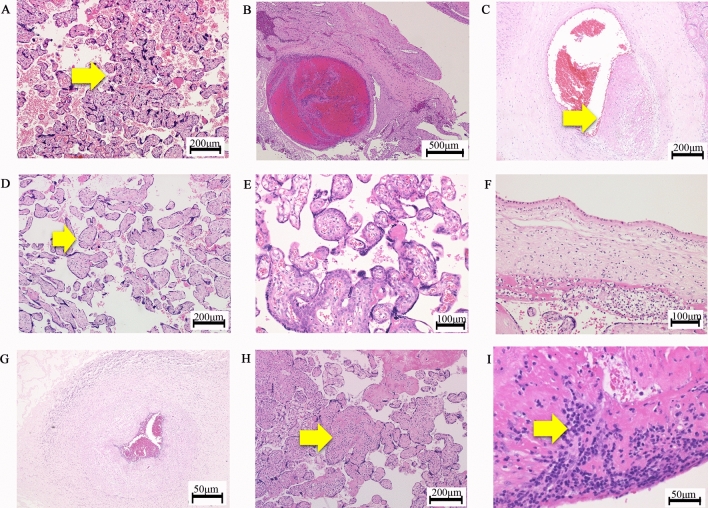
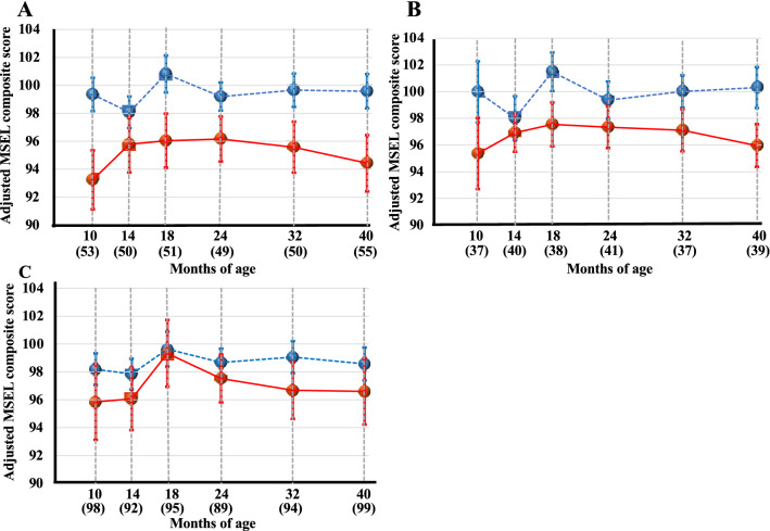
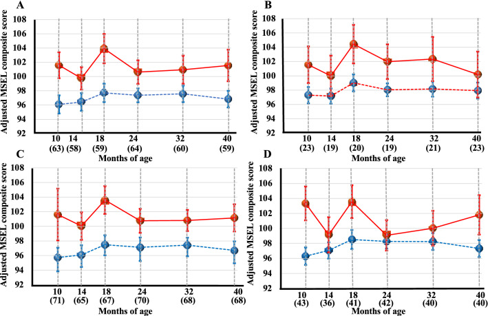
The aim of present study was to investigate the association of placental pathological findings with infantile neurodevelopment during the early 40 months of life. 258 singleton infants were enrolled in the Hamamatsu Birth Cohort for Mothers and Children (HBC Study) whose placentas were saved in our pathological division. To assess the infantile neurodevelopment, we used Mullen Scales of Early Learning (gross motor, visual reception, fine motor, receptive language, expressive language) at 10, 14, 18, 24, 32, and 40 months. For obtaining placental blocks, we carried out random sampling and assessed eleven pathological findings using mixed modeling identified 'Accelerated villous maturation', 'Maternal vascular malperfusion', and 'Delayed villous maturation' as significant predictors of the relatively lower MSEL composite scores in the neurodevelopmental milestones by Mullen Scales of Early Learning. On the other hand, 'Avascular villi', 'Thrombosis or Intramural fibrin deposition', 'Fetal vascular malperfusion', and 'Fetal inflammatory response' were significant predictors of the relatively higher MSEL composite scores in the neurodevelopmental milestones by Mullen Scales of Early Learning. In conclusion, the present study is the first to report that some placental pathological findings are bidirectionally associated with the progression of infantile neurodevelopment during 10-40 months of age.


Conflict of interest statement

The authors declare no competing interests.

Figures

Figure 1
Figure 1
Representative pathological findings by HE staining of placentas. (A) ‘Accelerated villous maturation’; the yellow arrow indicates increases in the numbers of placental villi with the focal formation of tight adherent villous clusters with syncytial knots. (B) ‘Decidual arteriopathy’; A thrombus in decidual vessel. (C) ‘Thrombosis or Intramural fibrin deposition’; the yellow arrow indicates a fibrin cushion in the walls of stem villous vessels. (D) ‘Avascular villi’: the yellow arrow indicates a villus with hyalinized stroma, which is devoid of vessels. (E) ‘Delayed villous maturation’. (F) ‘Maternal inflammatory response’; the infiltration of neutrophils into the chorionic plate. (G) ‘Fetal inflammatory response’; the infiltration of neutrophils into the umbilical artery. (H) ‘VUE; villitis of unknown etiology’; the yellow arrow indicates lymphohistiocytic inflammation predominantly in the stroma of terminal villi. (I) ‘Deciduitis’; the yellow arrow indicates the infiltration of plasma cells.
Figure 2
Figure 2
Chronological changes of Mullen Scales of Early Learning composite scores in each placental pathological finding during 10 to 40 months; ‘Accelerated villous maturation’ (A), ‘Delayed villous maturation’ (B), and ‘Maternal vascular malperfusion’ (C) as significant predictors of the relatively lower MSEL composite scores in the infantile neurodevelopmental milestones by a mixed model analysis. Red and blue dots indicate MSEL composite scores with and without ‘Accelerated villous maturation’, ‘Maternal vascular malperfusion’, or ‘Delayed villous maturation’, respectively. Circles and error bars indicate the mean and the standard error of the mean.
Figure 3
Figure 3
Chronological changes of Mullen Scales of Early Learning composite scores in each placental pathological finding during 10 to 40 months; ‘Thrombosis or Intramural fibrin deposition’ (A), ‘Avascular villi’ (B), ‘Fetal vascular malperfusion’ (C), and ‘Fetal inflammatory response’ (D) as significant predictors of the relatively higher MSEL composite scores in the infantile neurodevelopmental milestones by a mixed model analysis. Red and blue dots indicate MSEL composite scores with and without ‘Thrombosis or Intramural fibrin deposition’, ‘Avascular villi’, ‘Fetal inflammatory response’, and ‘Fetal vascular malperfusion’. Circles and error bars indicate the mean and standard error of the mean.

References

    1. Gluckman PD, Hanson MA. Living with the past: Evolution, development, and patterns of disease. Science. 2004;305:1733–1736. doi: 10.1126/science.1095292. - DOI - PubMed
    1. Hanson MA, Gluckman PD. Early developmental conditioning of later health and disease: Physiology or pathophysiology? Physiol. Rev. 2014;94:1027–1076. doi: 10.1152/physrev.00029.2013. - DOI - PMC - PubMed
    1. Itoh H, Kanayama N. Nutritional conditions in early life and risk of non-communicable diseases (NCDs); the perspective of preemptive medicine in perinatal care. Hypertens. Res. Pregnancy. 2015;3:1–12. doi: 10.14390/jsshp.3.1. - DOI
    1. Itoh H, Kanayama N. Developmental Origins of Health and Diseases (DOHaD); Perspective toward Preemptive Medicine. Springer Nature; 2017. pp. 237–250.
    1. Gluckman PD, Hanson MA. Developmental Origins of Health and Disease. Cambridge University Press; 2006.

Publication types