. 2022 Jan 1;18(3):1008-1021.
doi: 10.7150/ijbs.68028. eCollection 2022.

Mir-484 contributes to diminished ovarian reserve by regulating granulosa cell function via YAP1-mediated mitochondrial function and apoptosis

Affiliations

Mir-484 contributes to diminished ovarian reserve by regulating granulosa cell function via YAP1-mediated mitochondrial function and apoptosis

Huiying Li et al. Int J Biol Sci. .

Erratum in

Abstract

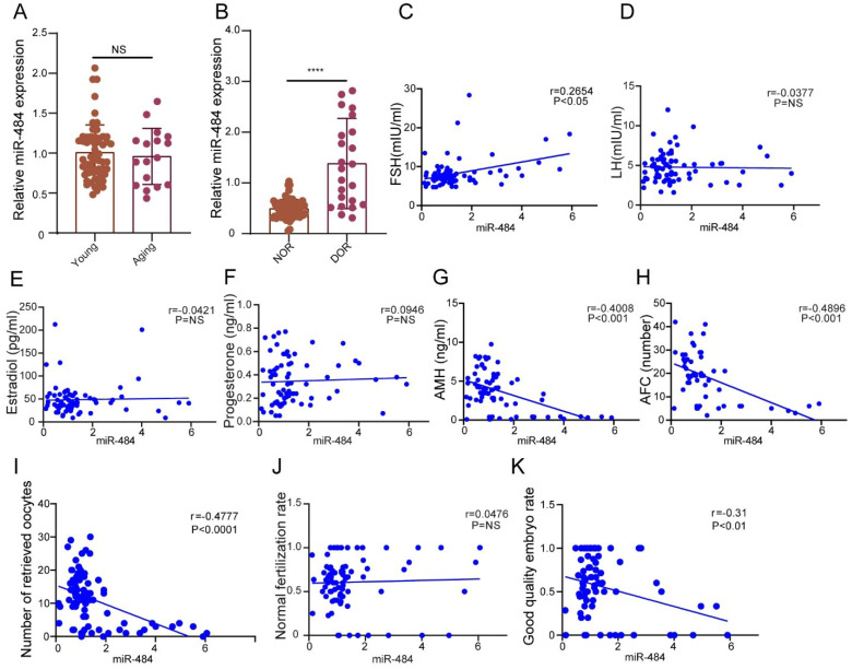
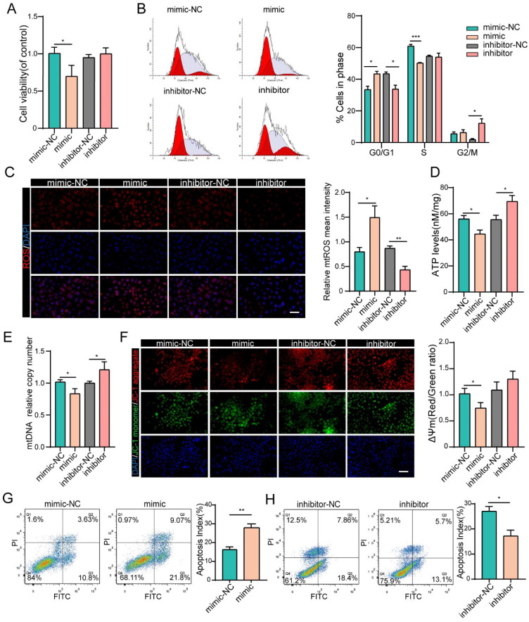
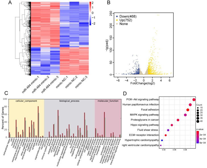
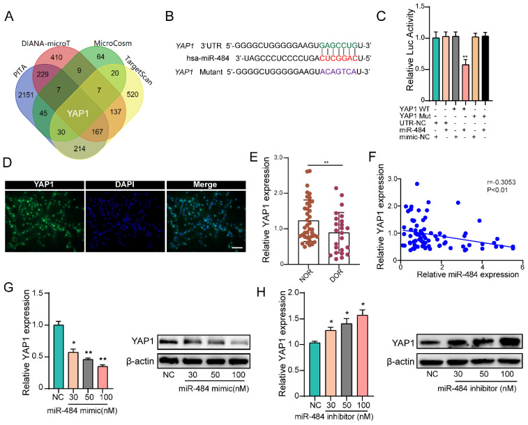
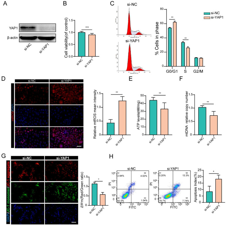
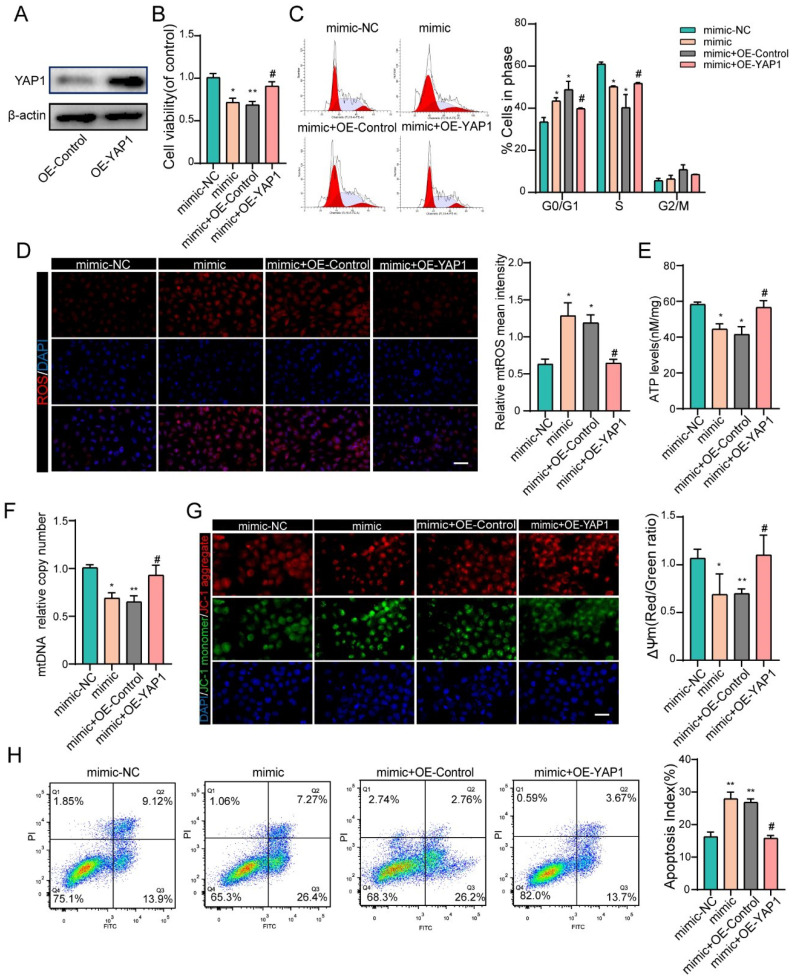
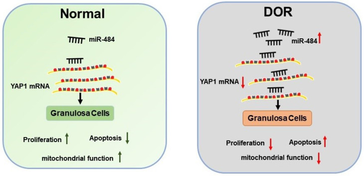
Women with diminished ovarian reserve (DOR) have reduced fertility, but the underlying regulation of ovarian function remains unknown. Although differential microRNA (miRNA) expression has been described in several ovarian disorders, little is known about the role of miRNAs in the pathogenesis of DOR. In this study, we investigated the expression levels of miR-484 in granulosa cells (GCs) derived from human follicular fluid, and explored their correlation with female ovarian reserve function as well as clinical outcomes of assisted reproduction technology (ART). Additionally, we investigated the effects of miR-484 on the biological functions of GC cell lines in vitro. We found that miR-484 was highly expressed in GCs from DOR patients and was correlated with decreasing AMH levels and AFC, as well as increasing FSH levels, but not with LH, progesterone, or estradiol. Additionally, miR-484 was negatively related to the number of retrieved oocytes and the ratio of high-quality embryos. Moreover, we found that miR-484 repressed the proliferation of GCs and induced apoptosis, which can in part be attributed to mitochondrial dysfunction. Conversely, silencing miR-484 had the opposite effect. Multiple approaches, including bioinformatic analysis, RNA-seq, qPCR, immunofluorescence, western blotting and luciferase reporter assays, identified YAP1 as a direct target of miR-484 in GCs. Additionally, reintroduction of YAP1 rescued the effects of miR-484 in GCs. The present study indicates that miR-484 can directly target the mRNA of YAP1, induce mitochondrial dysfunction, and consequently reduce the viability and promote the apoptosis of granulosa cells, which contributes to the pathogenesis of DOR.


Conflict of interest statement

Competing Interests: The authors have declared that no competing interest exists.

Figures

Figure 1
Figure 1
The expression level of miR-484 in GCs from human follicular fluid is associated with the ovarian reserve and assisted reproductive technology outcomes. (A-B) The relative expression levels of miR-484 in females of different age and different ovarian reserve groups. (C-H) Correlations between the levels of miR-484 and reproductive hormones (FSH, LH, estradiol, progesterone, AMH) and antral follicle counts (AFC). (I, J, K) Correlation analysis of the expression level of miR-484 with the number of retrieved oocytes, normal fertilization rate and ratio of high-quality embryos. According to the consensus of experts of the Society for Assisted Reproductive Technology (SART), the normal fertilization rate was calculated as the number of 2PN/number of fertilized oocytes; the ratio of high-quality embryos was calculated as the number of high-quality embryos/number of 2PN embryos. Each bar in the figure represents the mean ±SD. Young group (≤35 years old, n=65) and Aging group (>35 years old, n=17). Normal ovarian reserve group (NOR, n=65) and Diminished ovarian reserve group (DOR, n=23). NS, not statistically significant; *, P < 0.05; **, P < 0.01; ***, P < 0.001; ****, P < 0.0001.
Figure 2
Figure 2
miR-484 affects GC function by regulating mitochondrial function and related apoptosis. (A-B) Cell viability changes and cell cycle alterations were quantified. mtROS (red) in GCs were measured and quantified (C). (D-E) ATP and mtDNA levels were measured. (F) The mitochondrial membrane potential (ΔΨm) was measured using the JC-1 assay. (G-H) Apoptotic cells were detected by flow cytometry. Bar graphs illustrating the percentage of viable (Q3), early apoptotic (Q4), late apoptotic (Q2), and necrotic (Q1) cells according to Annexin V FITC/PI staining. Each bar in the figure represents the mean ± SD from three replicates. Bar =50 µm. *, P < 0.05; **, P < 0.01; ***, P < 0.001 compared with the NC group.
Figure 3
Figure 3
Effects of miR-484 on GCs on a transcriptome-wide scale. (A) Heatmap of the DEGs in GCs overexpressing miR-484 according to RNA-seq. (B) Scatter chart of all the expressed genes in GCs overexpressing miR-484. (C) Gene Ontology analysis of transcripts from GCs overexpressing miR-484. (D) Pathway analysis of transcripts from GCs overexpressing miR-484.
Figure 4
Figure 4
YAP1 is a direct target of miR-484 in GCs. (A) Predicted targets of miR-484 according to four algorithms. (B) The miR-484 binding site in the 3'UTR of YAP1 was predicted using TargetScan. (C) The luciferase reporter assay showed direct binding between the miR-484 and the YAP1 3' UTR. (D) Expression and localization of YAP1 in human GCs. (E-F) GCs were transfected with increasing amounts of the miR-484 mimic and inhibitor, followed by quantitative analysis of changes in the mRNA and protein expression of YAP1. Bar: 25 µm. *, P < 0.05; **, P < 0.01; ***, P < 0.001 compared with the si‐NC group.
Figure 5
Figure 5
YAP1 positively regulated GC functions. (A) Transfection effectively knocked down YAP1 expression. (B) Proliferation was inhibited by YAP1 knockdown in GCs. (C) cell cycle alterations were analyzed. (D) mtROS (red) in GCs were measured and quantified. (E-F) ATP and mtDNA levels were measured following YAP1 knockdown in GCs. (G) The mitochondrial membrane potential (ΔΨm) was measured using the JC-1 assay. (H) Flow cytometry showed that apoptosis was induced by YAP1 knockdown in GCs. Each bar in the figure represents the mean ± SD from three replicates. Bar: 50 µm. *, P < 0.05; **, P < 0.01; ***, P < 0.001 compared with the si‐NC group.
Figure 6
Figure 6
YAP1 rescued the effects of miR-484 in GCs. (A) Western blot showing that YAP1 was overexpressed in GCs. (B) YAP1 rescued the miR-484-induced repression of proliferation in GCs. (C) Cell cycle distribution was analyzed using flow cytometry. (D) mtROS (red) in GCs were measured and quantified. (E-F) ATP and mtDNA levels were measured. (G) The mitochondrial membrane potential (ΔΨm) was measured using the JC-1 assay. (H) YAP1 rescued miR-484-induced apoptosis in GCs. Each bar in the figure represents the mean ± SD from three replicates. Bar: 50 µm. *, P < 0.05; **, P < 0.01; ***, P < 0.001 compared with the miR‐NC group, #p < 0.05 compared with the mimic+ OE-NC group.
Figure 7
Figure 7
Schematic diagram demonstrating the molecular mechanisms underlying miR-484 in DOR. Mir-484 contributes to diminished ovarian reserve by regulating granulosa cell function via YAP1-mediated mitochondrial function and apoptosis.

References

    1. Bellver J, Donnez J. Introduction: Infertility etiology and offspring health. Fertil Steril. 2019;111:1033–5. - PubMed
    1. Zhou Z, Zheng D, Wu H, Li R, Xu S, Kang Y. et al. Epidemiology of infertility in China: a population-based study. BJOG. 2018;125:432–41. - PubMed
    1. Bunnewell SJ, Honess ER, Karia AM, Keay SD, Al Wattar BH, Quenby S. Diminished ovarian reserve in recurrent pregnancy loss: a systematic review and meta-analysis. Fertil Steril. 2020;113:818–27. e3. - PubMed
    1. Steiner AZ, Pritchard D, Stanczyk FZ, Kesner JS, Meadows JW, Herring AH. et al. Association Between Biomarkers of Ovarian Reserve and Infertility Among Older Women of Reproductive Age. JAMA. 2017;318:1367–76. - PMC - PubMed
    1. Boucret L, Chao de la Barca JM, Moriniere C, Desquiret V, Ferre-L'Hotellier V, Descamps P. et al. Relationship between diminished ovarian reserve and mitochondrial biogenesis in cumulus cells. Hum Reprod. 2015;30:1653–64. - PubMed

Publication types