. 2022 Jan 31:13:790712.
doi: 10.3389/fphar.2022.790712. eCollection 2022.

CB2R Deficiency Exacerbates Imiquimod-Induced Psoriasiform Dermatitis and Itch Through the Neuro-Immune Pathway

Affiliations

CB2R Deficiency Exacerbates Imiquimod-Induced Psoriasiform Dermatitis and Itch Through the Neuro-Immune Pathway

Li Li et al. Front Pharmacol. .

Abstract

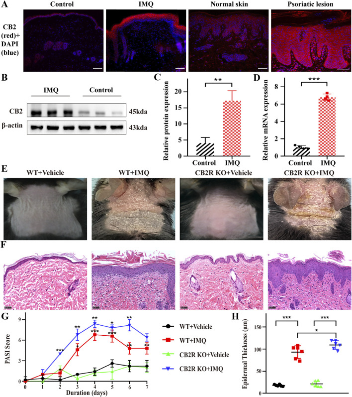
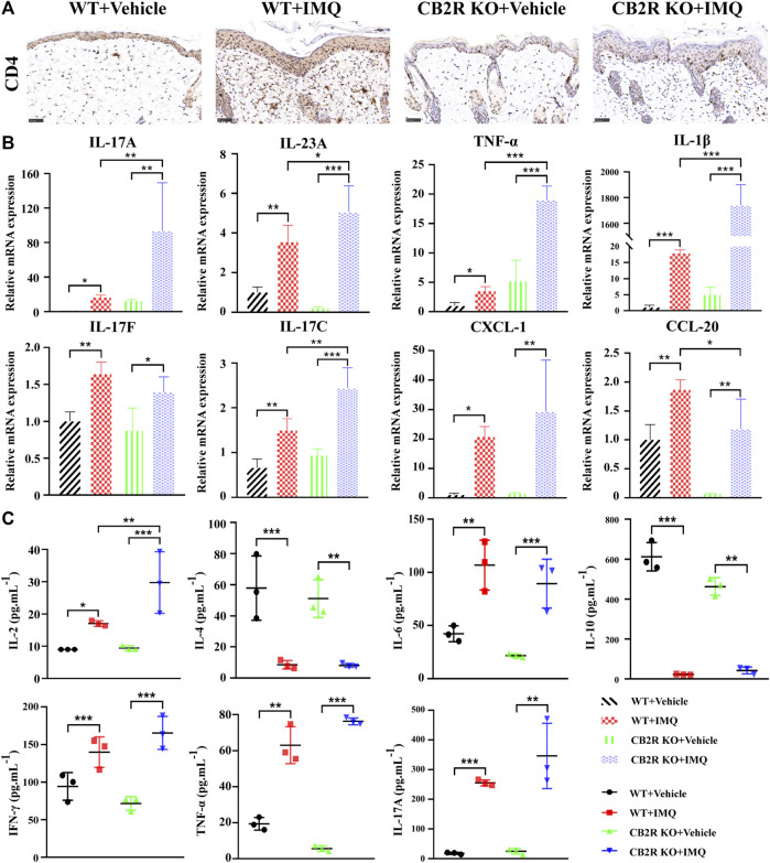
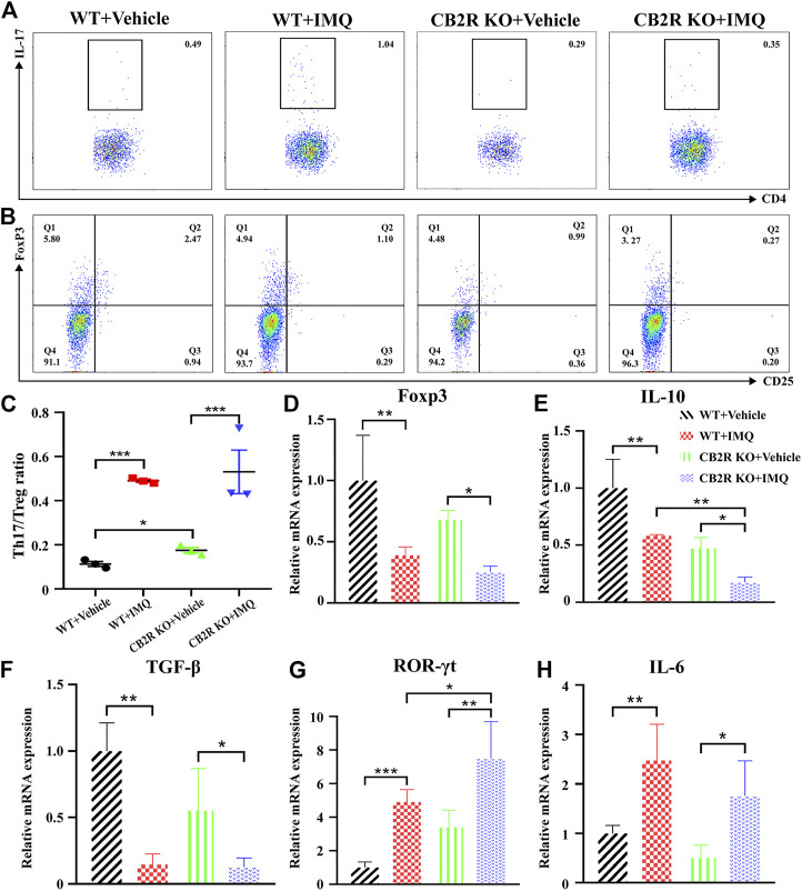
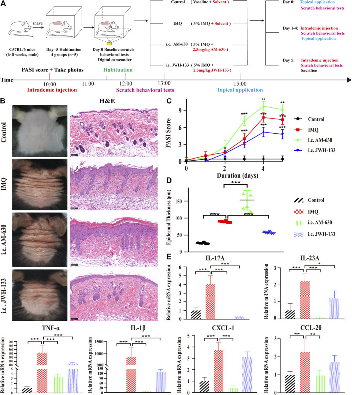
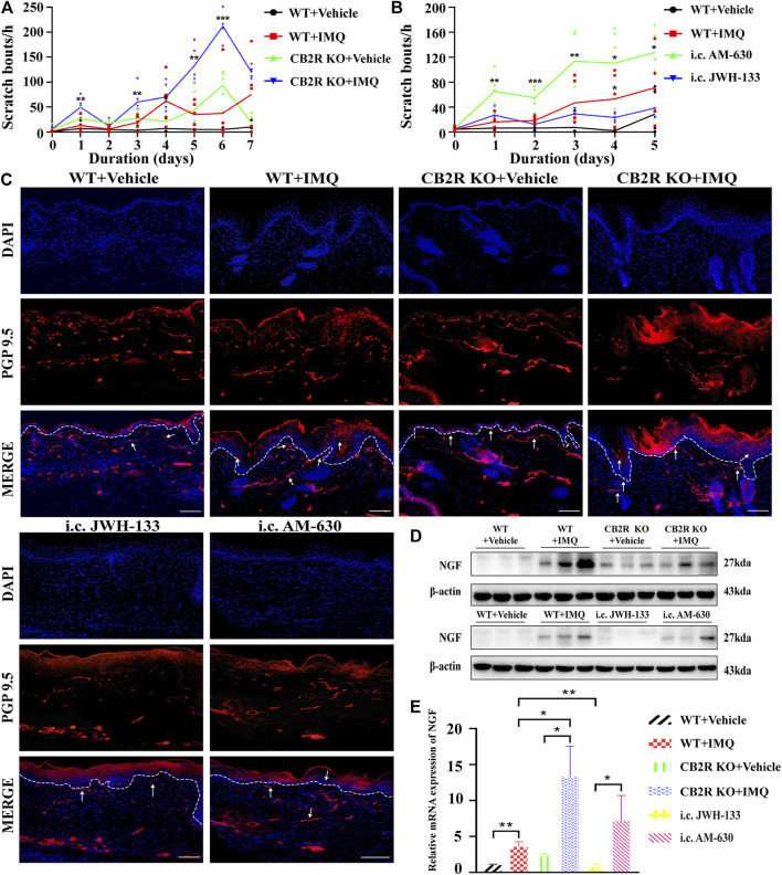
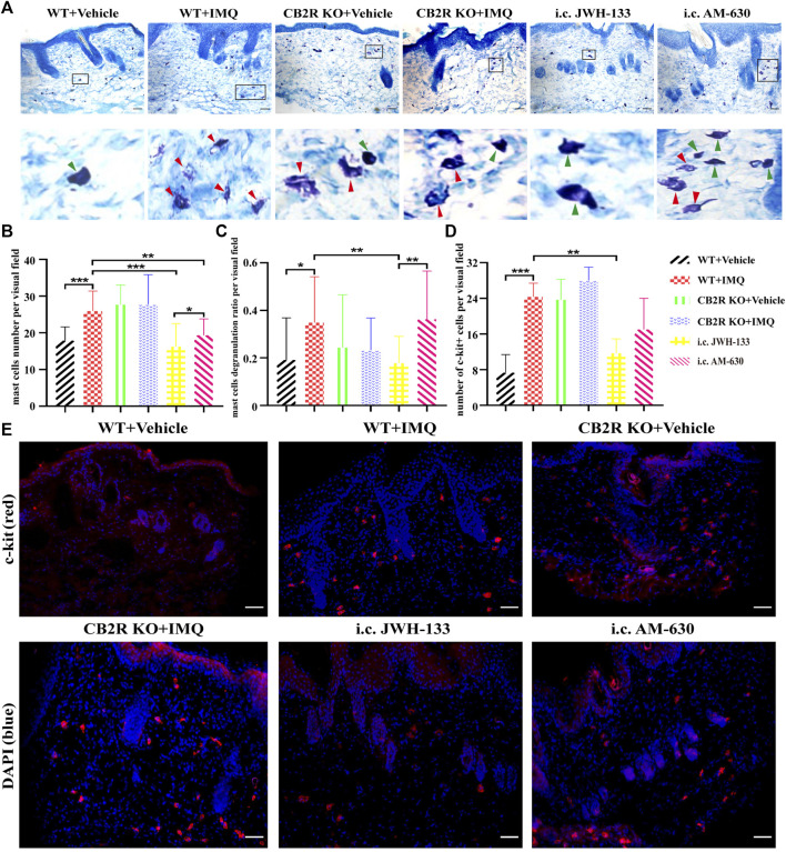
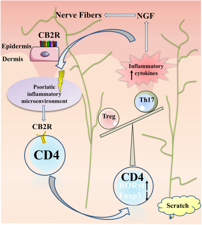
Background: Cannabinoid receptor 2 (CB2R) is a potential target for anti-inflammatory and pain therapeutics given its significant immunomodulatory and analgesic effects. However, the role of CB2R in imiquimod (IMQ)-induced psoriasiform dermatitis (PsD) and itch is poorly understood. Objective: To investigate the function and mechanism of CB2R in PsD and itch in mice. Methods: Following daily treatment with topical IMQ cream for 5-7 consecutive days in C56BL/6 wild-type (WT) and CB2R gene knockout (KO) mice, we assessed the Psoriasis Area and Severity Index (PASI) scores and the scratch bouts every day, and hematoxylin and eosin (H&E) staining, toluidine blue staining were used to observe the histological changes. mRNA levels were analyzed by quantitative real-time polymerase chain reaction (qRT-PCR). Protein levels were detected by western blotting (WB), immunohistochemistry (IHC), immunofluorescence (IF) and cytometric bead array (CBA). Flow cytometry (FCM) was used to examine the proportion of Th17/Treg cells. Results: We found that CB2R expression levels were increased in mice with psoriasis. Compared with WT mice, CB2R deficiency exacerbated IMQ-induced PsD and scratching bouts and upregulated the expression of proinflammatory cytokines by increasing the infiltration of CD4+ T cells and the Th17/Treg ratio. Obvious proliferation and prolongation of nerve fibers and high expression of nerve growth factor (NGF) were observed in PsD and CB2R KO mice. Pretreatment with the CB2R agonist, JWH-133 significantly reversed inflammation and scratching bouts. CB2R didn't participate in the induction of itch in psoriasis by regulating the expression of IL-31, thymic stromal lymphopoietin (TSLP) and mast cells in mouse skins. Conclusion: Our results demonstrate that CB2R plays a pivotal role in the pathophysiology of psoriasis, providing a new potential target for anti-inflammatory and antipruritic drugs.

Keywords: inflammation; itch (pruritus); nerve fiber; psoriasis; the cannabinoid receptor 2.


Conflict of interest statement

The authors declare that the research was conducted in the absence of any commercial or financial relationships that could be construed as a potential conflict of interest.

Figures

FIGURE 1
FIGURE 1
CB2R was upregulated throughout the epidermis, and CB2R deficiency exacerbated the morphological and histological features of psoriasiform dermatitis induced by IMQ treatment. (A) Immunofluorescence was used to detect the protein level of CB2R in psoriasiform dermatitis, human normal skin and psoriasis lesions (scale bar: 100 μm). blue, DAPI; red, CB2R. (B, C) Protein levels of CB2R were quantified by western blotting. β-actin was used as a loading control. Relative protein expression was normalized to that of the internal control. (D) mRNA levels of CB2R in skin tissues from the normal and psoriasis groups were analyzed by real-time quantitative PCR. (E) Representative phenotype manifestation of WT and CB2R KO mouse back skin induced by IMQ application at day 7. (F) H&E staining of cross-sectional slices of the dorsal skin of C57BL/6 mice on the seventh day. Scale bar represents 50 μm. (G) Clinical scores for disease severity were calculated daily of epidermal erythema, scales, and thickening of the dorsal skin. The PASI score was calculated by adding the scores of three independent criteria (ranging from 0 to 12). (H) The epidermal thickness of the dorsal skin on the seventh day was measured by four randomly selected fields per section of each mouse. Results are representative of three independent experiments. Error bars represent mean ± SD. (n = 5 for each group). *p < 0.05, **p < 0.01, and ***p < 0.001 when compared.
FIGURE 2
FIGURE 2
Effects of CB2R on the infiltration of CD4+ T cell and expression levels of inflammatory cytokines in mice with IMQ-induced psoriasiform dermatitis. (A) The representative immunohistochemical staining images of IMQ- and vehicle- treated skin in WT and CB2R KO mice at day 7. Scale bar represents 50 μm.(B) Real-time quantitative PCR was performed to quantify the mRNA level of IL-17A, IL-23A, TNF-ɑ, IL-1β, IL-17F, IL-17C, CXCL-1, CCL-20 in the skin biopsies from mice. Data are obtained from duplicate samples from five mice in each group. (C) CBA assay was performed to quantify the protein level of IL-2, IL-4, IL-6, IL-10, IFN-γ, TNF-ɑ and IL-17A in the serum from mice. All the assays were repeated three times with consistent results. Error bars represent mean ± SD. *p < 0.05, **p < 0.01, and ***p < 0.001 when compared.
FIGURE 3
FIGURE 3
Effects of CB2R deficiency on differentiation proportion of Th17/Treg in IMQ-induced PsD. (A) Flow cytometric analysis of the proportion of Th17 cells (CD4+, IL-17A+ cells) in single cell suspensions of spleen of IMQ- and vehicle- treated skin in WT and CB2R KO mice. (B) Flow cytometric analysis of the proportion of Treg cells (CD4+, CD25+, and Foxp3+ cells) in single cell suspensions of spleen of IMQ- and vehicle- treated skin in WT and CB2R KO mice. (C) The ratio of Th17/Treg cells. (D-H) The mRNA expression levels of Foxp3 (D), IL-10 (E), TGF-β (F), ROR-γt (G), IL-6 (H). Error bars represent mean ± SD. *p < 0.05, **p < 0.01, and ***p < 0.001 when compared.
FIGURE 4
FIGURE 4
The selective agonist of CB2R, JWH-133, could alleviate PsD in IMQ-induced mice. (A) Schematic diagram of the animal experiment protocol for the control, IMQ, i.c. AM-630 and i.c. JWH-133 groups (n = 5). (B) Representative macroscopic view and H&E staining of cross-sectional slices of the dorsal skin of C57BL/6 mice on the fifth day. Scale bar represents 100 μm. (C) Daily assessment of epidermal erythema, scales, and thickening of the dorsal skin. The PASI score was calculated by adding the scores of three independent criteria (ranging from 0 to 12). (D) The epidermal thickness of the dorsal skin on the fifth day was measured by four randomly selected fields per section of each mouse. (E) The mRNA expression levels of IL-17A, IL-23A, TNF-α, IL-1β, CXCL-1 and CCL-20 were measured by qRT-PCR. Error bars represent mean ± SD. (n = 5 for each group). *p < 0.05, **p < 0.01, and ***p < 0.001 when compared. All the assays were repeated three times with consistent results.
FIGURE 5
FIGURE 5
Effects of CB2R on the scratching behavior and expression of nerve fibers in IMQ-induced PsD. (A, B) The behavior results of mice in different groups. ( C) PGP 9.5 stained (red fluorescence) nerve fibers in the skin biopsies from mice, co-stained with DAPI (blue fluorescence) to detect the nucleus. Scale bar represents 100 μm. (D) Protein level of NGF was analyzed by western blotting in the six groups of mice. β-actin served as the loading control. ( E) The mRNA expression levels of NGF was measured by qRT-PCR. Error bars represent mean ± SD. (n = 5 for each group). *p < 0.05, **p < 0.01, and ***p < 0.001 when compared. All the assays were repeated three times with consistent results.
FIGURE 6
FIGURE 6
Regulation of mast cells number and degranulation by CB2R in IMQ-induced psoriasis. (A–C) Toluidine bule staining to detect the number and the degranulation of mast cells, green arrows pointed to non-degranulated mast cells, red arrows pointed to degranulated mast cells. (D, E) Immunofluorescence to analysis of c-kit (mast cell marker) expression and quantification of mast cells number in the skin. Error bars represent mean ± SD. *p < 0.05, **p < 0.01, and ***p < 0.001 when compared. Scale bar represents 100 μm.
FIGURE 7
FIGURE 7
Summary of CB2R actions in psoriatic inflammation and itch. Under the psoriatic inflammatory microenvironment, CB2R regulated proliferation and differentiation of CD4+ T cell, increased ROR-γt expression and decreased Foxp3 expression, resulting in imbalance of Th17 and Treg cells, further aggravating the inflammatory microenvironment of psoriasis. The local high expression of a variety of inflammatory factors stimulated the keratinocytes secreting NGF for nerve fiber hyperplasia, resulting in pruritus paresthesia.

Similar articles

Cited by

References

    1. Agarwala M. K., George R., Pramanik R., McGrath J. A. (2016). Olmsted Syndrome in an Indian Male with a New De Novo Mutation in TRPV3. Br. J. Dermatol. 174 (1), 209–211. 10.1111/bjd.13910 - DOI - PubMed
    1. Armstrong A. W., Read C. (2020). Pathophysiology, Clinical Presentation, and Treatment of Psoriasis: A Review. JAMA 323 (19), 1945–1960. 10.1001/jama.2020.4006 - DOI - PubMed
    1. Avila C., Massick S., Kaffenberger B. H., Kwatra S. G., Bechtel M. (2020). Cannabinoids for the Treatment of Chronic Pruritus: A Review. J. Am. Acad. Dermatol. 82 (5), 1205–1212. 10.1016/j.jaad.2020.01.036 - DOI - PubMed
    1. Boehncke W.-H., Schön M. P. (2015). Psoriasis. The Lancet 386 (9997), 983–994. 10.1016/S0140-6736(14)61909-7 - DOI - PubMed
    1. Bovenschen H. J., van de Kerkhof P. C., van Erp P. E., Woestenenk R., Joosten I., Koenen H. J. (2011). Foxp3+ Regulatory T Cells of Psoriasis Patients Easily Differentiate into IL-17A-producing Cells and Are Found in Lesional Skin. J. Invest. Dermatol. 131 (9), 1853–1860. 10.1038/jid.2011.139 - DOI - PubMed