Imaging β-Cell Function Using a Zinc-Responsive MRI Contrast Agent May Identify First Responder Islets
- PMID: 35173681
- PMCID: PMC8842654
- DOI: 10.3389/fendo.2021.809867
Imaging β-Cell Function Using a Zinc-Responsive MRI Contrast Agent May Identify First Responder Islets
Abstract
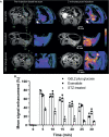
An imaging method for detecting β-cell function in real-time in the rodent pancreas could provide new insights into the biological mechanisms involving loss of β-cell function during development of type 2 diabetes and for testing of new drugs designed to modulate insulin secretion. In this study, we used a zinc-responsive MRI contrast agent and an optimized 2D MRI method to show that glucose stimulated insulin and zinc secretion can be detected as functionally active "hot spots" in the tail of the rat pancreas. A comparison of functional images with histological markers show that insulin and zinc secretion does not occur uniformly among all pancreatic islets but rather that some islets respond rapidly to an increase in glucose while others remain silent. Zinc and insulin secretion was shown to be altered in streptozotocin and exenatide treated rats thereby verifying that this simple MRI technique is responsive to changes in β-cell function.
Keywords: glucose stimulated zinc secretion (GSZS); glucose-stimulated insulin secretion (GSIS); magnetic resonance imaging; metabolic imaging; pancreatic β-cell function; zinc-responsive contrast agent.
Copyright © 2022 Thapa, Suh, Parrott, Khalighinejad, Sharma, Chirayil and Sherry.
Conflict of interest statement
The authors declare that the research was conducted in the absence of any commercial or financial relationships that could be construed as a potential conflict of interest.
Figures


Similar articles
-
Imaging Insulin Secretion from Mouse Pancreas by MRI Is Improved by Use of a Zinc-Responsive MRI Sensor with Lower Affinity for Zn2+ Ions.J Am Chem Soc. 2018 Dec 19;140(50):17456-17464. doi: 10.1021/jacs.8b07607. Epub 2018 Dec 11. J Am Chem Soc. 2018. PMID: 30484648 Free PMC article.
-
MRI Methods for Imaging Beta-Cell Function in the Rodent Pancreas.Methods Mol Biol. 2023;2592:101-111. doi: 10.1007/978-1-0716-2807-2_7. Methods Mol Biol. 2023. PMID: 36507988 Free PMC article.
-
Imaging Beta-Cell Function in the Pancreas of Non-Human Primates Using a Zinc-Sensitive MRI Contrast Agent.Front Endocrinol (Lausanne). 2021 May 26;12:641722. doi: 10.3389/fendo.2021.641722. eCollection 2021. Front Endocrinol (Lausanne). 2021. PMID: 34122330 Free PMC article.
-
Pancreatic Thyrotropin Releasing Hormone and Mechanism of Insulin Secretion.Cell Physiol Biochem. 2018;50(1):378-384. doi: 10.1159/000494013. Epub 2018 Oct 4. Cell Physiol Biochem. 2018. PMID: 30286449 Review.
-
Metabolic signaling of insulin secretion by pancreatic β-cell and its derangement in type 2 diabetes.Eur Rev Med Pharmacol Sci. 2014;18(15):2215-27. Eur Rev Med Pharmacol Sci. 2014. Retraction in: Eur Rev Med Pharmacol Sci. 2016;20(3):391. PMID: 25070829 Retracted. Review.
Cited by
-
Zinc Chloride Enhances the Antioxidant Status, Improving the Functional and Structural Organic Disturbances in Streptozotocin-Induced Diabetes in Rats.Medicina (Kaunas). 2022 Nov 10;58(11):1620. doi: 10.3390/medicina58111620. Medicina (Kaunas). 2022. PMID: 36363577 Free PMC article.
-
Impact of dietary zinc on stimulated zinc secretion MRI in the healthy and malignant mouse prostate.Npj Imaging. 2024;2(1):47. doi: 10.1038/s44303-024-00051-1. Epub 2024 Nov 6. Npj Imaging. 2024. PMID: 39525279 Free PMC article.
-
Relationship between imaging changes of the pancreas and islet beta-cell function.World J Radiol. 2024 Dec 28;16(12):717-721. doi: 10.4329/wjr.v16.i12.717. World J Radiol. 2024. PMID: 39801662 Free PMC article.
-
Complementary Approaches to Interrogate Mitophagy Flux in Pancreatic β-Cells.J Vis Exp. 2023 Sep 15;(199):10.3791/65789. doi: 10.3791/65789. J Vis Exp. 2023. PMID: 37782087 Free PMC article.
References
-
- Huynh J, Yamada J, Beauharnais C, Wenger JB, Thadhani RI, Wexler D, et al. . Type 1, Type 2 and Gestational Diabetes Mellitus Differentially Impact Placental Pathologic Characteristics of Uteroplacental Malperfusion. Placenta (2015) 36:1161–6. doi: 10.1016/j.placenta.2015.08.004 - DOI - PMC - PubMed
-
- Baynest HW. Classification, Pathophysiology, Diagnosis and Management of Diabetes Mellitus. J Diabetes Metab (2015) 6:1–9. doi: 10.4172/2155-6156.1000541 - DOI
Publication types
MeSH terms
Substances
LinkOut - more resources
Full Text Sources
Medical

