

Scheimpflug Corneal Densitometry Values and Severity of Guttae in Relation to Visual Acuity in Fuchs Endothelial Corneal Dystrophy
- PMID: 35175018
- PMCID: PMC8857507
- DOI: 10.1097/ICO.0000000000002762
Scheimpflug Corneal Densitometry Values and Severity of Guttae in Relation to Visual Acuity in Fuchs Endothelial Corneal Dystrophy
Abstract
Purpose: The purpose of this study was to investigate the association between corneal densitometry (CD) values from Scheimpflug tomography imaging, severity of guttae, and visual acuity in eyes with Fuchs endothelial corneal dystrophy (FECD).
Methods: This was a retrospective, cross-sectional study. Patients with FECD were examined at the Bascom Palmer Eye Institute from January 2015 to September 2019. We extracted CD values at central annuli of 0-2, 2-6, 6-10 and 10-12 mm from Scheimpflug tomography images. We investigated the association of corrected distance visual acuity (CDVA) with CD values, severity of guttae, central corneal thickness (CCT), cataract grade, refractive error, corneal edema grade, age, and gender using multivariate generalized estimating equation regression models.
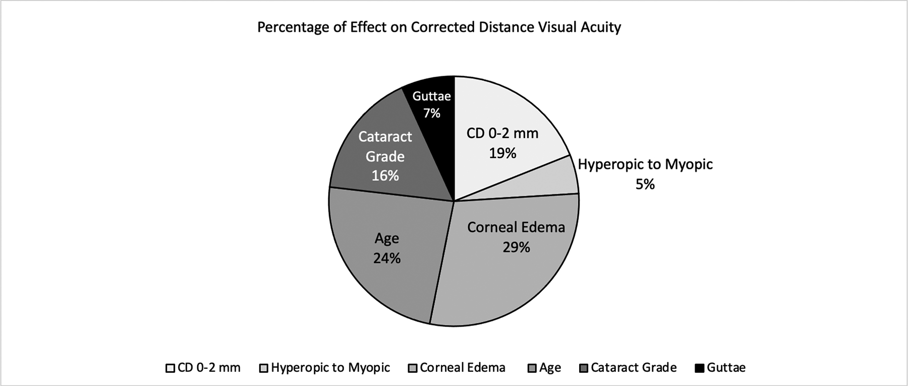
Results: One hundred ninety-two eyes from 110 patients were included in this study. Increase in central CD values at the 0 to 2 mm zone (P < 0.001), severity of guttae (P = 0.046), age (P < 0.001), cataract grade (P < 0.001), corneal edema grade (P < 0.001), and type of refractive error (P = 0.008) were significantly associated with decreased CDVA. Central corneal thickness, sex, and the peripheral CD values (2-6, 6-10, and 10-12 mm) were not significantly associated with CDVA (P > 0.05) in the final multivariate regression model.
Conclusions: Our study demonstrates that central CD values at 0 to 2 mm and severity of guttae are each associated with decreased CDVA in FECD. These findings carry implications for patients with FECD considering surgical intervention for phacoemulsification alone, Descemet stripping only, or endothelial cell transplantation and provide a multifactorial perspective on vision loss in FECD.
Copyright © 2021 Wolters Kluwer Health, Inc. All rights reserved.
Conflict of interest statement
The authors have no conflicts of interest to disclose.
Figures
References
-
- Eye Bank Association of America, 2019. Eye Banking Statistical Report. www.restoresight.org. Accessed December 26, 2020.
-
- Otri AM, Fares U, Al-Aqaba MA, Dua HS. Corneal densitometry as an indicator of corneal health. Ophthalmology. 2012;119:501–508. - PubMed
-
- Tekin K, Sekeroglu MA, Kiziltoprak H, Yilmazbas P. Corneal Densitometry in Healthy Corneas and Its Correlation With Endothelial Morphometry. Cornea. 2017;36:1336–1342. - PubMed
-
- Gorovoy MS. Descemet-stripping automated endothelial keratoplasty. Cornea. 2006;25:886–889. - PubMed
-
- Melles GRJ, Ong TS, Ververs B, van der Wees J. Descemet membrane endothelial keratoplasty (DMEK). Cornea. 2006;25:987–990. - PubMed
MeSH terms
Grants and funding
LinkOut - more resources
Full Text Sources
Medical
