. 2022 Feb 17;479(4):463-477.
doi: 10.1042/BCJ20210280.

Beyond protein modification: the rise of non-canonical ADP-ribosylation

Affiliations

Beyond protein modification: the rise of non-canonical ADP-ribosylation

Marion Schuller et al. Biochem J. .

Abstract

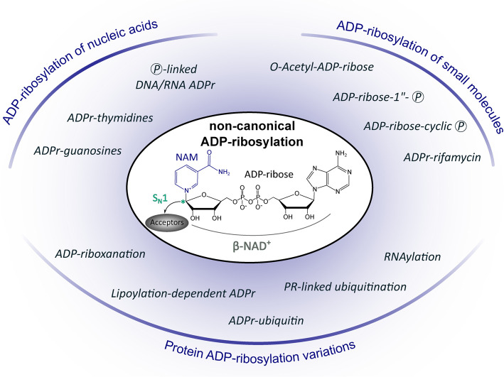
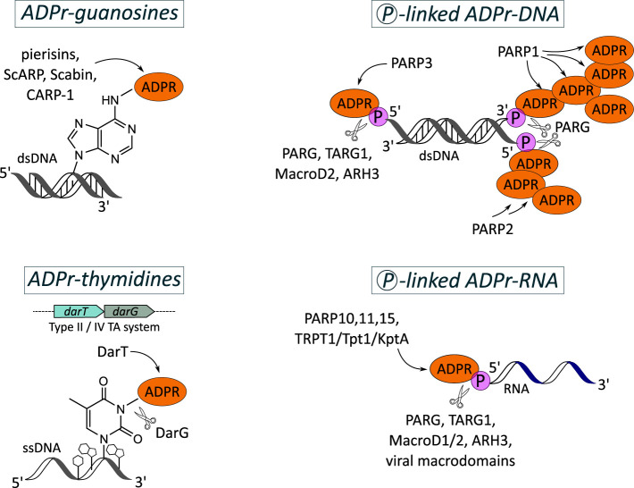
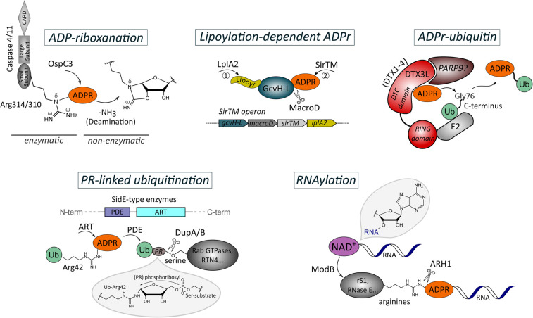
ADP-ribosylation has primarily been known as post-translational modification of proteins. As signalling strategy conserved in all domains of life, it modulates substrate activity, localisation, stability or interactions, thereby regulating a variety of cellular processes and microbial pathogenicity. Yet over the last years, there is increasing evidence of non-canonical forms of ADP-ribosylation that are catalysed by certain members of the ADP-ribosyltransferase family and go beyond traditional protein ADP-ribosylation signalling. New macromolecular targets such as nucleic acids and new ADP-ribose derivatives have been established, notably extending the repertoire of ADP-ribosylation signalling. Based on the physiological relevance known so far, non-canonical ADP-ribosylation deserves its recognition next to the traditional protein ADP-ribosylation modification and which we therefore review in the following.

Keywords: ADP-ribosylation; PARP; nucleic acids; protein modification.


Conflict of interest statement

The authors declare that there are no competing interests associated with the manuscript.

Figures

Figure 1.
Figure 1.. Overview of non-canonical ADP-ribosylation reactions discussed in this review.
(ADPr: ADP-ribosylated/ADP-ribosylation; NAM: Nicotinamide; Ⓟ: phosphate; PR: phosphoribosyl; SN1: Nucleophilic Substitution, First Order — reaction mechanism for catalysing ADP-ribosylation which involves an oxocarbenium ion generated by NAD+ cleavage for nucleophilic attack of acceptors; * anomeric carbon linking ADP-ribose to acceptors).
Figure 2.
Figure 2.. ADP-ribosylation of nucleic acids.
(Top, left) Irreversible mono-ADP-ribosylation of guanosine bases by pierisins, ScARP, Scabin and CARP-1. (Bottom, left) Mono-ADP-ribosylation of thymidine bases in ssDNA by the toxin DarT which is reversible by its antitoxin partner, DarG. (Top, right) Mono- and poly-ADP-ribosylation of the DNA backbone via terminal phosphate-linkage catalysed by PARP1–3. The modifications are reversible by macrodomain-containing proteins (PARG, TARG1, MacroD2) and ARH3. (Bottom, right) Mono-ADP-ribosylation of the RNA backbone via phosphate-linkage catalysed by PARP family members and TRPT1/TPT1/KptA. (ADPR: ADP-ribose; ADPr: ADP-ribosylated; Ⓟ: phosphate; TA: Toxin-Antitoxin system).
Figure 3.
Figure 3.. ADP-ribosylation of small molecules.
(Top, right) De-acetylation of proteins by sirtuins results in generation of O-acetyl-ADP-ribose which is cleaved to ADP-ribose and acetate by the hydrolases MacroD1/2, TARG1 and ARH3. (Top, left) De-phosphorylation of tRNA as intermediate step in tRNA splicing by Tpt1 family members releases mature tRNA and ADP-ribose-cyclic phosphate. The latter is cleaved to ADP-ribose and phosphate by the hydrolases Poa1P and MacroD1/2. (Bottom, left) The toxin MbcT of the type II TA system MbcTA functions as a NAD+ phosphorylase, thereby generating ADP-ribose-1″-phosphate. Binding of the antitoxin MbcA to MbcT inhibits toxin activity that is stimulated by inorganic phosphate. (Bottom, right) Mono-ADP-ribosylation of rifamycin by Arr enzymes leads to loss of the antibiotic activity of the molecule by inhibiting the binding of rifamycin to its target, i.e. the DNA-dependent RNA polymerase. (Ac: Acetate; ADPR: ADP-ribose; Ⓟ: phosphate; TA: Toxin-Antitoxin system).
Figure 4.
Figure 4.. Non-canonical protein ADP-ribosylation.
(Top, left) The type III secretion system effector of Shigella flexneri, OspC3, ADP-ribosylates the caspase-conserved arginine residues in caspase 4/11 (Arg314/Arg310) which is followed by non-enzymatic internal deamination — a process termed ADP-riboxanation. The modification is understood to be irreversible and to provide means for the pathogen to escape the inflammatory response of the host. (Top, middle) In Staphylococcus aureus and Streptococcus pyogenes, ADP-ribosylation activity of SirTM is dependent on prior lipoylation of its specific target, the lipoyl-carrier protein GcvH-L, by the lipoate-protein ligase A (LplA2). The modification is reversed by a MacroD hydrolyse which is encoded within the same operon as SirTM, GcvH-L and LplA2. (Top, right) The DTC domain of DTX1-4 ADP-ribosylates the C-terminus of ubiquitin at Gly76 which is recruited to site of action by the interaction of the RING domain of DTX1–4 with E2 ligase. PARP9 forms a complex with the family member DTX3L, yet its precise role in this ADP-ribosylation reaction is still unclear. (Bottom, left) In a two-step reaction, SidE-type bacterial effectors first ADP-ribosylate with their ART domain ubiquitin at Arg42. The pyrophosphate of the ADP-ribose modification is then cleaved by their PDE domain resulting in phosphoribosylated ubiquitin (PR-Ub) which is conjugated to serine residues in substrate proteins. The PR-UB modification can be removed from the serine residues by the hydrolases DupA/B. (Bottom, right) The ARTC-class member produced by T4 bacteriophage, ModB, attaches RNA chains to arginine residues of host acceptor proteins via a diphosphoriboside linkage by utilising NAD+-capped RNAs as substrate. The modification can be reversed by ARH1. Amino acid identifier refer to human proteins in the ADP-riboxanation, ADPr-ubiquitin and PR-linked ubiquitination panels. (ADPR: ADP-ribose; ADPr: ADP-ribosylation; ART: ADP-ribosyltransferase; DTC: ‘Deltex carboxyl-terminal’ domain; PDE: Phosphodiesterase; PR: phosphoribosyl; Ub: ubiquitin).

References

    1. Lüscher, B., Bütepage, M., Eckei, L., Krieg, S., Verheugd, P. and Shilton, B.H. (2018) ADP-ribosylation, a multifaceted posttranslational modification involved in the control of cell physiology in health and disease. Chem. Rev. 118, 1092–1136 10.1021/acs.chemrev.7b00122 - DOI - PubMed
    1. Challa, S., Stokes, M.S. and Kraus, W.L. (2021) Marts and marylation in the cytosol: biological functions, mechanisms of action, and therapeutic potential. Cells 10, 313 10.3390/cells10020313 - DOI - PMC - PubMed
    1. Palazzo, L., Mikolčević, P., Mikoč, A. and Ahel, I. (2019) ADP-ribosylation signalling and human disease. Open Biol. 9, 190041 10.1098/rsob.190041 - DOI - PMC - PubMed
    1. Mikolčević, P., Hloušek-Kasun, A., Ahel, I. and Mikoč, A. (2021) ADP-ribosylation systems in bacteria and viruses. Comput. Struct. Biotechnol. J. 19, 2366–2383 10.1016/j.csbj.2021.04.023 - DOI - PMC - PubMed
    1. Crawford, K., Oliver, P.L., Agnew, T., Hunn, B.H.M. and Ahel, I. (2021) Behavioural characterisation of macrod1 and macrod2 knockout mice. Cells 10, 1–24 10.3390/cells10020368 - DOI - PMC - PubMed

Publication types

MeSH terms