. 2022 Mar 22;23(4):e136-e161.
doi: 10.1093/ehjci/jeab292.

Clinical applications of cardiac computed tomography: a consensus paper of the European Association of Cardiovascular Imaging-part II

Affiliations

Clinical applications of cardiac computed tomography: a consensus paper of the European Association of Cardiovascular Imaging-part II

Gianluca Pontone et al. Eur Heart J Cardiovasc Imaging. .

Abstract

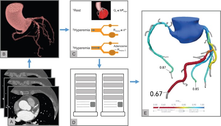
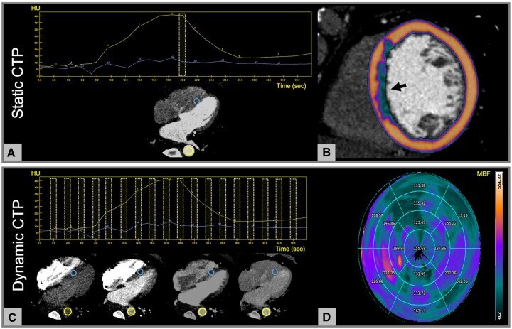
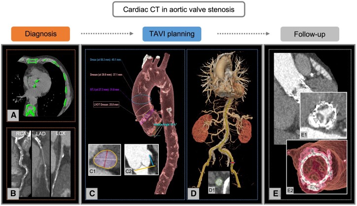
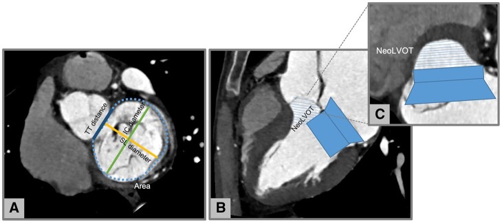
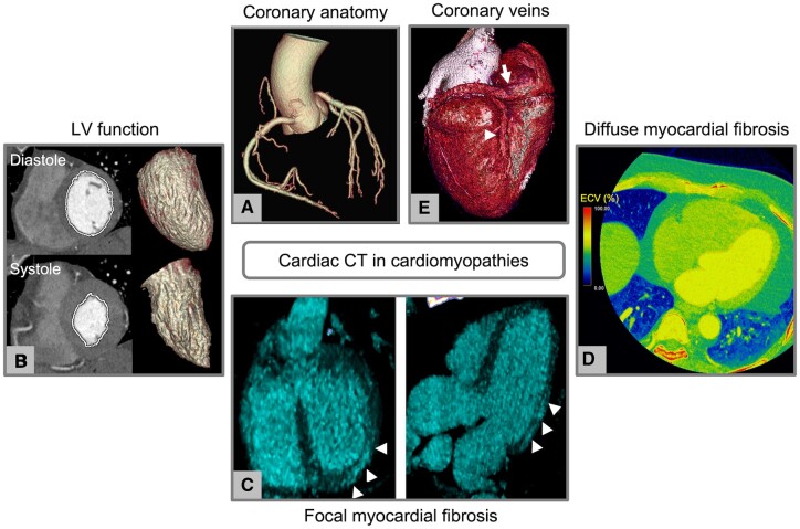
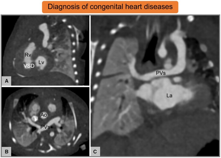
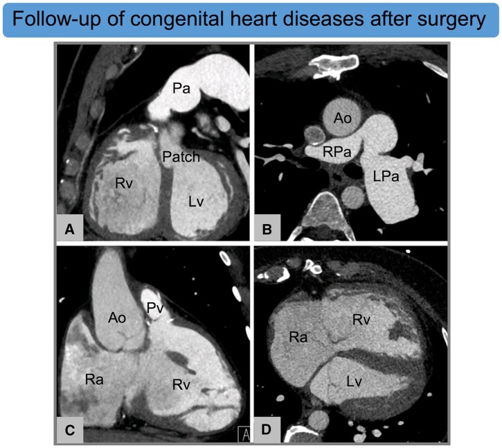
Cardiac computed tomography (CT) was initially developed as a non-invasive diagnostic tool to detect and quantify coronary stenosis. Thanks to the rapid technological development, cardiac CT has become a comprehensive imaging modality which offers anatomical and functional information to guide patient management. This is the second of two complementary documents endorsed by the European Association of Cardiovascular Imaging aiming to give updated indications on the appropriate use of cardiac CT in different clinical scenarios. In this article, emerging CT technologies and biomarkers, such as CT-derived fractional flow reserve, perfusion imaging, and pericoronary adipose tissue attenuation, are described. In addition, the role of cardiac CT in the evaluation of atherosclerotic plaque, cardiomyopathies, structural heart disease, and congenital heart disease is revised.

Keywords: CT perfusion imaging; FFRCT; artificial intelligence; cardiac computed tomography; cardiomyopathy; coronary computed tomography angiography (CCTA); fractional flow reserve; myocardial ischaemia; plaque imaging; radiomics; structural heart disease.


Figures

None
Clinical applications of cardiac CT. For more details, please refer to Table 1 which summarizes the main applications of cardiac CT. CAD, coronary artery disease; CT, computed tomography; CTP, computed tomography perfusion; FFRCT, CT-derived fractional flow reserve; LAA, left atrial appendage; TAVI, transcatheter aortic valve implantation.
Figure 1
Figure 1
Plaque characterization and quantitative assessment using semi-automated software. (A–E) Plaque of the proximal LAD. Straight (A) and curved (B) MPRs demonstrate a non-calcified plaque of the proximal LAD. The cross-section (C) shows a low-attenuation component of the plaque associated with positive remodelling and a rim of higher attenuation corresponding to the napkin-ring sign. The semi-automate software delineates inner (green) and outer (blue) vessel walls and automatically identifies components with attenuation <30 HU (blue), 31–312 HU (pink), and >312 HU (yellow). The lumen is highlighted in light blue (B and D). Quantitative data on area stenosis severity and volume of plaque subcomponents are provided in (E). HU, Hounsfield unit; LAD, left anterior descending artery; MPR, multiplanar reconstruction.
Figure 2
Figure 2
FFRCT. (A–E) Calculation of FFRCT by using computational fluid dynamics principles. (A and B) Standard rest CCTA images (A) are used to create a 3-dimensional anatomic model of the coronary arteries (B). (C) A physiological model of the coronary microcirculation is derived according to the following principles: resting coronary flow is proportional to myocardial mass; microvascular resistance is inversely proportional to vessel size; and microvascular resistance is reduced to simulate maximal hyperaemia. (D and E) An off-site supercomputer is used to compute coronary blood flow by applying the physical laws of fluid dynamics (D) and calculated FFRCT throughout the entire coronary tree (E). FFRCT, CT-derived fractional flow reserve.
Figure 3
Figure 3
CTP imaging during pharmacological stress. (A and B) Static CTP consists of a single acquisition of the entire heart at the time of maximum myocardial enhancement as shown by the yellow dotted box in (A). The image assessment is qualitative and relies on the visual comparison between the hypo-perfused area and a normal (remote) region of the myocardium. In this example, a colour-coded map representing the myocardial distribution of HU has been overlayed to the myocardium showing a subendocardial perfusion defect of the septum (arrow, B). (C and D) Dynamic CTP protocols are based on the acquisition of serial CT datasets of the whole myocardium at different time points, which allows the construction of time attenuation curves of the descending aorta (yellow circle) and the myocardium (blue circle, C). MBF is calculated in absolute terms for each single voxel of the myocardium by applying dedicated mathematical models to the time-attenuation curves. A representative polar map of the MBF (mL/100g/min) distribution based on 17-segment AHA model is shown in (D) demostrating reduced perfusion in the anterior and antero-lateral walls of the left ventricle. The numbers indicate the average perfusion value for each myocardial segment. AHA, American Heart Association; CT, computed tomography; CTP, CT perfusion; HU, Hounsfield unit; MBF, myocardial blood flow.
Figure 4
Figure 4
Role of cardiac CT in patients with aortic valve stenosis. (AE) Overview of the applications of cardiac CT in patients with aortic valve stenosis. (A and B) Diagnostic work-up of aortic valve stenosis. The severity of aortic valve stenosis can be estimated by the calcium load of aortic valve calculated according to the Agatston method on non-enhanced cardiac CT (A). In addition, CCTA can be used for CAD evaluation. In this example, curves MPRs show diffuse calcifications of RCA, LAD, and LCX (B). (C and D) Pre-procedural planning, device selection, and access sites prior to TAVI. Cardiac CT provides information on aortic root and thoracic aorta measurements (C), aortic annulus sizing with determination of maximum (white), and minimum (red) diameters, area (purple), and perimeter (orange, C1), as well as distance of the coronary ostia from the aortic valve plane (C2). In addition, CT allows the assessment of abdominal aorta and peripheral vascular access. In this case, a transfemoral vascular approach has been evaluated (D) with sizing of maximum and minimum diameters of the left femoral artery (D1). (E) Follow-up after TAVI. Cross-section (E1) and volume rendering (E2) of the prosthetic valve show an example of a patient with HALT (arrowheads). CAD, coronary artery disease; CCTA, computed tomography coronary angiography; CT, computed tomography; HALT, hypo-attenuated leaflet thickening; MPR, multiplanar reconstruction; LAD, left anterior descending coronary artery; LCX, left circumflex coronary artery; RCA, right coronary artery; TAVI, transcatheter aortic valve implantation.
Figure 5
Figure 5
Neo LVOT assessment in native mitral regurgitation with CT. (A) Measurement of mitral annulus size: area, SL, IC, and TT diameters. (B) Virtual valve implantation and (C) identification of the neo LVOT. Images by courtesy of Antonio Esposito and Anna Palmisano, San Raffaele IRCCS, Milan (Italy). CT, computed tomography; IC, inter-commissural; LVOT, left ventricular outflow tract; SL, septal–lateral; TT, trigone to trigone.
Figure 6
Figure 6
Cardiac CT in cardiomyopathies. (A–E) Overview of the potential information derived from cardiac CT in patients with cardiomyopathies. (A) Evaluation of coronary anatomy; (B) functional assessment with calculation of ventricular volumes and ejection fraction; (C) identification of focal myocardial fibrosis with late iodine enhancement technique (arrowheads); (D) assessment of diffuse fibrosis using extracellular volume measurement; and (E) assessment of the coronary venous system prior to cardiac resynchronization therapy (arrow, coronary sinus; arrowhead, posterior interventricular vein). CT, computed tomography.
Figure 7
Figure 7
Cardiac CT in neonate with suspected congenital heart disease. (A–C) Ten-day-old female patient with partial anomalous pulmonary venous return and a small muscular VSD. Images by courtesy of Francesco Bianco, Ospedale Riuniti Ancona, Ancona (Italy). Ao, aorta; CT, computed tomography; La, left atrium; Lv, left ventricle; PVs, pulmonary veins; Rv, right ventricle; VSD, ventricular septal defect.
Figure 8
Figure 8
Cardiac CT for the postoperative follow-up of congenital heart disease. (A–D) Thirty-one-year-old male with repaired Tetralogy of Fallot, postsurgical LPa aneurism and Rv dilatation. Ventricular septal defect has been repaired by using a patch (A). Images by courtesy of Francesco Bianco, Ospedale Riuniti Ancona, Ancona (Italy). Ao, aorta; CT, computed tomography; LPa, left pulmonary artery; Lv, left ventricle; Pa, pulmonary artery; Pv, pulmonary vein; Ra, right atrium; Rpa, right pulmonary artery; Rv, right ventricle.

References

    1. Chang HJ, Lin FY, Lee SE, Andreini D, Bax J, Cademartiri F et al. Coronary atherosclerotic precursors of acute coronary syndromes. J Am Coll Cardiol 2018;71:2511–22. - PMC - PubMed
    1. SCOT-HEART Investigators; Newby DE, Adamson PD, Berry C, Boon NA, Dweck MR, Flather M et al. Coronary CT angiography and 5-year risk of myocardial infarction. N Engl J Med 2018;379:924–33. - PubMed
    1. Hoffmann U, Ferencik M, Udelson JE, Picard MH, Truong QA, Patel MR et al. Prognostic value of noninvasive cardiovascular testing in patients with stable chest pain: insights from the PROMISE trial (Prospective Multicenter Imaging Study for Evaluation of Chest Pain). Circulation 2017;135:2320–32. - PMC - PubMed
    1. Min JK, Shaw LJ, Devereux RB, Okin PM, Weinsaft JW, Russo DJ et al. Prognostic value of multidetector coronary computed tomographic angiography for prediction of all-cause mortality. J Am Coll Cardiol 2007;50:1161–70. - PubMed
    1. Mushtaq S, De Araujo Goncalves P, Garcia-Garcia HM, Pontone G, Bartorelli AL, Bertella E et al. Long-term prognostic effect of coronary atherosclerotic burden: validation of the computed tomography-Leaman score. Circ Cardiovasc Imaging 2015;8:e002332. - PubMed

Publication types

MeSH terms