

The behaviour of T2* and T2 relaxation time in extrinsic foot muscles under continuous exercise: A prospective analysis during extended running
- PMID: 35176114
- PMCID: PMC8893273
- DOI: 10.1371/journal.pone.0264066
The behaviour of T2* and T2 relaxation time in extrinsic foot muscles under continuous exercise: A prospective analysis during extended running
Abstract
Objectives: Previous studies on T2* and T2 relaxation time of the muscles have shown that exercise leads to an initial increase, presumably representing different intramuscular physiological processes such as increase in intracellular volume or blood oxygenation level dependent effects with a subsequent decrease after cessation of exercise. Their behaviour during prolonged exercise is still unknown but could provide important information for example about the pathophysiology of overuse injuries. The aim of this study was to evaluate the temporal course of T2* and T2 relaxation time in extrinsic foot muscles during prolonged exercise and determine the optimal mapping technique.
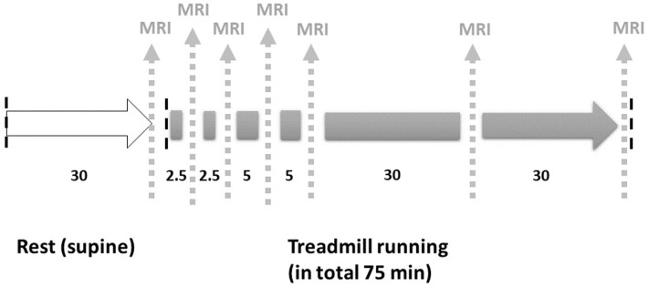
Methods: Ten participants had to run a total of 75 minutes at their individual highest possible running speed, with interleaved MR scans at baseline and after 2.5, 5, 10, 15, 45 and 75 minutes. The examined extrinsic foot muscles were manually segmented, and relaxation time were analysed regarding its respective time course.
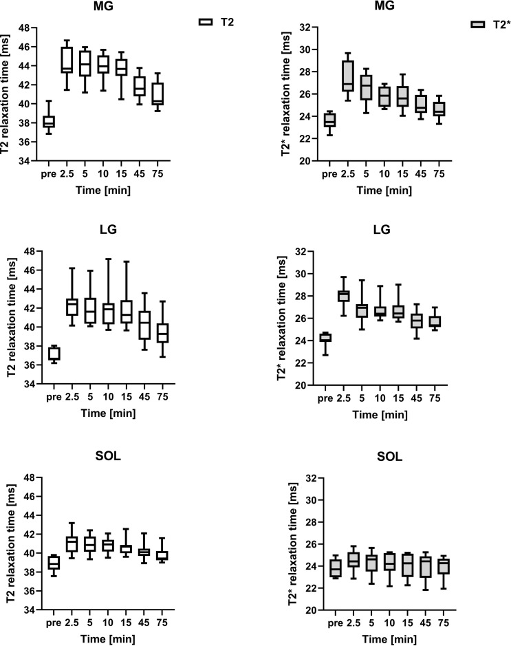
Results: T2* and T2 relaxation time showed an initial increase, followed by a plateau phase between 2.5 and 15 minutes and a subsequent decrease. For the T2* relaxation time, this pattern was also apparent, but less pronounced, with more muscles not reaching significance (p<0.05) when comparing different time points.
Conclusions: T2* and T2 relaxation time showed a similar course with an initial rapid increase, a plateau phase and a subsequent decrease under prolonged exercise. Moderate but long-term muscular activity appears to have a weaker effect on T2* relaxation time than on T2 relaxation time.
Conflict of interest statement
I have read the journal’s policy and the authors of this manuscript have the following competing interests: David Maintz is on the speakers’ bureau for Philips Healthcare. The author Gert-Peter Brüggemann currently works for the running shoe company True Motion Running GmbH. The remaining authors have no potential conflict of interest to declare.
Figures

Similar articles
-
Insights into extrinsic foot muscle activation during a 75 min run using T2 mapping.Sci Rep. 2021 Apr 1;11(1):7331. doi: 10.1038/s41598-021-86810-1. Sci Rep. 2021. PMID: 33795777 Free PMC article.
-
Damage and recovery of the intrinsic and extrinsic foot muscles from running a full marathon.Scand J Med Sci Sports. 2023 Aug;33(8):1486-1493. doi: 10.1111/sms.14377. Epub 2023 Apr 27. Scand J Med Sci Sports. 2023. PMID: 37102625
-
Probing muscle recovery following downhill running using precise mapping of MRI T2 relaxation times.Magn Reson Med. 2023 Nov;90(5):1990-2000. doi: 10.1002/mrm.29765. Epub 2023 Jun 22. Magn Reson Med. 2023. PMID: 37345717
-
Contributions to the understanding of gait control.Dan Med J. 2014 Apr;61(4):B4823. Dan Med J. 2014. PMID: 24814597 Review.
-
The effect of running on foot muscles and bones: A systematic review.Hum Mov Sci. 2019 Apr;64:75-88. doi: 10.1016/j.humov.2019.01.006. Epub 2019 Jan 22. Hum Mov Sci. 2019. PMID: 30682645
Cited by
-
Imaging of muscle injuries in soccer.Skeletal Radiol. 2025 Apr;54(4):655-667. doi: 10.1007/s00256-023-04514-1. Epub 2023 Nov 22. Skeletal Radiol. 2025. PMID: 37991553 Review.
-
Molecular imaging of viral pathogenesis and opportunities for the future.Npj Imaging. 2025;3(1):3. doi: 10.1038/s44303-024-00056-w. Epub 2025 Jan 24. Npj Imaging. 2025. PMID: 39872292 Free PMC article. Review.
-
Sports Radiology: A Key Driver of Clinical Decision-Making.Diagnostics (Basel). 2025 Aug 7;15(15):1977. doi: 10.3390/diagnostics15151977. Diagnostics (Basel). 2025. PMID: 40804941 Free PMC article.
-
[Imaging of muscle injuries in sports medicine].Radiologie (Heidelb). 2023 Apr;63(4):249-258. doi: 10.1007/s00117-023-01118-7. Epub 2023 Feb 16. Radiologie (Heidelb). 2023. PMID: 36797330 Review. German.
References
-
- Meyer RA, Prior BM. Functional magnetic resonance imaging of muscle. Exerc Sport Sci Rev. 2000;28(2):89–92. - PubMed
