. 2022 Apr 19;61(17):e202202302.
doi: 10.1002/anie.202202302. Epub 2022 Feb 26.

Spontaneous Membranization in a Silk-Based Coacervate Protocell Model

Affiliations

Spontaneous Membranization in a Silk-Based Coacervate Protocell Model

Zhuping Yin et al. Angew Chem Int Ed Engl. .

Abstract

Molecularly crowded coacervate micro-droplets are useful protocell constructs but the absence of a physical membrane limits their application as cytomimetic models. Auxiliary surface-active agents have been harnessed to stabilize the coacervate droplets by irreversible shell formation but endogenous processes of reversible membranization have received minimal attention. Herein, we describe a dynamic alginate/silk coacervate-based protocell model in which membrane-less droplets are reversibly reconfigured and inflated into semipermeable coacervate vesicles by spontaneous self-organization of amphiphilic silk polymers at the droplet surface under non-neutral charge conditions in the absence of auxiliary agents. We show that membranization can be reversibly controlled endogenously by programming the pH within the protocells using an antagonistic enzyme system such that structural reconfigurations in the protocell microstructure are coupled to the trafficking of water-soluble solutes. Our results open new perspectives in the design of hybrid protocell models with dynamical structural properties.

Keywords: Coacervates; Protocells; Silk.


Conflict of interest statement

The authors declare no conflict of interest.

Figures
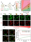
Figure 1
Figure 1
Preparation and diversity of alginate/CSF coacervate‐based protocells. a) Scheme showing cationization of silk fibroin (SF) via EDC‐activated coupling of 1,6‐hexanediamine at pH 6.5 (left). Carboxylic acid groups (‐COOH) of SF are derivatized with primary amines (‐NH2), which ionize in water to produce positively charged cationized silk fibroin (CSF). Addition of alginate (ALG, M w=140–160 kDa) to CSF results in a diversity of microscale objects depending on the [COOH] (alginate) : [NH2] (CSF) charge ratio. Labels; homogeneous near‐neutral coacervate droplets (C), positive multi‐compartmentalized coacervate droplets (PMC), negative multi‐compartmentalized coacervate droplets (NMC), positive coacervate vesicles (PCV), negative coacervate vesicles (NCV). b) Grid of LSCM images showing variation in silk‐containing coacervate‐based protocell constructs with changes in alginate (COOH) concentration at a constant CSF (NH2) concentration of 4 mM. Green (FITC‐CSF) and red (RITC‐alginate) fluorescence images are shown. Increasing the RITC‐alginate concentration gives the following sequence: discrete and hemi‐fused PCVs ([COOH], 2–4 mM), PMC droplets (COOH, 4–6 mM), C micro‐droplets ([COOH], 10 mM), NMC droplets ([COOH], 15 mM) and aggregates of hemi‐fused NCVs ([COOH], 20 mM). In each case, CSF (green) and alginate (red) are co‐distributed homogeneously throughout the polyelectrolyte‐rich coacervate phase and are excluded from the water‐filled sub‐compartments associated with the PCV, PMC, NMC and NCV microstructures. Graphics and labels for coacervate microstructures are as given in (a). Scale bars, 10 μm. c) Diagram showing mapping of silk‐based coacervate microstructures onto alginate [COOH] and CSF [NH2] concentrations used for sample preparations. Graphics for coacervate microstructures are as given in (a). No coacervate phase was observed at alginate : CSF ratios greater than 20 : 1 (upper left red zone). d) LSCM time‐dependent images of a giant coacervate vesicle (GCV) produced after centrifugation‐induced (1000 rpm, 2 min) fusion of a positively charged alginate/FITC‐CSF coacervate vesicle (F‐CSF, green fluorescence) with a positively charged alginate/RITC‐CSF coacervate vesicle (R‐CSF, red fluorescence). Images recorded at 5, 20, 40, 60 min after fusion. Red/green merged images are shown. Localized regions labelled 1 and 2 correspond to initially unmixed regions of segregated R‐CSF and F‐CSF. Scale bars, 10 μm. e) Corresponding time‐dependent changes in fluorescence intensity ratio derived from measurements of the green (F‐CSF) or red fluorescence (R‐CSF) (gray values) intensities recorded at positions 1 and 2 in (d). The R‐CSF (spot 1) : R‐CSF (spot 2) ratios decrease to a constant value after 20 min of centrifugation‐induced fusion due to diffusion of R‐CSF from spot 1 to 2. Diffusion of F‐CSF from spot 2 reaches a close to steady state ratio after approximately 60 min.
Figure 2
Figure 2
Membrane structuration in alginate/CSF coacervate vesicles. a, b) High magnification LSCM images of localized regions of the outer membrane of a positively charged coacervate vesicle (a, PCV) and negatively charged coacervate vesicle (b, NCV) stained with FITC‐dextran (Mw ≈250 kDa, green fluorescence, hydrophilic) and Nile red (red fluorescence, hydrophobic) showing alternate arrangement of two sets of hydrophilic and hydrophobic double nanolayers. Scale bars, 25 μm. c) SAXS profiles of membraneless coacervate droplets (C, red profile) and positively charged vesicles (PCV, green profile) showing weak Bragg reflection at ca. 0.015 A−1 only in the PCVs. Samples were prepared under identical component concentrations (COOH : NH2=1.0; [NH2]=4 mM; [COOH]=4 mM) but at different pH values (PCV, pH 6; C, pH 9). d, e) Structural models showing spontaneous ordering of hydrophobic/hydrophilic domains of CSF in the surface layers of a PCV (d) and NCV (e) prepared in the presence of excess free CSF or alginate, respectively.
Figure 3
Figure 3
Enzyme‐mediated reconfiguration and molecular trafficking in silk‐based coacervate protocells. a) Scheme showing endogenous enzyme‐mediated transitions between positively charged vesicles (PCV), positively charged multi‐compartmentalized coacervate droplets (PMC) and homogeneous coacervate droplets (C). b) Corresponding time‐dependent LSCM images of a single alginate/RITC‐CSF PCV (pH 6.5) undergoing urease‐mediated reconfiguration to C within 10 minutes of adding urea (final pH 8.5) (top row). RITC‐CSF accumulates initially at the vacuole/coacervate interface, after which the biopolymer is dispersed homogeneously in the coacervate droplet. Addition of glucose reverses the pH‐induced transformation within 20 min via a PMC intermediate microstructure (bottom row). c) Enzyme‐mediated pH‐induced trafficking of partitioned molecular dyes associated with urease‐mediated PCV to PMC to C reversible transformations. Reconfiguration of the vesicles to homogeneous droplets results in expulsion of calcein and uptake of positively charged rhodamine (Rho.) 123, rhodamine (Rho.) 6G and hydrophobic Nile Red. d) Plot showing time‐dependent changes in the fluorescence intensity partitioning constants of calcein (top row), rhodamine 123 (middle row) and rhodamine 6G (bottom row) during urease‐mediated PCV (pH 6.5) to C (pH 8.5) transformation. Fluorescence (Fluo.) partition constants were determined by measuring the fluorescence intensity (gray value) of the dyes within the coacervate phase and external water‐rich phase as determined from LSCM images. e) As for (d) but during GOx‐mediated C (pH 8.5) to PMC (pH 7.5) to PCV (pH 6.5) reconfiguration. Scale bars, 10 μm.

References

    1. Keating C. D., Acc. Chem. Res. 2012, 45, 2114–2124. - PMC - PubMed
    1. Crowe C. D., Keating C. D., Interface Focus 2018, 8, 20180032. - PMC - PubMed
    1. Koga S., Williams D. S., Perriman A. W., Mann S., Nat. Chem. 2011, 3, 720–724. - PubMed
    1. Drobot B., Iglesias-Artola J. M., Le Vay K., Mayr V., Kar M., Kreysing M., Mutschler H., Tang T.-Y. D., Nat. Commun. 2018, 9, 3643. - PMC - PubMed
    1. Strulson C. A., Molden R. C., Keating C. D., Bevilacqua P. C., Nat. Chem. 2012, 4, 941–946. - PubMed

Publication types

LinkOut - more resources