. 2022 May:102:110-123.
doi: 10.1016/j.bbi.2022.02.008. Epub 2022 Feb 14.

Palmitoylethanolamide dampens neuroinflammation and anxiety-like behavior in obese mice

Affiliations

Palmitoylethanolamide dampens neuroinflammation and anxiety-like behavior in obese mice

Adriano Lama et al. Brain Behav Immun. 2022 May.

Abstract

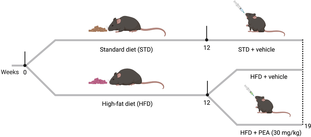
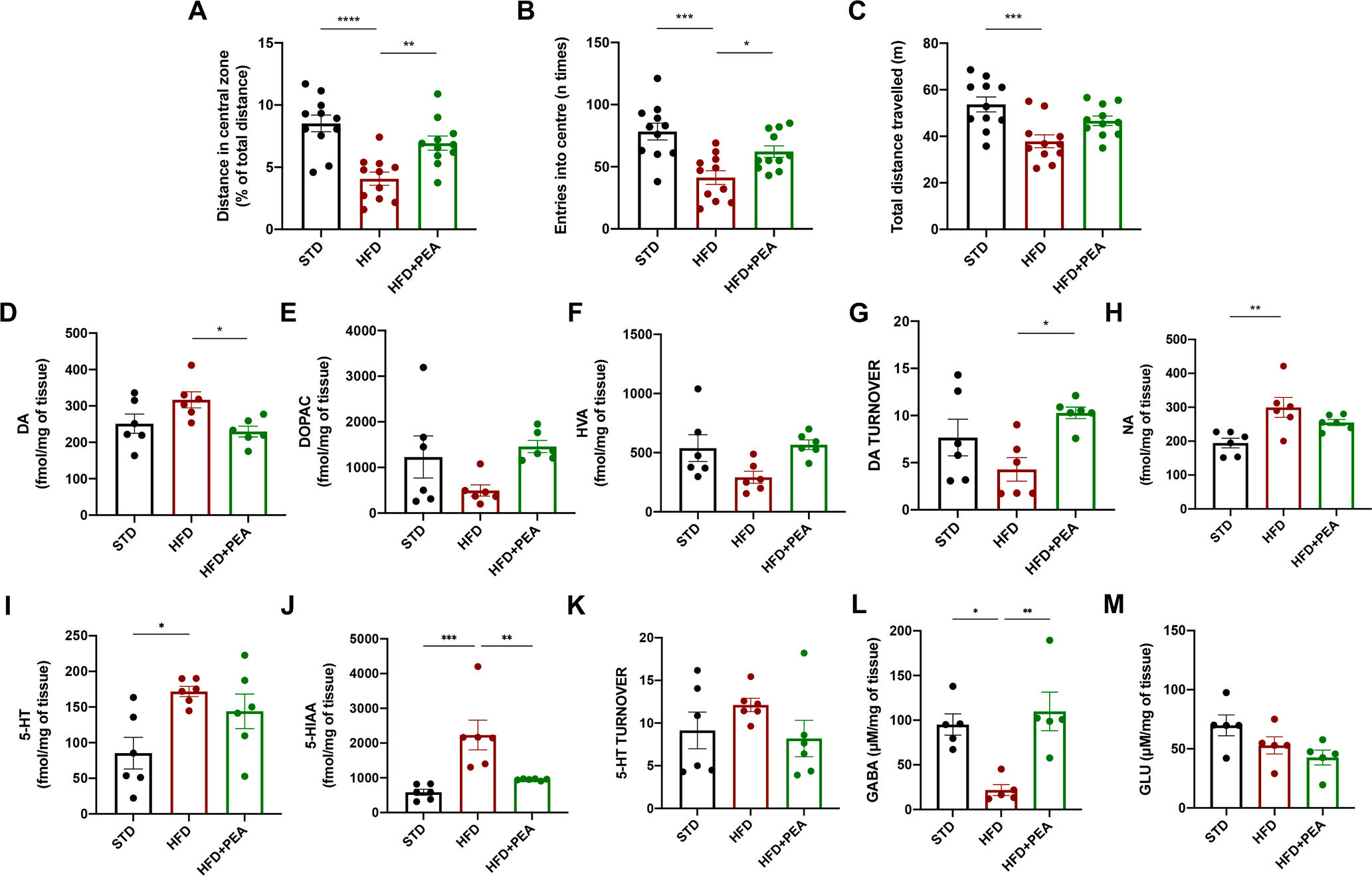
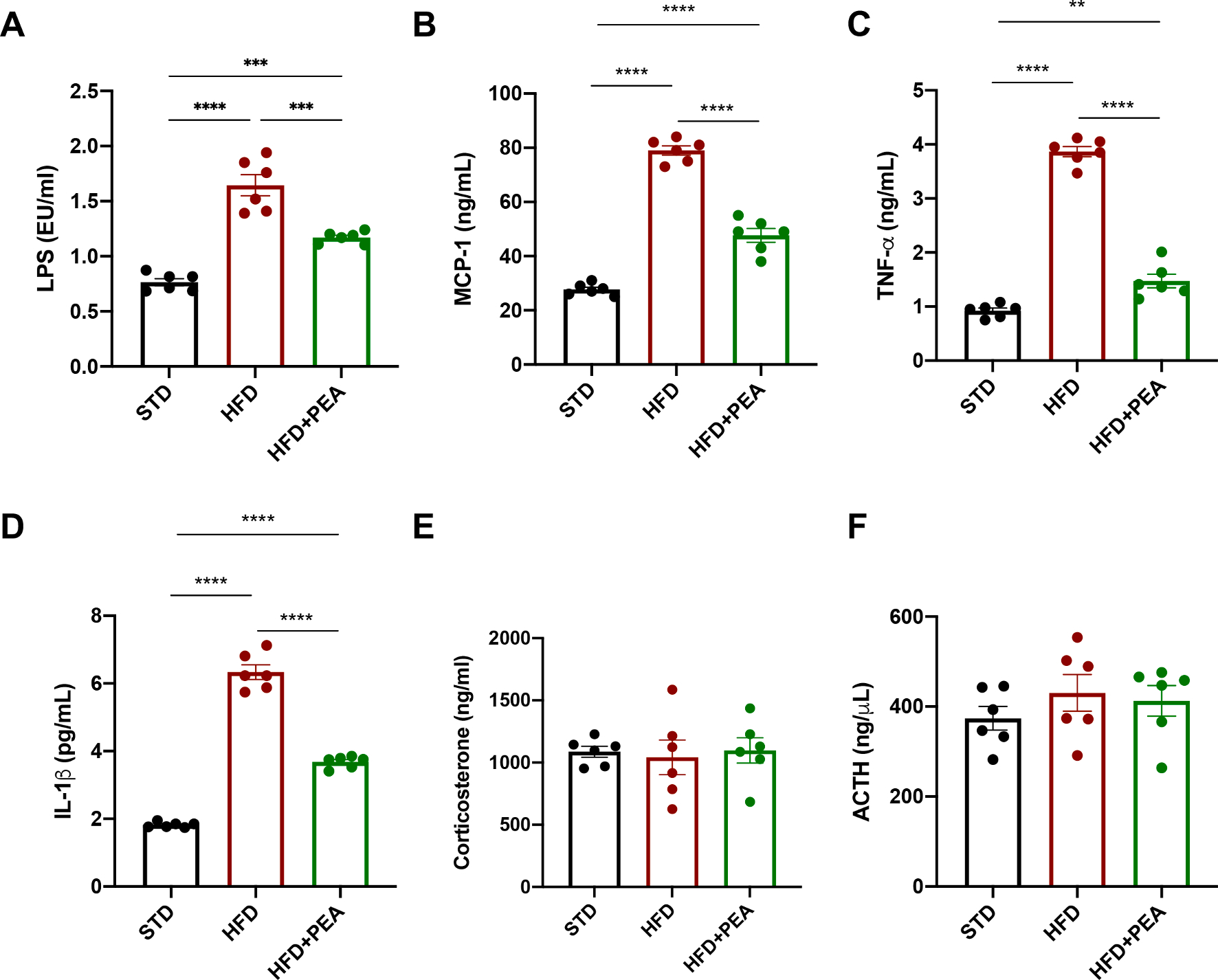
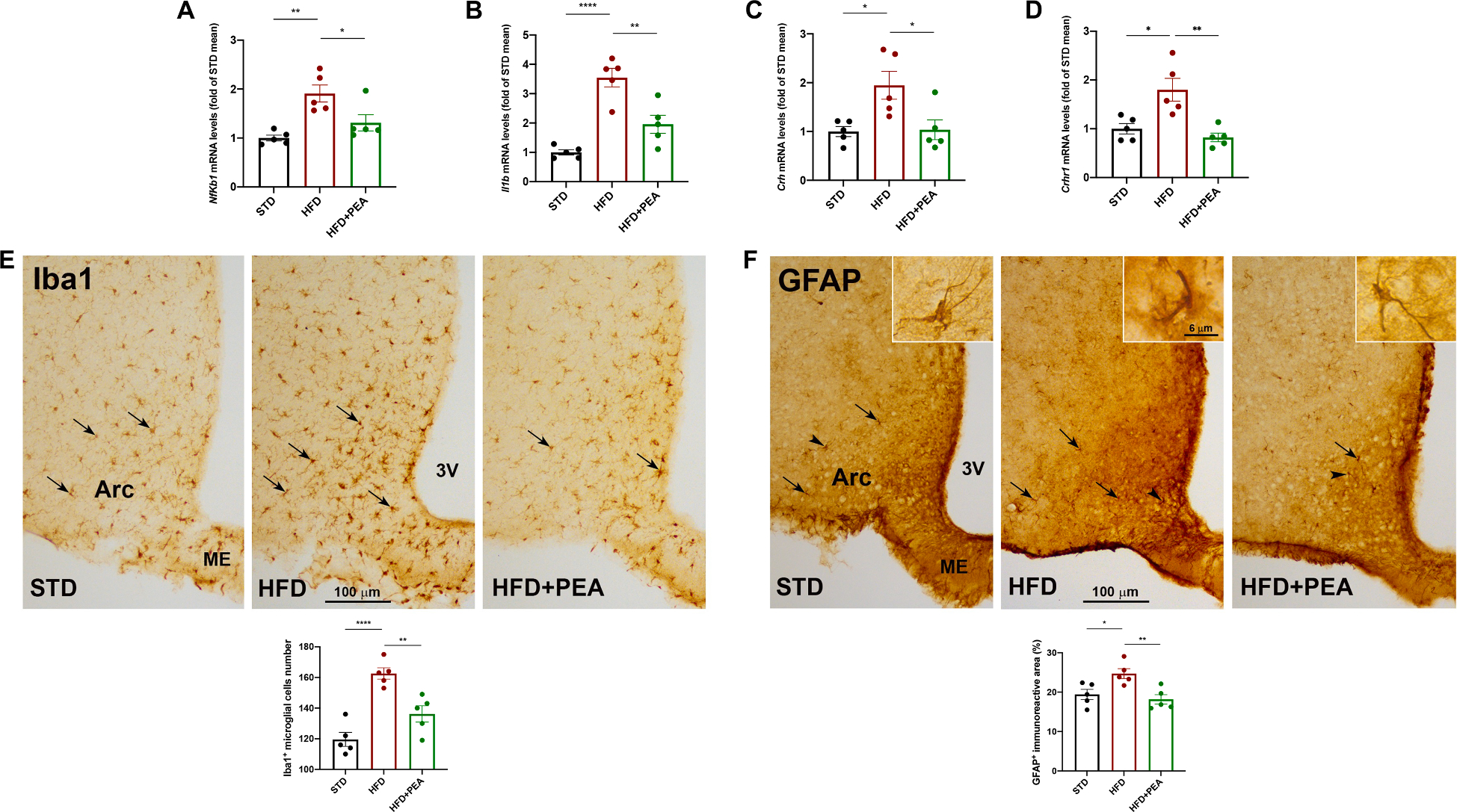
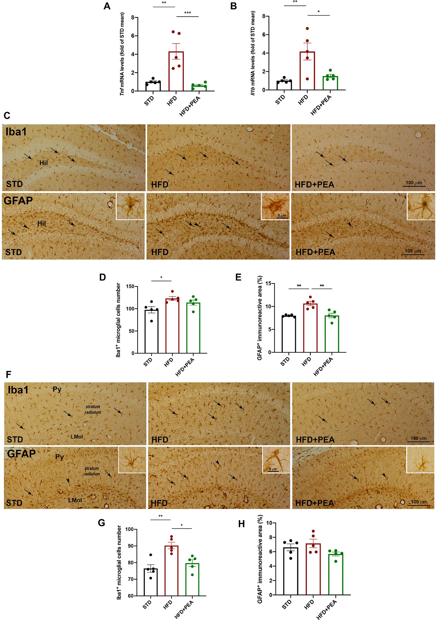
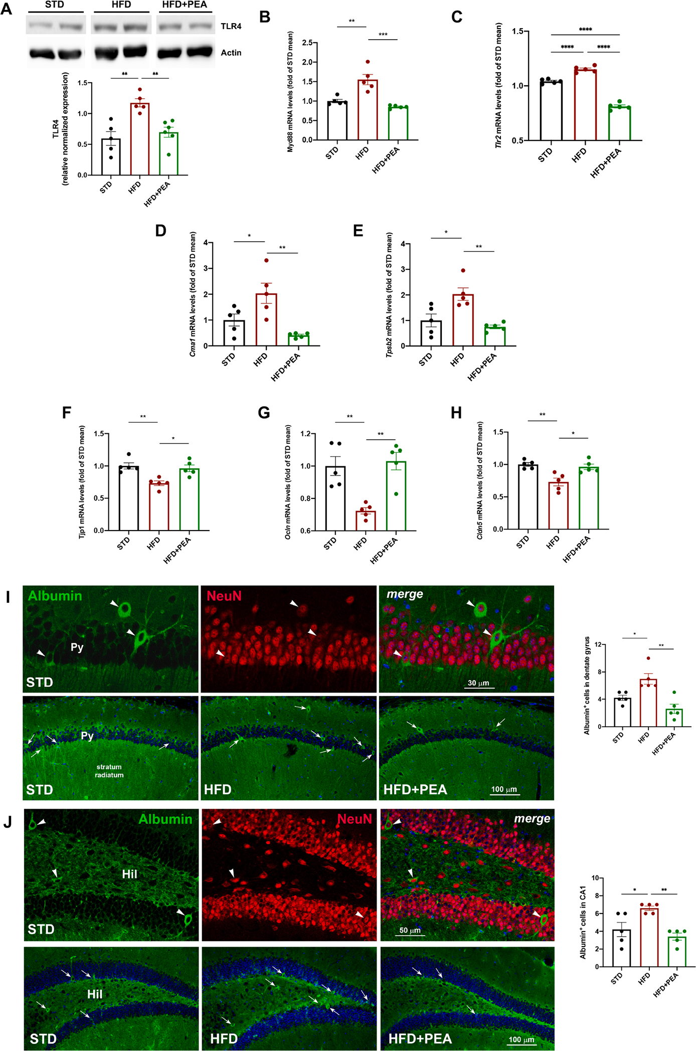
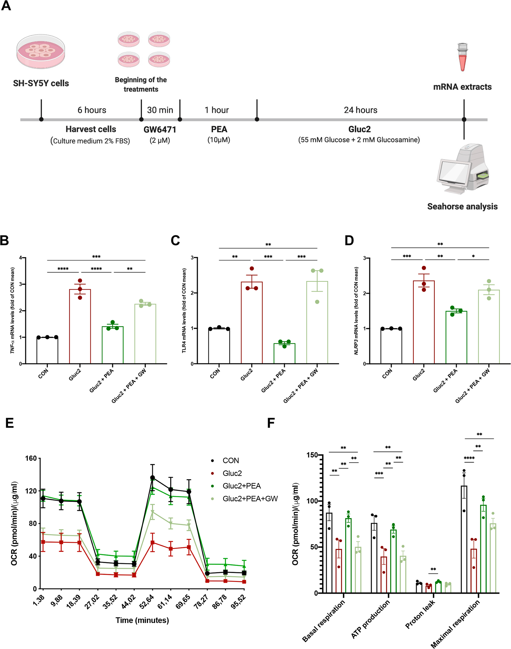
High-fat diet (HFD) consumption leads to obesity and a chronic state of low-grade inflammation, named metainflammation. Notably, metainflammation contributes to neuroinflammation due to the increased levels of circulating free fatty acids and cytokines. It indicates a strict interplay between peripheral and central counterparts in the pathogenic mechanisms of obesity-related mood disorders. In this context, the impairment of internal hypothalamic circuitry runs in tandem with the alteration of other brain areas associated with emotional processing (i.e., hippocampus and amygdala). Palmitoylethanolamide (PEA), an endogenous lipid mediator belonging to the N-acylethanolamines family, has been extensively studied for its pleiotropic effects both at central and peripheral level. Our study aimed to elucidate PEA capability in limiting obesity-induced anxiety-like behavior and neuroinflammation-related features in an experimental model of HFD-fed obese mice. PEA treatment promoted an improvement in anxiety-like behavior of obese mice and the systemic inflammation, reducing serum pro-inflammatory mediators (i.e., TNF-α, IL-1β, MCP-1, LPS). In the amygdala, PEA increased dopamine turnover, as well as GABA levels. PEA also counteracted the overactivation of HPA axis, reducing the expression of hypothalamic corticotropin-releasing hormone and its type 1 receptor. Moreover, PEA attenuated the immunoreactivity of Iba-1 and GFAP and reduced pro-inflammatory pathways and cytokine production in both the hypothalamus and hippocampus. This finding, together with the reduced transcription of mast cell markers (chymase 1 and tryptase β2) in the hippocampus, indicated the weakening of immune cell activation underlying the neuroprotective effect of PEA. Obesity-driven neuroinflammation was also associated with the disruption of blood-brain barrier (BBB) in the hippocampus. PEA limited the albumin extravasation and restored tight junction transcription modified by HFD. To gain mechanistic insight, we designed an in vitro model of metabolic injury using human neuroblastoma SH-SY5Y cells insulted by a mix of glucosamine and glucose. Here, PEA directly counteracted inflammation and mitochondrial dysfunction in a PPAR-α-dependent manner since the pharmacological blockade of the receptor reverted its effects. Our results strengthen the therapeutic potential of PEA in obesity-related neuropsychiatric comorbidities, controlling neuroinflammation, BBB disruption, and neurotransmitter imbalance involved in behavioral dysfunctions.

Keywords: Astrogliosis; Blood–brain barrier permeability; High-fat diet; Inflammation; Mastocytosis; Metabolic impairment; Microgliosis; Mood disorders; N-acylethanolamines; Peroxisome proliferator-activated receptor-α.


Figures

Fig. 1.
Fig. 1.. The experimental protocol.
Mice of the experimental HFD groups received HFD for 12 weeks, while the STD group was fed with a standard chow diet. After 12 weeks on diet, the STD group received the vehicle, while HFD groups received either vehicle or ultra-micronized PEA (30 mg/kg daily p.o.) for seven weeks along with HFD.
Fig. 2.
Fig. 2.. PEA limits the anxiety-like behavior of obese mice.
(A) Total distance in the centre, (B) the number of entries into the centre, and (C) the total distance travelled by mice in the open field test (n = 11 per group). The levels of (D) DA, (E) DOPAC, (F) HVA, and (G) DA turnover, (H) NA, (I) 5-HT, (J) 5-HIAA, and (K) 5-HT turnover (n = 6 per group) in AMY of all mice. The levels of (L) GABA and (M) GLU in AMY of all experimental groups (n = 5 per group). All data are shown as mean ± S.E.M. *p < 0.05, **p < 0.01, ***p < 0.001, and ****p < 0.0001.
Fig. 3.
Fig. 3.. PEA improves systemic parameters altered by HFD.
Serum levels of (A) LPS, (B) MCP-1, (C) TNF-α, (D) IL-1β, (E) corticosterone and (F) ACTH (n = 6 per group). All data are shown as mean ± S.E.M. **p < 0.01, ***p < 0.001, and ****p < 0.0001.
Fig. 4.
Fig. 4.. PEA counteracts hypothalamic injury in HFD-induced obese mice.
The mRNA transcription of (A) Nfkb1, (B) Il1b, (C) Crh, and (D) Crhr1 in the hypothalamus (n = 5 per group). Immunohistochemical evaluations and morphometric analyses of (E) the number of IBA1-positive microglial cells and (F) GFAP immunoreactive area in the hypothalamic arcuate nucleus (Arc) (n = 5 per group). 3 V, third ventricle, ME, median eminence. All data are shown as mean ± S.E.M. *p < 0.05, **p < 0.01, and ****p < 0.0001.
Fig. 5.
Fig. 5.. PEA limits neuroinflammation, astrogliosis and microgliosis in the hippocampus of HFD mice.
The mRNA expression of (A) Tnf and (B) Il1b in the hippocampus of all experimental groups (n = 5 per group). The immunohistochemical analyses of Iba1-positive microglial cells and GFAP-positive astrocytes in (C) the DG and (F) stratum radiatum (n = 5 per group). The count of the number of positive cells for (D, G) Iba1 and (E, H) of GFAP immunoreactive in DG and stratum radiatum, respectively (n = 5 per group). Hil, hilus of the DG; Py, pyramidal cell layer; LMol, lacunosum molecular layer. All data are shown as mean ± S.E.M. *p < 0.05, **p < 0.01, and ***p < 0.001.
Fig. 6.
Fig. 6.. The impact of PEA on immune activation, mast cell degranulation, and BBB integrity in the hippocampus of HFD mice.
Western blot for (A) TLR4, and Real-Time PCR analysis of (B) Myd88, and (C) Tlr2, (D) Cma1, (E) Tpsb2 (F) Tjp1, (G) Ocln, and (H) Cldn5 in the hippocampus of all experimental groups (at least n = 5 per group for western blot; n = 5 per group for Real-Time PCR). Morphometric analysis of albumin-immunoreactive cells in hippocampal (I) DG and (J) CA1 subfield. As shown in the upper panels of I and J, most albumin-positive cells (green, arrowheads) are also positive for the neuronal marker NeuN (red, arrowheads). The lower panels of I and J represent images depicting albumin staining in the DG and, respectively, CA1 subfield for each experimental condition. Hil, hilus; Py, pyramidal cell layer. In the merged images, cell nuclei are stained with TO-PRO3. The semiquantitative evaluation of albumin-positive neuronal-like profiles (n = 5 per group) is also shown. All data are shown as mean ± S.E.M. *p < 0.05, **p < 0.01, ***p < 0.001, ****p < 0.0001.
Fig. 7.
Fig. 7.. The role of PPAR-α in PEA effects on inflammation and mitochondrial function in glucose þ glucosamine (Gluc2)-insulted SH-SY5Y cells.
(A) The experimental protocol on human SH-SY5Y cells. The mRNA extracts of all treated cells for (B) TNF-α, (C) TLR4, and (D) NLRP3. (E) SH-SY5Y cells were undergone to Cell Mito Stress Test in Seahorse analyzer, evaluating the basal oxygen consumption rate (OCR), after the addition of oligomycin, the uncoupler FCCP, rotenone, and antimycin A. (F) Basal respiration, ATP production, proton leak, and maximal respiration were determined. OCR was normalized with the protein amount by Bradford assay. Data are the mean ± SEM of three different experiments with three replicates. All data are shown as mean ± S.E.M. *p < 0.05, **p < 0.01, ***p < 0.001, and **** p < 0.0001.

References

    1. Annunziata C, Lama A, Pirozzi C, Cavaliere G, Trinchese G, Di Guida F, Nitrato Izzo A, Cimmino F, Paciello O, De Biase D, Murru E, Banni S, Calignano A, Mollica MP, Mattace Raso G, Meli R, 2020. Palmitoylethanolamide counteracts hepatic metabolic inflexibility modulating mitochondrial function and efficiency in diet-induced obese mice. FASEB J. 34 (1), 350–364. 10.1096/fj.201901510RR. - DOI - PubMed
    1. Araujo JR, Tomas J, Brenner C, Sansonetti PJ, 2017. Impact of high-fat diet on the intestinal microbiota and small intestinal physiology before and after the onset of obesity. Biochimie 141, 97–106. 10.1016/j.biochi.2017.05.019. - DOI - PubMed
    1. Artamonov M, Zhukov O, Shuba I, Storozhuk L, Khmel T, Klimashevsky V, Mikosha A, Gula N, 2005. Incorporation of labelled N-acylethanolamine (NAE) into rat brain regions in vivo and adaptive properties of saturated NAE under x-ray irradiation. Ukr. Biokhim. Zh. 1999 (77), 51–62. - PubMed
    1. Baker KD, Loughman A, Spencer SJ, Reichelt AC, 2017. The impact of obesity and hypercaloric diet consumption on anxiety and emotional behavior across the lifespan. Neurosci. Biobehav. Rev. 83, 173–182. 10.1016/j.neubiorev.2017.10.014. - DOI - PubMed
    1. Beggiato Sarah, Tomasini Maria Cristina, Cassano Tommaso, Ferraro Luca, 2020. Chronic oral palmitoylethanolamide administration rescues cognitive deficit and reduces neuroinflammation, oxidative stress, and glutamate levels in a transgenic murine model of Alzheimer’s Disease. Journal of Clinical Medicine 9(2), 428. 10.3390/jcm9020428. - DOI - PMC - PubMed

Publication types