. 2022 Mar;13(3):5421-5433.
doi: 10.1080/21655979.2021.2024386.

Down-regulated long non-coding RNA LHFPL3 antisense RNA 1 inhibits the radiotherapy resistance of nasopharyngeal carcinoma via modulating microRNA-143-5p/homeobox A6 axis

Affiliations

Down-regulated long non-coding RNA LHFPL3 antisense RNA 1 inhibits the radiotherapy resistance of nasopharyngeal carcinoma via modulating microRNA-143-5p/homeobox A6 axis

Weifeng Wang et al. Bioengineered. 2022 Mar.

Abstract

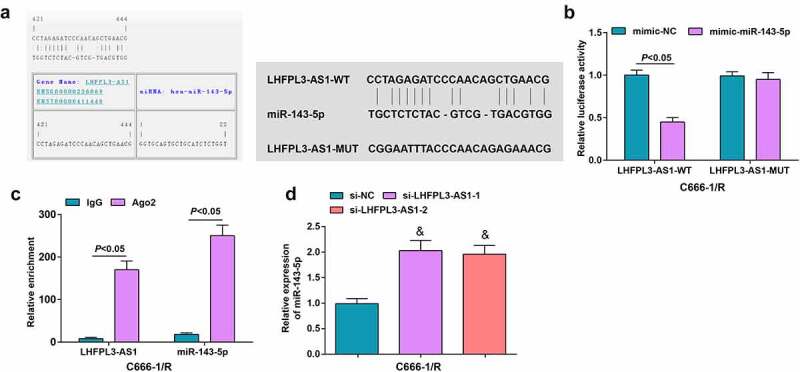
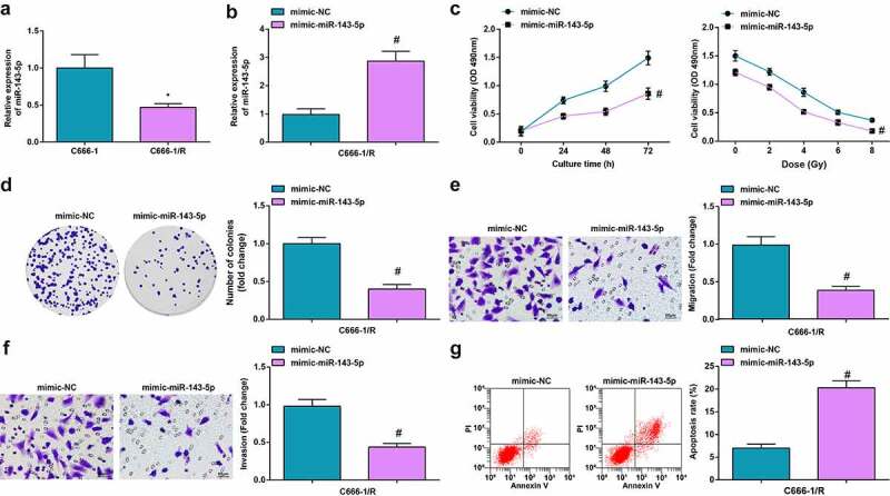
The function of long non-coding RNA LHFPL3 antisense RNA 1 (LHFPL3-AS1) in cancer progression has been studied, while its role in nasopharyngeal carcinoma (NPC) remains unclear. This study aims to unravel the effects of LHFPL3-AS1 on NPC progression via microRNA (miR)-143-5p/homeobox A6 (HOXA6) axis. NPC tissues were collected and NPC cells were cultured. NPC cells were subjected to radiation therapy to construct the radiation therapy resistance NPC cell line. The levels of LHFPL3-AS1, miR-143-5p and HOXA6 in NPC cells and tissues were examined. LHFPL3-AS1, miR-143-5p or HOXA6 expression was changed and then transfected into radiation-resistant NPC cells to detect cell proliferation, colony formation, migration, invasion and cell apoptosis in vitro. The tumorigenesis in nude mice in vivo was conducted to detect tumor growth. The targeting relations among LHFPL3-AS1, miR-143-5p and HOXA6 were validated. It was discovered that LHFPL3-AS1 and HOXA6 expression was elevated while the miR-143-5p level was depleted in radiation-resistant NPC cells and NPC tissues. The silenced LHFPL3-AS1 or augmented miR-143-5p repressed the proliferation, colony formation, migration and invasion of radiation-resistant NPC cells, while accelerated cell apoptosis in vitro. Silenced LHFPL3-AS1 hindered tumor growth in vivo. MiR-143-5p deletion reversed the effects of reduced LHFPL3-AS1; while HOXA6 upregulation reversed the effects of enriched miR-143-5p. LHFPL3-AS1 sponged miR-143-5p that targeted HOXA6. It is concluded that the down-regulated LHFPL3-AS1 retards the development of radiation-resistant NPC cells via sponging miR-143-5p to modulate HOXA6. This study reveals novel therapeutic targets for NPC treatment.

Keywords: Nasopharyngeal carcinoma; apoptosis; homeobox A6; long non-coding RNA LHFPL3 antisense RNA 1; microRNA-143-5p; proliferation; radiation resistance.


Conflict of interest statement

No potential conflict of interest was reported by the author(s).

Figures

Figure 1.
Figure 1.
LHFPL3-AS1 is high-expressed in NPC tissues and cells and is associated with shorter OS. (a) LHFPL3-AS1 level in NPC tissues was detected by RT-qPCR; (b) LHFPL3-AS1 levels in NPC cell lines were examined by RT-qPCR; (c) Relation curve between LHFPL3-AS1 expression and OS of NPC patients. The data was expressed as mean ± standard deviation. * P < 0.05 vs. the NP69 cells.
Figure 2.
Figure 2.
Silenced LHFPL3-AS1 inhibits radiation-resistant NPC cell growth in vitro and tumor growth in vivo. (a) RT-qPCR was used to detect the expression of LHFPL3-AS1 in both parent and resistant strains; (b). cell proliferation ability of parental and resistant strains was evaluated by CCK-8 assay; (c) LHFPL3-AS1 expression in NPC cells after the down-regulation of LHFPL3-AS1 was detected by RT-qPCR; (d) Proliferation of NPC cells after the down-regulation of LHFPL3-AS1 was examined by CCK-8 assay; (e) Colony formation ability of NPC cells after the down-regulation of LHFPL3-AS1 was assessed by colony formation assay; (f,g) Migration and invasion of NPC cells after the down-regulation of LHFPL3-AS1 were detected by Transwell assay; (h) Apoptosis of NPC cells after the down-regulation of LHFPL3-AS1 was detected by flow cytometry; (i) The weight and volume of the tumor were detected in nude mice. The data iwas expressed as mean ± standard deviation. * P < 0.05 vs. C666-1 cells; # P < 0.05 vs. the si-NC group.
Figure 3.
Figure 3.
LHFPL3-AS1 binds to miR-143-5p. (a) The presence of binding sites between LHFPL3-AS1 and miR-143-5p was predicted by Jefferson website; (b) Binding relation between LHFPL3-AS1 and miR-143-5p was validated by dual luciferase reporter gene assay; (c) enrichment levels of LHFPL3-AS1 and miR-143-5p were detected by RIP assay; (d) miR-143-5p expression in NPC cell lines after the silencing of LHFPL3-AS1 was examined by RT-qPCR. The data was expressed as mean ± standard deviation. & P < 0.05 vs. the si-NC group.
Figure 4.
Figure 4.
Up-regulated miR-143-5p retards the biological function of NPC cells. (a) miR-143-5p expression in NPC cells after X-ray irradiation treatment was detected by RT-qPCR; (b) miR-143-5p expression in NPC cells after the up-regulation of miR-143-5p was detected by RT-qPCR; (c) proliferation of NPC cells after the up-regulation of miR-143-5p was examined by CCK-8 assay; (d) colony formation ability of NPC cells after the up-regulation of miR-143-5p was assessed by colony formation assay; (e,f) Migration and invasion of NPC cells after the up-regulation of miR-143-5p were detected by Transwell assay; (g) Apoptosis of NPC cells after the up-regulation of miR-143-5p was detected by flow cytometry. The data was expressed as mean ± standard deviation.# P < 0.05 vs. the mimic-NC group; * P < 0.05 vs. C666-1 cells.
Figure 5.
Figure 5.
MiR-143-5p targets HOXA6. (a) Binding sites between miR-143-5p and HOXA6 were predicted by Targetscan; (b) Targeting relation between miR-143-5p and HOXA6 was validated by dual luciferase reporter gene assay; (c) HOXA6 expression was detected byWestern blot analysis. The data was expressed as mean ± standard deviation. % P < 0.05 vs. the mimic-NC group; $ P < 0.05 vs. the si-NC group.
Figure 6.
Figure 6.
Down-regulated LHFPL3-AS1 hinders the development of NPC via miR-143-5p/HOXA6 axis. (a) Proliferation of NPC cells was examined by CCK-8 assay; (b) Colony formation ability of NPC cells was assessed by colony formation assay; (c,d) Migration and invasion of NPC cells were detected by Transwell assay; E, apoptosis of NPC cells was detected by flow cytometry. The data was expressed as mean ± standard deviation.# P < 0.05 vs. the si-LHFPL3-AS1 +inhibitor NC group; * P < 0.05 vs. the miR-143-5p mimic + oe-NC group.

Similar articles

Cited by

References

    1. Chen Y-P, Chan ATC, Le Q-T, et al. Nasopharyngeal carcinoma. Lancet. 2019;394(10192):64–80. - PubMed
    1. Dai W, Zheng H, Cheung AKL, et al. Genetic and epigenetic landscape of nasopharyngeal carcinoma. Chin Clin Oncol. 2016;5(2):16. - PubMed
    1. Lam WKJ, Chan JYK.. Recent advances in the management of nasopharyngeal carcinoma. F1000Res. 2018;7:1829. - PMC - PubMed
    1. Wang KH, Austin, SA, Chen, SH, et al. Nasopharyngeal carcinoma diagnostic challenge in a nonendemic setting: our experience with 101 patients. Perm J. 2017;21:16–180. - PMC - PubMed
    1. Dong Q, Zhou L, Liu F, et al. Long non-coding RNAs in the development, diagnosis and prognosis of nasopharyngeal carcinoma. Int J Clin Exp Pathol. 2017;10(8):8098–8105. - PMC - PubMed