. 2022 Feb 17;41(1):66.
doi: 10.1186/s13046-022-02243-2.

RelB upregulates PD-L1 and exacerbates prostate cancer immune evasion

Affiliations

RelB upregulates PD-L1 and exacerbates prostate cancer immune evasion

Yanyan Zhang et al. J Exp Clin Cancer Res. .

Abstract

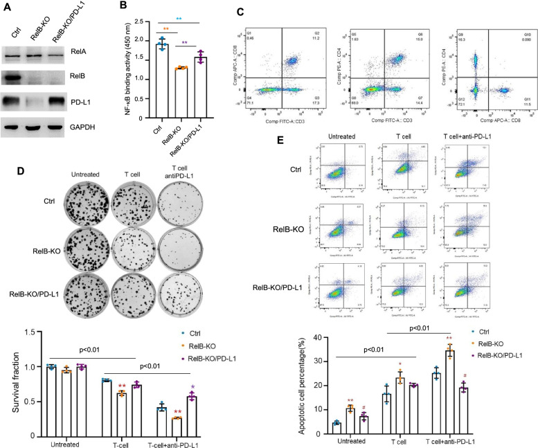
Background: The interaction between programmed death receptor (PD-1) and its ligand (PD-L1) is essential for suppressing activated T-lymphocytes. However, the precise mechanisms underlying PD-L1 overexpression in tumours have yet to be fully elucidated. Here, we describe that RelB participates in the immune evasion of prostate cancer (PCa) via cis/trans transcriptional upregulation of PD-L1.

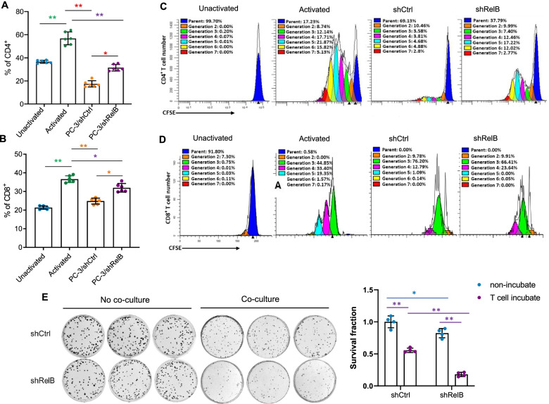
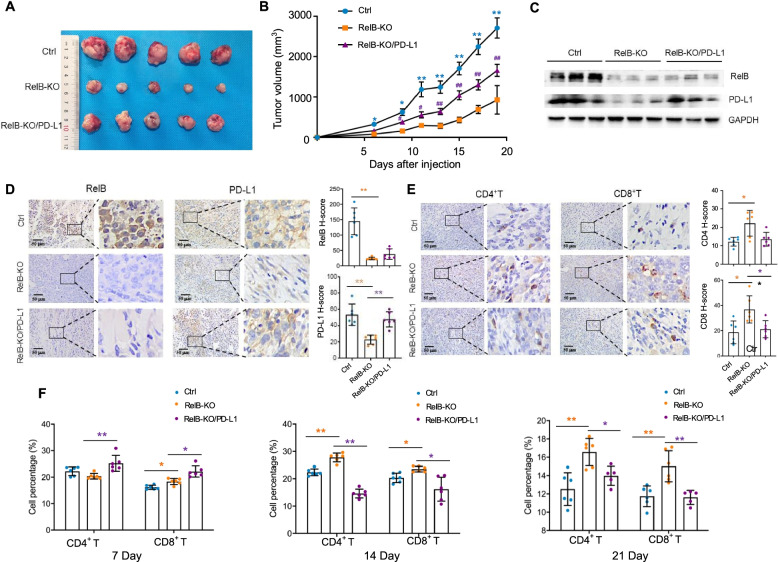
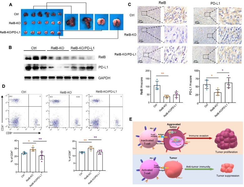
Methods: Based on transcriptome results, RelB was manipulated in multiple human and murine PCa cell lines. Activated CD4+ and CD8+ T cells were cocultured with PCa cells with different levels of RelB to examine the effect of tumourous RelB on T cell immunity. Male mice were injected with murine PCa cells to validate the effect of RelB on the PD-1/PD-L1-mediated immune checkpoint using both tumour growth and metastatic experimental models.

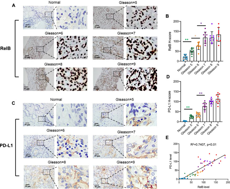
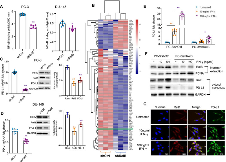
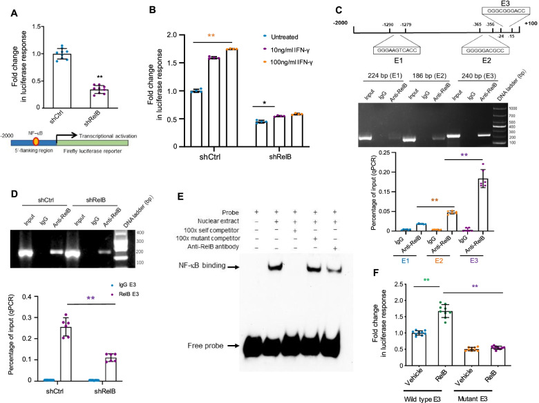
Results: PD-L1 is uniquely expressed at a high level in PCa with high constitutive RelB and correlates with the patients' Gleason scores. Indeed, a high level of PD-L1 is associated with RelB nuclear translocation in AR-negative aggressive PCa cells. Conversely, the silencing of RelB in advanced PCa cells resulted in reduced PD-L1 expression and enhanced susceptibility of PCa cells to the T cell immune response in vitro and in vivo. Mechanistically, a proximal NF-κB enhancer element was identified in the core promoter region of the human CD274 gene, which is responsible for RelB-mediated PD-L1 transcriptional activation. This finding provides an informative insight into immune checkpoint blockade by administering RelB within the tumour microenvironment.

Conclusion: This study deciphers the molecular mechanism by which tumourous RelB contributes to immune evasion by inhibiting T cell immunity via the amplification of the PD-L1/PD-1-mediated immune checkpoint.


Conflict of interest statement

The authors declare no competing interests.

Figures

Fig. 1
Fig. 1
PD-L1 is correlated with RelB in PCa progression. a-b Normal prostate and PCa tissues were examined to determine the association between RelB expression and patients’ Gleason scores by IHC. c-d Correspondingly, the association between PD-L1 expression and patients’ Gleason scores was analysed. e Linear regression analysis determined the correlation of RelB and PD-L1 in PCa progression. The H-scores of IHC were plotted and *(p < 0.05) and **(p < 0.01) show significance between the two groups as indicated
Fig. 2
Fig. 2
RelB regulates PD-L1 in PCa cells. a RelB was silenced in PC-3 and DU-145 cells using a lentiviral shRNA specifically targeting endogenic RelB. The NF-κB binding activities in RelB-silenced cells (shRelB) vs. scramble sequence (shCtrl) were measured using an ELISA kit with a standard probe containing the consensus NF-κB element. b mRNA expression profiles of RelB-silenced cells vs. control cells were examined by RNA sequencing. The relative levels of inflammatory and immune responsive transcripts were analysed, as indicated by the heatmap. The signature of CD274 gene expression was indicated in a green box. c-d The levels of RelA, RelB and PD-L1 mRNA and proteins were quantified using RT-qPCR and immunoblots by normalizing to GAPDH. e PC-3 cells were treated with different doses of IFN-γ as indicated. The stimulated PD-L1 mRNA expression was quantified by RT-qPCR with GAPDH normalization. f After IFN-γ treatment, the levels of nuclear RelB and relative cytosolic PD-L1 in PC-3 cells were measured by immunoblots. PCNA served as an internal control for the nuclear fraction, and GAPDH served as a loading control for the cytosolic fraction. g Accordingly, RelB nuclear translocation and relative PD-L1 in the cytosol were confirmed using a confocal microscope. *(p < 0.05) and **(p < 0.01) show significance between the two groups as indicated
Fig. 3
Fig. 3
RelB regulates PD-L1 in PC-3 cells. a A 2000-bp flanking region of the human CD274 gene containing putative NF-κB elements and a core promoter was cloned to drive the luciferase reporter gene, as indicated in the lower panel. The reporter construct was transfected into PC-3 cells with different levels of RelB and the transcriptional activity was estimated by β-gal-normalized reporter responses. b After cell transfection, the cells were treated with IFN-γ as indicated. Τhe IFN-γ-mediated transcriptional induction was analysed by the reporter assay. c Chromatin was precipitated using a RelB antibody, and DNA fragments containing three putative RelB elements (E1, E2, E3) were quantified by regular PCR (upper panel) and quantitative PCR (lower panel). Chromatins without the IP procedure were amplified as input controls. IgG was used as a negative antibody control. d Chromatins derived from RelB-silenced cells were pulled down and the reduction in the E3 fragment was quantified. e A 24-bp double-stranded DNA fragment containing the E3 element was synthesized for the preparation of an EMSA probe with biotin-labelling. Nuclear extract was incubated with the probe and the specific NF-κB binding was determined by EMSA with a self competitor. Additionally, the NF-κB element in the E3 fragment was mutated as a mutant competitor (shown in Fig. S3a). Furthermore, the RelB antibody was used to specifically reduce the E3 binding activity. f The mutated E3 binding site was further cloned and its effect on RelB-mediated transcriptional activation was assessed by the reporter assay. *(p < 0.05) and **(p < 0.01) show significance between the two groups as indicated
Fig. 4
Fig. 4
Silencing RelB in PC-3 cells enhances the immunities of CD4+ and CD8+ T cells. a-b PC-3 cells were cocultured with activated T cells derived from human PBMCs. CD4+ and CD8+ T cells were quantified by flow cytometry using the relevant antibodies. c-d Subsequently, the proliferation of CD4+ and CD8+ T cells was analysed by flow cytometry. The percentage of each generation (cell division) was plotted. e After coculture with activated T cells, the survival rate of PC-3 cells was measured using a clonogenic assay. *(p < 0.05) and **(p < 0.01) show significance between the two groups as indicated
Fig. 5
Fig. 5
RelB deprivation in mouse PCa RM-1 cells restores mouse T cell function. a RelB was knocked out in RM-1 cells using the CRISPR/Cas9-based gene-editing system and further PD-L1 was enforcedly expressed in RelB-deprived cells to restore the immunocompromise. The cellular levels of RelA, RelB and PD-L1 proteins in these cell lines were measured by immunoblots. b Subsequently, the NF-κB binding activities in the established cell lines were quantified. **(p < 0.01) shows significance between the two groups as indicated. c T cells derived from mouse spleen tissues were activated and CD4+ and CD8+ T cells were quantified by flow cytometry. d RM-1 cells were treated with an anti-PD-L1 mAb prior to coculture with activated T cells, and the RM-1 cell survival was measured using a clonogenic assay. The significance between the two groups as indicated on the tap. Within the same groups, *(p < 0.05), **(p < 0.01) shows significance in RelB-KO and RelB-KO/PD-L1 cells compared to control cells. e Correspondingly, the apoptosis of RM-1 cells was further analysed using flow cytometry. The significance between the two groups as indicated on the tap. Within the same groups, *(p < 0.05), **(p < 0.01) show significance in RelB-KO cells vs. the control cells, and #(p < 0.05) shows significance in RelB-KO/PD-L1 cells compared to control and RelB-KO cells
Fig. 6
Fig. 6
RelB deprivation reduces mouse xenograft tumour growth. a RM-1 (control), RM-1:RelB-KO and RM-1:RelB-KO/PD-L1 cell lines were cultured and then subcutaneously injected into male C57BL/6 mice for tumour formation. After tumours reached the maximal volume, the mice were sacrificed and the excised tumour tissues were photographed. b Tumour volume was measured every other day, and the tumour growth rate was determined as plotted in each group. *(p < 0.05), **(p < 0.01) show the significance in RelB-KO cells and RelB-KO/PD-L1 compared to the control cells; and #(p < 0.05), ##(p < 0.01) indicate the significances in RelB-KO/PD-L1 cell vs. RelB-KO cells. c The expression levels of RelB and PD-L1 in the tumour tissues were measured by immunoblots. d-e The levels of RelB, PD-L1, CD4 and CD8 proteins in the tumour tissues were quantified by IHC and the relative H-scores were plotted. *(p < 0.05), **(p < 0.01) show significance between the two groups as indicated. f Blood samples were collected from mice on different days after cell injection as indicated to analyse the percentages of CD4+ and CD8+ T cells by flow cytometry. *(p < 0.05), **(p < 0.01) show significance between the two groups as indicated
Fig. 7
Fig. 7
RelB deprivation decreases mouse xenograft tumour lung metastasis. a RM-1 and its generated cell lines were intravenously injected into male C57BL/6 mice. Lung tissues with tumours in the control group and without tumours in the RelB-KO groups were excised and examined. b The expression levels of RelB and PD-L1 in the excised tissues were measured by immunoblots. c Additionally, the expression of RelB and PD-L1 was further quantified by IHC and the relative H-scores were plotted. d The percentages of CD4+ and CD8+ T cells derived from mouse blood samples were analysed by flow cytometry. *(p < 0.05), **(p < 0.01) show significance between the two groups as indicated. e Depiction of the suggested mechanism underlying RelB-mediated immune evasion of PCa cells

Similar articles

Cited by

References

    1. Culp MB, Soerjomataram I, Efstathiou JA, Bray F, Jemal A. Recent global patterns in prostate Cancer incidence and mortality rates. Eur Urol. 2020;77:38–52. - PubMed
    1. Miller KD, Nogueira L, Mariotto AB, Rowland JH, Yabroff KR, Alfano CM, Jemal A, Kramer JL, Siegel RL. Cancer treatment and survivorship statistics, 2019. CA Cancer J Clin. 2019;69:363–385. - PubMed
    1. Criscuolo D, Morra F, Giannella R, Cerrato A, Celetti A. Identification of novel biomarkers of homologous recombination defect in DNA repair to predict sensitivity of prostate Cancer cells to PARP-inhibitors. Int J Mol Sci. 2019;20. - PMC - PubMed
    1. Morra F, Merolla F, Criscuolo D, Insabato L, Giannella R, Ilardi G, Cerrato A, Visconti R, Staibano S, Celetti A. CCDC6 and USP7 expression levels suggest novel treatment options in high-grade urothelial bladder cancer. J Exp Clin Cancer Res. 2019;38:90. - PMC - PubMed
    1. Criscuolo D, Morra F, Giannella R, Visconti R, Cerrato A, Celetti A. New combinatorial strategies to improve the PARP inhibitors efficacy in the urothelial bladder Cancer treatment. J Exp Clin Cancer Res. 2019;38:91. - PMC - PubMed

Substances