Review
. 2022 Feb;226(2S):S844-S866.
doi: 10.1016/j.ajog.2021.11.1356.

The etiology of preeclampsia

Affiliations
Review

The etiology of preeclampsia

Eunjung Jung et al. Am J Obstet Gynecol. 2022 Feb.

Abstract

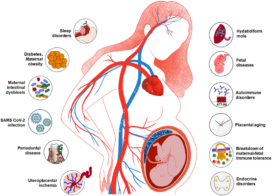
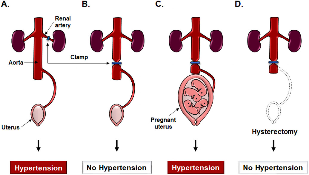
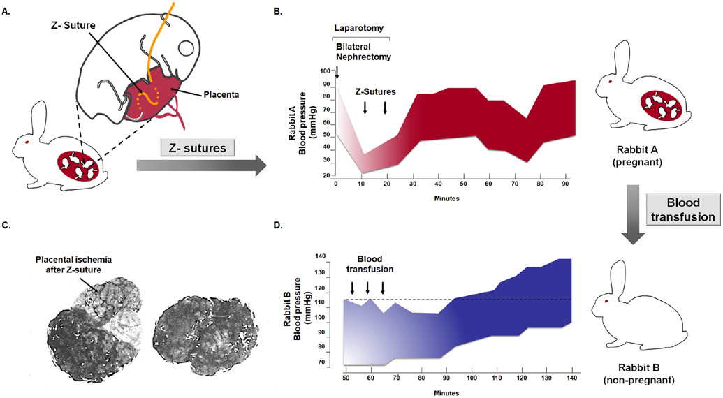
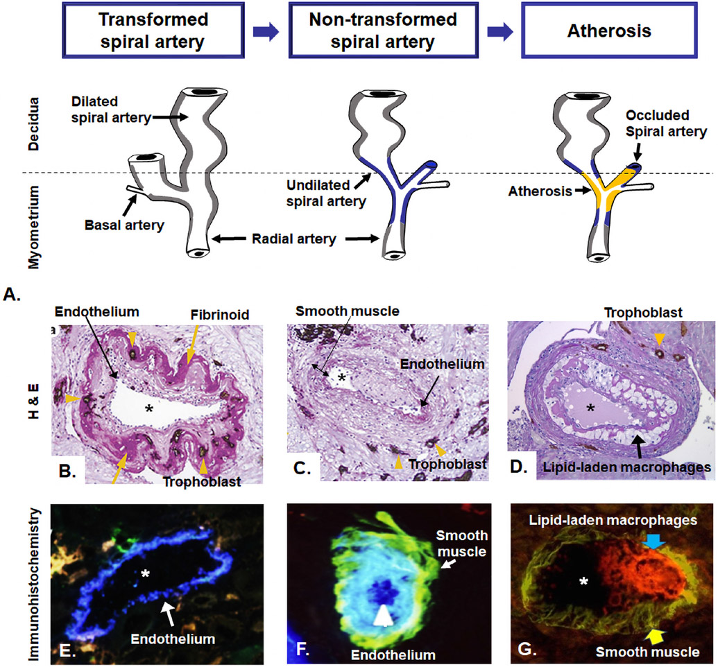
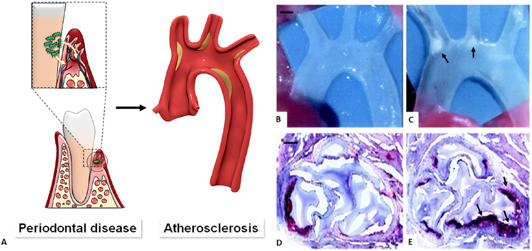
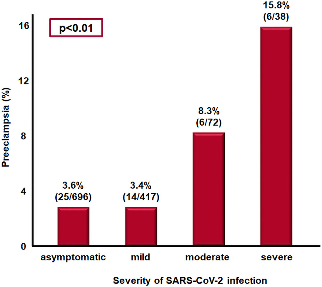
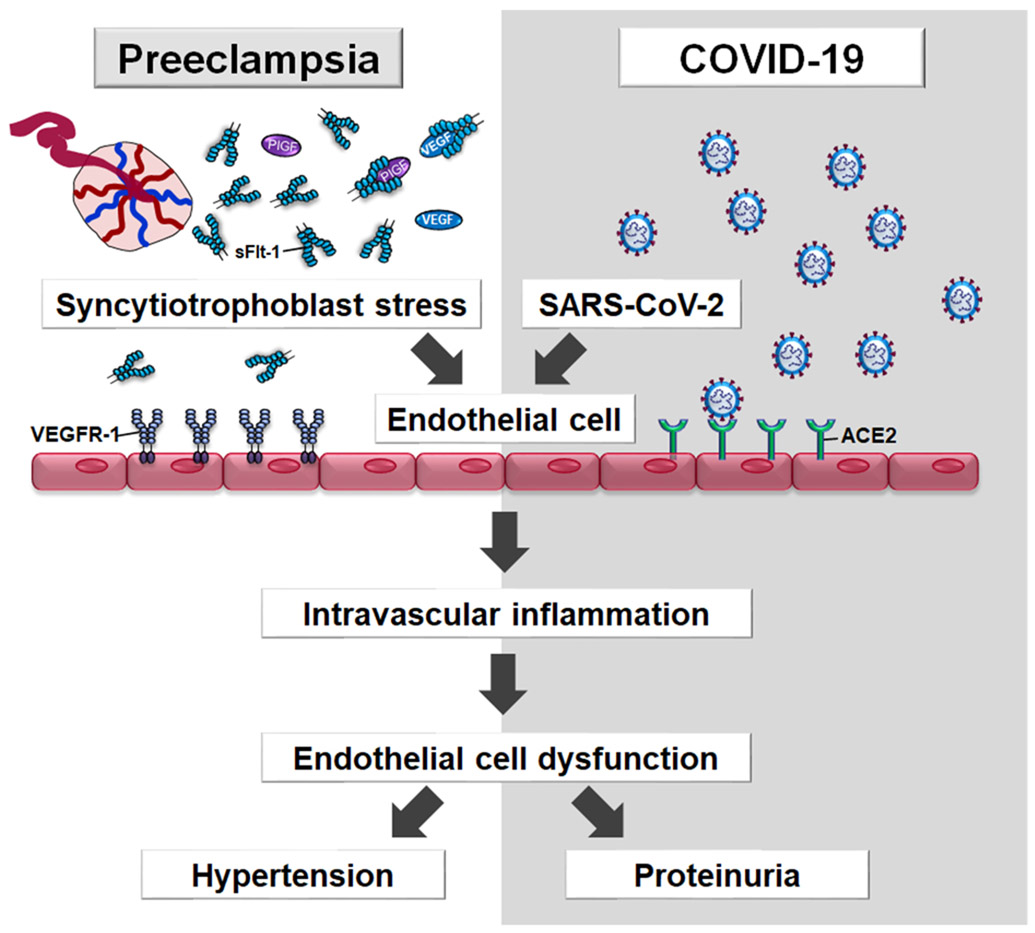
Preeclampsia is one of the "great obstetrical syndromes" in which multiple and sometimes overlapping pathologic processes activate a common pathway consisting of endothelial cell activation, intravascular inflammation, and syncytiotrophoblast stress. This article reviews the potential etiologies of preeclampsia. The role of uteroplacental ischemia is well-established on the basis of a solid body of clinical and experimental evidence. A causal role for microorganisms has gained recognition through the realization that periodontal disease and maternal gut dysbiosis are linked to atherosclerosis, thus possibly to a subset of patients with preeclampsia. The recent reports indicating that SARS-CoV-2 infection might be causally linked to preeclampsia are reviewed along with the potential mechanisms involved. Particular etiologic factors, such as the breakdown of maternal-fetal immune tolerance (thought to account for the excess of preeclampsia in primipaternity and egg donation), may operate, in part, through uteroplacental ischemia, whereas other factors such as placental aging may operate largely through syncytiotrophoblast stress. This article also examines the association between gestational diabetes mellitus and maternal obesity with preeclampsia. The role of autoimmunity, fetal diseases, and endocrine disorders is discussed. A greater understanding of the etiologic factors of preeclampsia is essential to improve treatment and prevention.

Keywords: Ballantyne syndrome; COVID-19; Cushing’s syndrome; SARS-CoV-2; angiotensin receptor II; atherosclerosis; autoantibodies; body mass index; endothelial cell dysfunction; genetic incompatibility; gestational diabetes mellitus; hydatidiform mole; hydrops fetalis; hyperaldosteronism; hyperparathyroidism; hypertension; infection; inflammation; insulin resistance; intestinal dysbiosis; maternal antifetal rejection; metabolic syndrome; mirror syndrome; molar pregnancy; obesity; placental aging; placental ischemia; placental lesions of maternal vascular malperfusion; primipaternity; proteinuria; sleep disorders; sleep-disordered breathing; snoring; tolerance.


Conflict of interest statement

Disclosure: The authors declare no conflicts of interest.

Figures

Figure 1.
Figure 1.
Multiple etiologies implicated in preeclampsia. Uteroplacental ischemia, maternal infection and inflammation (eg periodontal disease, urinary tract infection, COVID-19), maternal intestinal dysbiosis, maternal obesity, sleep disorders, hydatidiform mole, fetal diseases (eg hydrops fetalis, viral infection, Trisomy 13, and unique complications of multiple gestation), autoimmune disorders, placental aging, breakdown of maternal-fetal immune tolerance, and endocrine disorders (eg hyperparathyroidism, Cushing’s syndrome, hyperaldosteronism).
Figure 2.
Figure 2.
A diagrammatic depiction of experiments demonstrating that uterine ischemia in pregnant animals, but not in non-pregnant animals, can cause hypertension. A. In the Goldblatt model of renovascular hypertension, clamping the renal artery leads to development of hypertension through renal ischemia and the release of renin in non-pregnant animals. B. By contrast, clamping the aorta below the renal arteries does not induce hypertension in non-pregnant animals. C. Clamping of the aorta in pregnant animals below the renal arteries leads to hypertension. D. The hypertension disappears after a hysterectomy has been performed; this suggests that the source of the signals leading to maternal systemic hypertension are derived from the gravid uterus. Modified from Chaiworapongsa T. et al. Pre-eclampsia part 1: current understanding of its pathophysiology. Nat Rev Nephrol. 2014 Aug; 10 (8):466-80.
Figure 3.
Figure 3.
Experimental demonstration that placental ischemia causes maternal hypertension and that a soluble factor in the blood of an animal with placental ischemia can induce hypertension in a non-pregnant animal. A. The Z suture is placed in the uterus to generate placental ischemia. B. Placental ischemia causes hypertension. Rabbit A had undergone a bilateral nephrectomy; therefore, the kidney is not a cause of the hypertension. After Z sutures were placed through several placentas, hypertension developed. C. Gross evidence that the suture has caused a placental infarction. The pale portion of the placenta with the arrow represents a large infarction. A control placenta of the same animal is illustrated on the right. D. Transfusions of blood from Rabbit A (a pregnant rabbit with placental ischemia) to a non-pregnant rabbit (Rabbit B) caused hypertension; this suggests that a circulating factor generated after placental ischemia is present in the maternal blood and that it can induce hypertension in a non-pregnant rabbit. Modified from Berger M. and Cavanagh D. Toxemia of pregnancy: The hypertensive effect of acute experimental placental ischemia. Am J Obstet Gynecol. 1963 Oct 1;87:293-305.
Figure 4.
Figure 4.
A. Diagram of maternal blood supply to the placenta. The spiral arteries undergo physiologic changes in normal pregnancy (grey). In preeclampsia, the myometrial segment of the spiral artery fails to undergo physiological transformation (blue), which is thought to explain the uteroplacental ischemia observed in preeclampsia. Moreover, non-transformed spiral arteries are prone to atherosis (yellow), characterized by the presence of lipid-laden macrophages within the lumen. Placental basal plate spiral arteries with hematoxylin-eosin stain (B, C, and D). B. Transformed spiral arteries are characterized by the presence of intramural trophoblasts (arrowheads) and fibrinoid degeneration (arrows) of the arterial wall. C. Non-transformed spiral arteries lack intramural trophoblasts and fibrinoid degeneration, and they retain smooth muscle. Arrowheads indicate the presence of trophoblasts in myometrium, but not in the wall of the spiral artery. D. Acute atherosis in decidual spiral artery. Many lipid-laden macrophages (arrows) are seen in the spiral artery with the lack of invasion of the trophoblast (arrowhead) into myometrial segment of the spiral artery. Images (B, C, and D) stained with cytokeratin 7 (brown) and periodic acid–Schiff (pink), original magnification ×200. Immunohistochemistry of placental basal plate spiral arteries (E, F, and G). E. Presence of endothelium (arrow, blue) in vessels with normal trophoblastic invasion, original magnification × 640. F. spiral artery with presence of endothelium (blue, arrowhead) and smooth muscle cells (green, arrow), original magnification ×640. G. Atherosis lesions show the presence of numerous macrophages CD36-reactive (red reactivity, blue arrow) and smooth muscle cells in the vessel wall (green reactivity, yellow arrow), original magnification x400. *lumen of spiral artery. Modified from McMaster-Fay RA. Uteroplacental vascular syndromes: theories, hypotheses and controversies. Clin Obstet Gynecol Reprod Med 2018;4:1–5. Espinoza J et al. Normal and abnormal transformation of the spiral arteries during pregnancy. J Perinat Med. 2006;34(6):447-58. Labarrere CA et al. Failure of physiologic transformation of spiral arteries, endothelial and trophoblast cell activation, and acute atherosis in the basal plate of the placenta. Am J Obstet Gynecol. 2017 Mar;216(3):287.e1-287.e16.
Figure 5.
Figure 5.
Acute atherosis on oil-red O staining. Fat droplets (arrows) in the non-transformed spiral artery are stained red. *Lumen of spiral artery. Cover, Am J Obstet Gynecol. November 2014 Yeon Mee Kim, Roberto Romero.
Figure 6.
Figure 6.
Periodontal diseases and association with atherosclerotic disease. A. Bacteria found in the periodontal space can enter the bloodstream (bacteremia) and eventually the heart, resulting in atherosclerotic plaque in the heart blood vessels. Compared with uninfected control (B), periodontal infection with Porphyromonas gingivalis causes atherosclerotic aortic arch plaques (C, arrow) in apoE-null mice. Scale bar =1 mm. Oil red O staining of cryosections at the aortic sinus shows few small fatty streaks (control, D), whereas atherosclerotic lesions were greater in number and size (arrow) in infected animals (E). Scale bar=50 μm. Modified from Hajishengallis G et al. Local and systemic mechanisms linking periodontal disease and inflammatory comorbidities. Nat Rev Immunol. 2021 Jul;21(7):426-440 and Lalla E. et al, Oral infection with a periodontal pathogen accelerates early atherosclerosis in apolipoprotein E-null mice. Arterioscler Thromb Vasc Biol. 2003 Aug 1;23(8):1405-11.
Figure 7.
Figure 7.
Association between SARS-CoV-2 infection severity and subsequent development of preeclampsia. The most severe COVID-19, the greater the risk of preeclampsia. Modified from Lai J et al. SARS-COV-2 and the subsequent development of preeclampsia and preterm birth: evidence of a dose response relationship supporting causality. Am J Obstet Gynecol. 2021 Aug 26:S0002-9378(21)00947-9.
Figure 8.
Figure 8.
Placental syncytiotrophoblast stress induces excessive sFlt-1 into the maternal circulation. sFlt-1 binds to free PlGF or VEGF (proangiogenic factors) with high affinity, thus preventing their interaction with their cell-surface receptors (i.e. VEGR-1) on the endothelial cells, leading to endothelial dysfunction. SARS-CoV-2 also targets the endothelium which normally express angiotensin-converting-enzyme 2 (ACE-2), one of the cell entry receptors for the virus, leading to endothelitis, which induces intravascular inflammation (i.e. cytokine storm) and endothelial dysfunction. ACE2; angiotensin-converting enzyme 2, COVID-19; Coronavirus disease 2019, SARS-CoV-2; severe acute respiratory syndrome coronavirus 2, sFlt-1; Soluble Fms-Like Tyrosine Kinase-1, VEGFR-1; vascular endothelial growth factor receptor-1
Figure 9.
Figure 9.
(A) Gut microbiota in patients with preeclampsia (red) and those with normal pregnancy (blue). The significantly different genera (Firmicutes, Bacteroidetes, Proteobacterias, and Actinobacteria) at the phylum level in the two groups. The boxes represent the interquartile range (IQR) between first and third quartiles and the line inside represents the median. *p < 0.05. (B) Maternal intestinal dysbiosis and preeclampsia. Mice that received fecal microbiota from patients with preeclampsia (red) had higher systolic blood pressure than mice that received fecal microbiota from normotensive pregnant women (blue) or those that received the phosphate-buffered saline (control, grey). Modified from Wang J et al. Gut Microbiota Dysbiosis and Increased Plasma LPS and TMAO Levels in Patients with Preeclampsia. Front Cell Infect Microbiol. 2019 Dec 3;9:409 and Chen X. et al. Gut dysbiosis induces the development of pre-eclampsia through bacterial translocation. Gut. 2020 Mar;69(3):513-522.
Figure 10.
Figure 10.
Probability of preeclampsia according to the pre-pregnancy body mass index in both nulliparous (top line, red) and multiparous (bottom line, blue) women. Modified from Catov JM et al. Risk of early or severe pre-eclampsia related to pre-existing conditions. Int J Epidemiol. 2007 Apr;36(2):412-9.
Figure 11.
Figure 11.
Interactions between maternal KIR and fetal HLA-C at the site of placentation. If the mother has a KIR BB genotype, which binds to trophoblast HLA-C1 molecules, this activates dNK cells, producing increased levels of cytokines such as GM-CSF that can enhance placentation. In contrast, when the mother has a KIR AA genotype and fetal HLA-C2 alleles, this inhibits dNK cells, leading to defective placentation. KIR; killer cell immunoglobulin like receptors, HLA; human leukocyte antigen, dNK; decidual natural killer cells, GM-CSF; granulocyte-macrophage colony-stimulating factor. Modified from https://www.ivi-rmainnovation.com/maternal-killer-immunoglobulin-receptors-predictive-live-birth-rate/
Figure 12.
Figure 12.
A. Optimal villi density for maximal oxygen uptake in the human placenta. Placentas with low villi density (rarefied villi) have low oxygen uptake, as fetal villi are rare. By contrast, in placentas with high villous density (villous overcrowding), there is no space for intervillous space for oxygen exchange. The optimal villi density for the oxygen uptake was 0.47 ± 0.06, calculated from histomorphometrical data for villi and intervillous volumes of placentas. B. Cross-section of the placenta: 1) rarefied villi in preeclampsia; 2) normal placenta; and 3) villous overcrowding in diabetes mellitus. H&E stained and scale represents 50 μm. Modified from Serov AS et al. Optimal villi density for maximal oxygen uptake in the human placenta. J Theor Biol. 2015 Jan 7;364:383-96.

References

    1. Young J The etiology of eclampsia and albuminuria and their relation to accidental hemorrhage. Transactions Edinburgh Obstetrical Society 1914;39:153–202. - PMC - PubMed
    1. Dixon WE, Taylor FE. An Epidiascopic Demonstration on "The Physiological Action of the Placenta.". Proc R Soc Med 1908;1:11–3. - PMC - PubMed
    1. Ogden E, Hildebrand G, Page EW. Rise of blood pressure during ischemia of the gravid uterus. Proc Soc Exp Biol Med 1940;43:49–51.
    1. Chaiworapongsa T, Chaemsaithong P, Yeo L, Romero R. Pre-eclampsia part 1: current understanding of its pathophysiology. Nature reviews Nephrology 2014;10:466–80. - PMC - PubMed
    1. Kumar D Chronic placental ischemia in relation to toxemias of pregnancy: a preliminary report. Am J Obstet Gynecol 1962;84:1323–29.

Publication types