

Hemivertebra Resection and Spinal Arthrodesis by Single-Stage Posterior Approach in Congenital Scoliosis and Kyphoscoliosis: Results at 9.6 Years Mean Follow-up
- PMID: 35177526
- PMCID: PMC9519081
- DOI: 10.14444/8188
Hemivertebra Resection and Spinal Arthrodesis by Single-Stage Posterior Approach in Congenital Scoliosis and Kyphoscoliosis: Results at 9.6 Years Mean Follow-up
Abstract
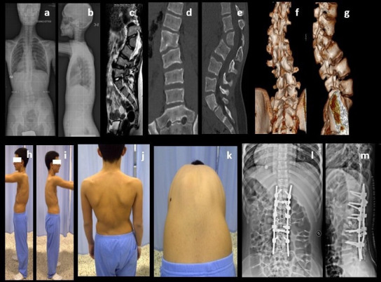
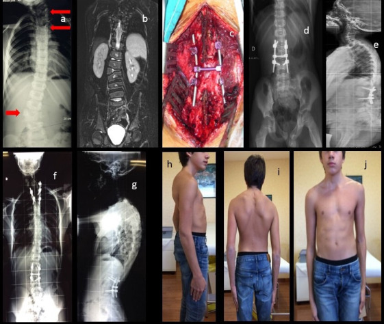
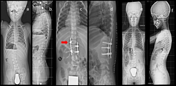
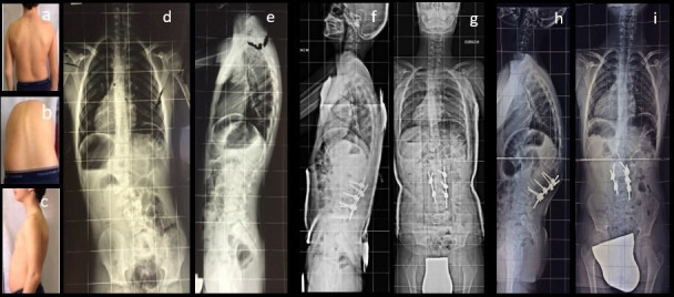
Background: Congenital kyphoscoliosis due to hemivertebra is generally treated surgically because of high risk of curve progression and high risk of nervous system complications. Modern posterior access surgical techniques, including total hemivertebra resection, can completely correct deformity without additional anterior access surgeries. The purpose of this study was to evaluate midterm results of hemivertebra resection and spinal arthrodesis; the hypothesis was that it is a safe, effective, and reproducible procedure.
Materials and methods: From 2006 to 2019, hemivertebra resection and instrumented spinal arthrodesis with pedicle screws was performed on 82 patients with congenital vertebral deformities (62 scoliosis and 20 kyphoscoliosis) by posterior approach. Mean age at surgery was 8.6 years, and 22 patients were under 10 years of age. After stabilization patients have been braced for a period from 3 to 5 months.
Results: Mean follow-up was 9.6 years (range 1.2-12.8 years); mean kyphosis curve after surgery was reduced to 20° Cobb; and mean scoliosis curve was reduced to 11° Cobb. We experienced no major complications (postsurgical infection, instrumentation failure, severe neurological impairment, severe blood loss) at latest follow-up .
Conclusion: We strongly advocate one-time posterior hemivertebra resection and arthrodesis as the most suitable surgical procedure for congenital scoliosis due to hemivertebra. Posterior approach interventions with pedicle screws instrumentation are less invasive than combined anterior-posterior approach interventions. We think that posterior approach procedures can lead to excellent deformity correction in both frontal and sagittal views, optimal stability, and low risk of nervous injury.
Clinical relevance: Congenital scoliosis treatment is one of the most challeging conditions a spine surgeon has to face. We advocate that a one-stage posterior approach for hemivertebrectomy and fusion is a reliable, safe tachnique, whom excellent results remain stable at a mid/long-term follow-up.
Level of evidence: Level 4.
Keywords: congenital scoliosis; hemivertebra resection; posterior vertebral arthrodesis.
This manuscript is generously published free of charge by ISASS, the International Society for the Advancement of Spine Surgery. Copyright © 2022 ISASS. To see more or order reprints or permissions, see http://ijssurgery.com.
Conflict of interest statement
Declaration of Conflicting Interests: The authors report no conflicts of interest related to this article.
Figures
References
LinkOut - more resources
Full Text Sources
Research Materials