. 2022 Feb 1;36(3-4):210-224.
doi: 10.1101/gad.349223.121. Epub 2022 Feb 17.

RBBP6 activates the pre-mRNA 3' end processing machinery in humans

Affiliations

RBBP6 activates the pre-mRNA 3' end processing machinery in humans

Vytaute Boreikaite et al. Genes Dev. .

Abstract

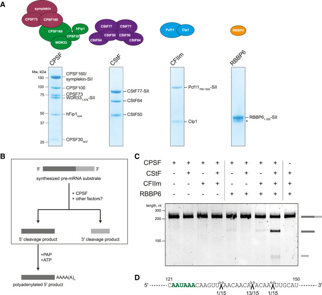
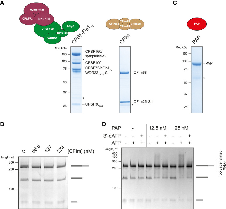
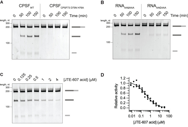
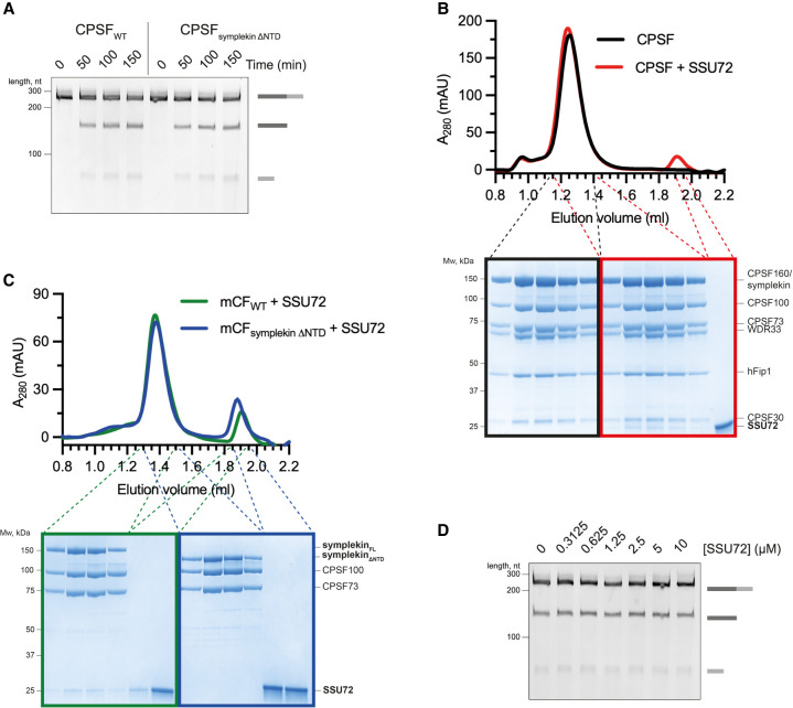
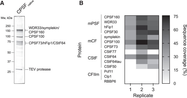
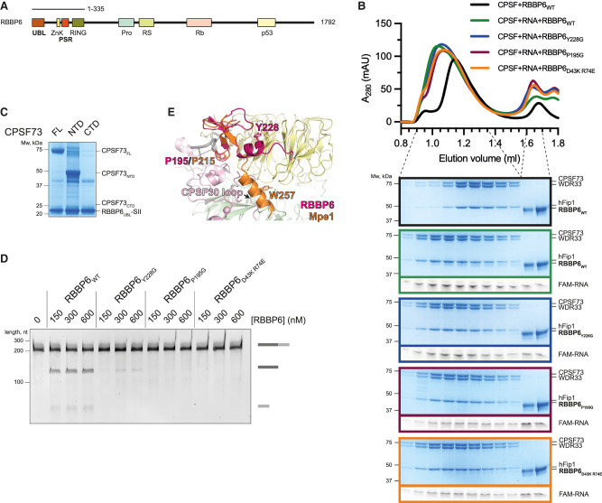
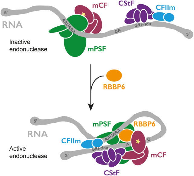
3' end processing of most human mRNAs is carried out by the cleavage and polyadenylation specificity factor (CPSF; CPF in yeast). Endonucleolytic cleavage of the nascent pre-mRNA defines the 3' end of the mature transcript, which is important for mRNA localization, translation, and stability. Cleavage must therefore be tightly regulated. Here, we reconstituted specific and efficient 3' endonuclease activity of human CPSF with purified proteins. This required the seven-subunit CPSF as well as three additional protein factors: cleavage stimulatory factor (CStF), cleavage factor IIm (CFIIm), and, importantly, the multidomain protein RBBP6. Unlike its yeast homolog Mpe1, which is a stable subunit of CPF, RBBP6 does not copurify with CPSF and is recruited in an RNA-dependent manner. Sequence and mutational analyses suggest that RBBP6 interacts with the WDR33 and CPSF73 subunits of CPSF. Thus, it is likely that the role of RBBP6 is conserved from yeast to humans. Overall, our data are consistent with CPSF endonuclease activation and site-specific pre-mRNA cleavage being highly controlled to maintain fidelity in mRNA processing.

Keywords: RNA; endonuclease; gene expression; polyadenylation.


Figures

Figure 1.
Figure 1.
CStF, CFIIm, and RBBP6 are required for activation of the CPSF endonuclease. (A) Schematic representations and SDS-PAGE analyses of the purified proteins used in the in vitro endonuclease assays. Residue boundaries and alternative isoforms are indicated for truncated proteins. An asterisk denotes degradation products. (SII) StrepII tag. (B) Schematic representation of the in vitro pre-mRNA 3′ end processing assay. The cleavage reaction is boxed out. The polyadenylation step was not assayed here. (C) Denaturing gel electrophoresis of the SV40 pre-mRNA substrate after incubation with various combinations of human 3′ end processing factors. The full-length and cleaved RNAs are shown schematically at the right. (D) Part of the sequence of the SV40 pre-mRNA substrate with the experimentally determined CPSF cleavage sites indicated (scissors). The frequency of a particular cleavage site identified by sequencing of 15 cleavage products is shown below. The polyadenylation signal (PAS) sequence is marked in green.
Figure 2.
Figure 2.
CFIm is not required for CPSF cleavage activity, and RNA is cleaved and polyadenylated in the presence of CPSF, RBBP6, CStF, CFIIm, and PAP. (A) SDS-PAGE analyses of purified CPSF containing full-length hFip1 (hFip1FL) and of purified CFIm complex. Asterisks denote degradation products. (SII) StrepII tag. (B) Cleavage assays of the SV40 pre-mRNA substrate with CPSF-hFip1FL in the presence of increasing concentrations of CFIm. CFIm does not substantially affect CPSF cleavage activity. (C) SDS-PAGE analysis of purified PAP. An asterisk denotes degradation products. (D) Coupled cleavage and polyadenylation assays of the SV40 pre-mRNA substrate at two different concentrations of PAP in the presence of either ATP or ATP and 3′-dATP together. 3′-dATP is also called cordycepin and is known to inhibit polyadenylation. The heterogeneous products that appear in the presence of ATP are largely absent when 3′-dATP is also added. This demonstrates that polyadenylation is responsible for the diffuse band. Some substrate RNAs may also get polyadenylated by free PAP.
Figure 3.
Figure 3.
Cleavage activity of purified recombinant CPSF is dependent on CPSF73 and a PAS. (A) Time-course cleavage assays of the SV40 pre-mRNA substrate comparing the activities of wild-type (CPSFWT) and nuclease-dead (CPSFCPSF73 D75N H76A) CPSF complexes. (B) Time-course cleavage assays of SV40 pre-mRNA substrates containing either a canonical PAS (RNAAAUAAA) or a mutant PAS (RNAAACAAA) sequence. (C) Cleavage assays in the presence of increasing concentrations of the JTE-607 acid compound. (D) Dose response curve of the CPSF cleavage activity as a function of the concentration of JTE-607 acid. Each dot represents a single measurement. At least three measurements were performed for each concentration of the drug, but some points overlap.
Figure 4.
Figure 4.
Canonical and histone pre-mRNA 3′ end processing complexes are activated by different mechanisms. (A) Time-course cleavage assays of the SV40 pre-mRNA substrate comparing wild-type CPSF (CPSFWT) and CPSF lacking the symplekin NTD (CPSFsymplekin ΔNTD). (B) Gel filtration chromatograms (top) and SDS-PAGE analyses (bottom) of CPSF in the presence (red) or absence (black) of SSU72. (C) Gel filtration chromatograms (top) and SDS-PAGE analyses (bottom) of wild-type mCF (mCFWT; green) and mCF lacking the NTD of symplekin (mCFsymplekin ΔNTD; blue) mixed with SSU72. (D) Cleavage assays in the presence of increasing concentrations of SSU72.
Figure 5.
Figure 5.
RBBP6 is not a stable subunit of CPSF purified from human cells. (A) SDS-PAGE analysis of the endogenous CPSF complex. The bands representing CPSF subunits are indicated. TEV protease was used to elute the complex from Strep-Tactin beads and remains present in the sample. The gel was stained with SYPRO Ruby. (B) Heat map representing the sequence coverage of each protein required for CPSF endonuclease activity in vitro in the endogenous CPSF preparations as detected by mass spectrometry. No RBBP6 peptides were detected across three independent experiments.
Figure 6.
Figure 6.
RBBP6 is a conserved activator of canonical pre-mRNA 3′ end cleavage. (A) Domain diagram of full-length human RBBP6 (1792 residues). The construct used in this study (residues 1–335) is indicated. (UBL) Ubiquitin-like domain, (ZnK) zinc knuckle, (PSR) pre-mRNA-sensing region, (Pro) proline-rich domain, (RS) arginine, serine-rich domain, (Rb) retinoblastoma protein-interacting region, (p53) p53-interacting region. (B) Gel filtration chromatograms of CPSF and RBBP6 in the presence or absence of a 5′-FAM fluorescently labeled 41-nt L3 RNA (top), and denaturing PAGE analysis of proteins and RNA from the indicated fractions (bottom). The gels are cropped and outlined in color to correspond with the colors of the chromatogram traces. (C) Pull-down of the SII-tagged UBL domain of RBBP6 in the presence of various constructs of CPSF73 from Sf9 insect cells. RBBP6 pulls down full-length CPSF73 and CPSF73-NTD. (FL) Full length, (NTD) N-terminal domain (residues 1–460), (CTD) C-terminal domain (residues 461–684). (D) Cleavage assays in the presence of various concentrations of either wild-type (RBBP6WT) or mutant (RBBP6Y228G, RBBP6P195G, and RBBP6D43K R74E) RBBP6. (E) Overlay of the experimental structure of the yeast Mpe1 PSR (orange) (Rodríguez-Molina et al. 2021) and an AlphaFold2 prediction of the structure of the equivalent region in human RBBP6 (magenta) overlaid on human mPSF (PDB 6BLL) (Sun et al. 2018). Residues of functional significance are indicated. A loop of CPSF30 would clash with the C-terminal helix of the Mpe1 PSR. (Yellow) WDR33, (pink) CPSF30, (green) CPSF160, (gray) PAS RNA.
Figure 7.
Figure 7.
Model for activation of CPSF cleavage. Coassembly of CPSF, CStF, CFIIm, and RBBP6 activates the endonuclease CPSF73 (star). Remodeling of protein and RNA may occur.

Similar articles

Cited by

References

    1. Adamson TE, Shutt DC, Price DH. 2005. Functional coupling of cleavage and polyadenylation with transcription of mRNA. J Biol Chem 280: 32262–32271. 10.1074/jbc.M505532200 - DOI - PubMed
    1. Baejen C, Torkler P, Gressel S, Essig K, Söding J, Cramer P. 2014. Transcriptome maps of mRNP biogenesis factors define pre-mRNA recognition. Mol Cell 55: 745–757. 10.1016/j.molcel.2014.08.005 - DOI - PubMed
    1. Batra J, Hultquist JF, Liu D, Shtanko O, Von Dollen J, Satkamp L, Jang GM, Luthra P, Schwarz TM, Small GI, et al. 2018. Protein interaction mapping identifies RBBP6 as a negative regulator of Ebola virus replication. Cell 175: 1917–1930.e13. 10.1016/j.cell.2018.08.044 - DOI - PMC - PubMed
    1. Beaudoing E, Freier S, Wyatt JR, Claverie JM, Gautheret D. 2000. Patterns of variant polyadenylation signal usage in human genes. Genome Res 10: 1001–1010. 10.1101/gr.10.7.1001 - DOI - PMC - PubMed
    1. Buratowski S. 2005. Connections between mRNA 3′ end processing and transcription termination. Curr Opin Cell Biol 17: 257–261. 10.1016/j.ceb.2005.04.003 - DOI - PubMed

Publication types

MeSH terms