. 2022 Feb 1;36(3-4):195-209.
doi: 10.1101/gad.349217.121. Epub 2022 Feb 17.

Reconstitution of 3' end processing of mammalian pre-mRNA reveals a central role of RBBP6

Affiliations

Reconstitution of 3' end processing of mammalian pre-mRNA reveals a central role of RBBP6

Moritz Schmidt et al. Genes Dev. .

Abstract

The 3' ends of almost all eukaryotic mRNAs are generated in an essential two-step processing reaction: endonucleolytic cleavage of an extended precursor followed by the addition of a poly(A) tail. By reconstituting the reaction from overproduced and purified proteins, we provide a minimal list of 14 polypeptides that are essential and two that are stimulatory for RNA processing. In a reaction depending on the polyadenylation signal AAUAAA, the reconstituted system cleaves pre-mRNA at a single preferred site corresponding to the one used in vivo. Among the proteins, cleavage factor I stimulates cleavage but is not essential, consistent with its prominent role in alternative polyadenylation. RBBP6 is required, with structural data showing it to contact and presumably activate the endonuclease CPSF73 through its DWNN domain. The C-terminal domain of RNA polymerase II is dispensable. ATP, but not its hydrolysis, supports RNA cleavage by binding to the hClp1 subunit of cleavage factor II with submicromolar affinity.

Keywords: 3′ processing; CPSF; RBBP6; RNA cleavage; RNA processing; poly(A) polymerase; polyadenylation.


Figures

Figure 1.
Figure 1.
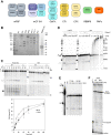
Reconstitution of pre-mRNA 3′ processing from 16 purified polypeptides. (A) Schematic representation of proteins participating in pre-mRNA cleavage. (B) Proteins used in this study were separated by SDS-PAGE and stained with Coomassie. CPSF160 and WDR33 are not resolved, and CPSF30 runs as a doublet. Two versions of mCF are shown (see the text). In the RBBP6 preparation, all major bands reacted with anti-RBBP6 in Western blotting. (C) Kinetics of cleavage of the SV40 late RNA in the presence of WT PAP and 3′-dATP. (Left) Reaction with purified proteins. (Right) Reaction in nuclear extract. The substrate RNA (wild type or mutant) is shortened by a few nucleotides from the 3′ end in nuclear extract. Downstream fragments are shown in a longer exposure of the bottom part of the same gel. Quantification is shown in the bottom panel (n = 3). (D) Kinetics of cleavage and polyadenylation with purified proteins in the presence of 3′-dATP versus ATP. Only the upstream cleavage product is shown. Reactions were analyzed directly or after RNase H/oligo(dT) treatment. Brackets indicate digestion products with oligo(A) stubs. (E) Analysis of 5′ cleavage fragment. A reaction with uncapped RNA reveals a single 5′ product, whereas capped RNA generates two products, presumably reflecting incomplete capping. Products comigrate with the one generated in nuclear extract. A partial alkaline hydrolysis ladder of an end-labeled RNA (OH) demonstrates single-nucleotide resolution. This lane was cut from the same gel, and contrast was enhanced to reveal individual bands. (F) Analysis of a 3′ cleavage fragment. Reactions were carried out with unlabeled substrate RNA and either wild-type (WT) mCF3 or mCF3 containing inactive CPSF73 (MUT). The cleavage site was mapped by primer extension. Products corresponding to uncleaved RNA and the major 3′ cleavage product are indicated. Minor products are labeled with asterisks. For the first lane, a synthetic 3′ cleavage fragment was used as template.
Figure 2.
Figure 2.
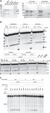
CF I is not an essential 3′ processing factor. (A) CF I is the only nonessential component of the reconstituted cleavage reaction. The SV40 late precursor RNA was processed in nuclear extract (NXT) or with a mixture of purified proteins (PM). Individual components listed at the top were left out. (B) Stimulation of cleavage by CF I depends on UGUA motifs. SV40 late and L3 RNAs with their UGUA motifs are sketched at the top. In L3-2xMUT and L3-3xMUT, the first two UGUA motifs or all three, respectively, were mutated. Titrations of CF I were carried out. (Right) Cleavage efficiencies (average of n = 3 ± SD). Efficiencies without CF I addition were 2.2% ± 0.3% for L3, 1.8% ± 0.4% for L3-2xMUT, and 1.9% ± 0.4% for L3-3xMUT.
Figure 3.
Figure 3.
Two structured domains of RBBP6 are sufficient for pre-mRNA cleavage. (A, top panel) Domain structure of RBBP6. Domain boundaries are indicated at the top, and deletion boundaries are indicated at the bottom. (Bottom panel) SDS–polyacrylamide gel analysis of RBBP6 preparations. (B) Titration of RBBP6 variants in cleavage assays. “Full-length” was MBP-RBBP6 with the MBP tag cleaved off; “full-length plus CF I” was RBBP6 from a coexpression with CF I. A quantification is shown at the bottom. (C) Cleavage activity of RBBP61–780 carrying point mutations. (D) RBBP61–780 was titrated in a filter binding assay with SV40 late RNA (cf. Supplemental Table S2). (E) RNA binding activity copurifies with RBBP6. (Top panel) UV profile and RNA binding activity of the final Resource Q column of an RBBP61–780 purification. (Bottom panel) SDS-PAGE of the same fractions. The main RBBP6 band is marked.
Figure 4.
Figure 4.
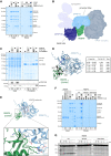
RBBP6 contacts the β-lactamase domain of CPSF73. (A) Coomassie-stained SDS-PAGE analysis of pull-down experiment showing that RBBP6 directly interacts with CPSF and, albeit more weakly, mCF. CPSF used here did not contain hFip1, but hFip1 was present in all cleavage assays (Fig. 1A,B; Supplemental Material). (B) Filtered and segmented cryo-EM map of the eight-subunit CPSF–RBBP6 complex. The proximal and distal lobes of mCF are indicated. (C) Coomassie-stained SDS-PAGE analysis of pull-down experiment showing that RBBP6 directly interacts with CPSF73 and not CPSF100. (D) Cross-links between CPSF731–458 and RBBP61–81 mapped onto the computationally predicted model of the complex. Intramolecular and intermolecular cross-links are in black and blue, respectively, with the thickness of the line indicating their score (thicker = higher score), and the respective residues indicated as spheres. A cross-link to an unmodeled region is shown in white (placed to the closest visible Cα atom). The table lists measured distances for visualized cross-links. (E) Computationally predicted structural model of the CPSF731–458–RBBP61–81 complex with close-up of the putative binding interface. The CPSF73 active site is indicated. Possible ionic interactions are shown with black dotted lines. Labeled positively charged RBBP6 residues were changed to aspartic acid. (F) Coomassie-stained SDS-PAGE analysis of pull-down experiment showing that reverse-charged mutations in the predicted RBBP6–CPSF73 interface disrupt the interaction of RBBP6 with mCF and CPSF. (G) RBBP61–335, either wild type or containing the same mutations as in F, was titrated into cleavage reactions.
Figure 5.
Figure 5.
Composition of CstF and mPSF, active in pre-mRNA cleavage. (A, left) Different versions of CstF and its subunits were analyzed by SDS-PAGE and Coomassie staining. “Canonical” subunits, including CstF64, are labeled with black arrows. CstF64τ is labeled with a gray arrow. (Right) CstF (64/τ) with a FLAG tag on CstF64τ and, as a control, CstF (64/64) without a FLAG tag were used in a FLAG pull-down experiment. (I) Input, (FT) flowthrough (E1 and E2) FLAG peptide eluates, (B) material remaining on the beads eluted with SDS sample buffer. Proteins were detected by Coomassie staining. The identity of the band running at the CstF64τ position in CstF (64/64) is unknown; based on Western blots, it is neither 64τ nor a fragment of CstF77. (B) Both CstF64 and 64τ are active in pre-mRNA cleavage, and CstF50 is essential. Proteins shown in A and their combinations were tested in cleavage assays. (C) Both mCF3 and mCF4 function in pre-mRNA cleavage. The two protein complexes, each with wild-type (+) or point-mutated (−) CPSF73, were combined with the remaining cleavage factors and tested in cleavage assays. The SV40 late Δ RNA served as negative control.
Figure 6.
Figure 6.
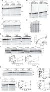
Pre-mRNA cleavage depends on ATP. (A) Creatine phosphate is not required for cleavage. Cleavage reactions were carried out with nuclear extract (NXT), a mixture of purified proteins (PM), or the same mixture plus creatine kinase (5 µg/mL). In the presence of 0.5 mM 3′-dATP, reactions were supplemented with inorganic phosphate (pH 8.0), creatine, a mixture of both, phosphocreatine, or phosphoserine as indicated, each at 20 mM. (B) ATP is essential for cleavage, but cleavage of phosphoanhydride bonds is not. Reactions contained the nucleotides indicated, each at 0.5 mM, and either WT PAP or PAP D115A. With WT PAP, cleavage in the presence of CTP, GTP, or ITP was not visible, presumably due to heterogeneous limited extension of the cleavage product. (C) A cleavage reaction lacking CF I is still ATP-dependent. Reactions were carried out with or without CF I as indicated. 3′-dATP was titrated between 3.9 and 1000 µM. (D) Mutations in the active site of PAP do not affect the ATP dependence of RNA cleavage. Reactions contained WT PAP or mutants as indicated. 3′-dATP was titrated between 2 and 1000 µM. (Right) Average of n = 3; highest ATP concentration was omitted. (E) Mutant hClp1 forms a stable complex with hPCF11. The Coomassie-stained SDS-PAGE shows the peak fraction from each Mono-Q column. Based on Western analysis of comparable wild-type preparations, most additional bands are breakdown products of hPcf11. (F) Mutations in the ATP binding site of hClp1 affect the RNA 5′ kinase activity of CF II. CF II preparations (shown in E) were tested in kinase assays with ATP or ITP. (Top) Representative time courses. (Bottom) Average of n = 3. (G) Mutations in the ATP binding site of hClp1 affect ATP dependence of RNA cleavage. CF II preparations (shown in E) were tested in cleavage assays with mutant PAP. ATP was titrated from 0.12 µM (WT) or 1.95 µM (mutants) to 500 µM. A representative experiment is shown. (H) A quantification of experiments as in G. The averages of n = 3 are plotted. (Top panel) Full concentration range. (Bottom panel) Enlargement of the low concentration range. (I) A mutation in the ATP binding site of hClp1 changes the nucleotide specificity of cleavage. The proteins indicated were tested as in G, but ITP was titrated. (Top) Representative experiment. (Bottom) Average of n = 3.
Figure 7.
Figure 7.
Model of the reconstituted pre-mRNA cleavage complex. RBBP6 and ATP are proposed to stabilize the activated conformation of the complex. CF I is not shown as part of the core complex, but it stimulates processing by interaction with hFip1 (Zhu et al. 2018).

Similar articles

Cited by

References

    1. Ahn SH, Kim M, Buratowski S. 2004. Phosphorylation of serine 2 within the RNA polymerase II C-terminal domain couples transcription and 3′ end processing. Mol Cell 13: 67–76. 10.1016/S1097-2765(03)00492-1 - DOI - PubMed
    1. Bienroth S, Keller W, Wahle E. 1993. Assembly of a processive messenger RNA polyadenylation complex. EMBO J 12: 585–594. 10.1002/j.1460-2075.1993.tb05690.x - DOI - PMC - PubMed
    1. Biyani N, Righetto RD, McLeod R, Caujolle-Bert D, Castano-Diez D, Goldie KN, Stahlberg H. 2017. Focus: the interface between data collection and data processing in cryo-EM. J Struct Biol 198: 124–133. 10.1016/j.jsb.2017.03.007 - DOI - PubMed
    1. Boreikaite V, Elliott TS, Chin JW, Passmore LA. 2022. RBBP6 activates the pre-mRNA 3′ end processing machinery in humans. Genes Dev (this issue). 10.1101/gad.349223.121 - DOI - PMC - PubMed
    1. Brown KM, Gilmartin GM. 2003. A mechanism for the regulation of pre-mRNA 3′ processing by human cleavage factor I-m. Mol Cell 12: 1467–1476. 10.1016/S1097-2765(03)00453-2 - DOI - PubMed

Publication types

MeSH terms

Substances