. 2022 Mar;43(3):422-428.
doi: 10.3174/ajnr.A7423. Epub 2022 Feb 17.

Characterization of Restenosis following Carotid Endarterectomy Using Contrast-Enhanced Vessel Wall MR Imaging

Affiliations

Characterization of Restenosis following Carotid Endarterectomy Using Contrast-Enhanced Vessel Wall MR Imaging

W Yang et al. AJNR Am J Neuroradiol. 2022 Mar.

Abstract

Background and purpose: Restenosis is an important determinant of the long-term efficacy of carotid endarterectomy. Our aim was to assess the role of high-resolution vessel wall MR imaging for characterizing restenosis after carotid endarterectomy.

Materials and methods: Patients who underwent vessel wall MR imaging after carotid endarterectomy were included in this study. Restenotic lesions were classified as myointimal hyperplasia or recurrent atherosclerotic plaques based on MR imaging features of lesion compositions. Imaging characteristics of myointimal hyperplasia were compared with those of normal post-carotid endarterectomy and recurrent plaque groups. Recurrent plaques were matched with primary plaques by categories of stenosis, and differences in plaque features were compared between the 2 groups.

Results: Twenty-two recurrent lesions from 18 patients (14 unilateral and 4 bilateral) were classified as myointimal hyperplasia or recurrent plaque. Myointimal hyperplasia showed no difference in enhancement compared with normal post-carotid endarterectomy vessels (5 unilateral) but showed stronger enhancement than recurrent plaques (80.10% [SD, 42.42%] versus 56.74% [SD, 46.54%], P = .042). A multivariate logistic regression model of plaque-feature detection in recurrent plaques compared with primary plaques adjusted for maximum wall thickness revealed that recurrent plaques were longer (OR, 4.27; 95% CI, 1.32-13.85; P = .015) and more likely to involve a flow divider and side walls (OR, 6.96; 95% CI, 1.37-35.28; P = .019). Recurrent plaques had a higher prevalence of intraplaque hemorrhage (61.5% versus 30.8%, P = .048) by a χ2 test, but compositional differences were not significant in the multivariate model.

Conclusions: Vessel wall MR imaging can distinguish recurrent plaques from myointimal hyperplasia and reveal features that may differ between primary and recurrent plaques, highlighting its value for evaluating patients with carotid restenosis.


Figures

FIG 1.
FIG 1.
Flow chart of patient inclusion. The flow chart details referral reasons and exclusion criteria for patients with VWMRI examinations. N indicates number of patients.
FIG 2.
FIG 2.
Progression from normal post-CEA appearance to MH. TOF-MRA (A) of a post-CEA carotid artery shows an expected vessel geometry 2 months after the operation. Pre- (B) and postcontrast (C) VWMRI acquired at the proximal ICA (indicated by white line in A) shows vessel wall enhancement but no abnormal wall thickening of the proximal ICA (long arrows). TOF-MRA acquired 10 months after CEA (D) shows luminal stenosis. Pre- (E) and postcontrast (F) VWMRIs acquired at the same location (indicated by white line in D) show circumferential wall thickening with mild, homogeneous enhancement compatible with MH (long arrows). Short arrows in B, C, E, and F indicate the external carotid artery. VWMRIs were acquired using an electrocardiogram-gated double inversion recovery turbo spin-echo sequence (TR/TE/echo-train, 1 RR/9 ms/10; resolution, 0.35 × 0.35 × 2 mm3).
FIG 3.
FIG 3.
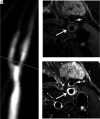
Representative images of MH. TOF-MRA (A) shows restenosis post-CEA extending from proximal to distal to the carotid bifurcation. Precontrast VWMRI (B) at the level of proximal ICA (indicated by white line in A) shows concentric homogeneous wall thickening (long arrow), indicative of MH. The lesion is enhanced on postcontrast VWMRI (C, long arrow). Short arrows in B and C indicate the external carotid artery. VWMRIs were acquired using an electrocardiogram-gated double inversion recovery turbo spin-echo sequence (TR/TE/echo-train, 1 RR/9 ms/10; resolution, 0.35 × 0.35 × 2 mm3).
FIG 4.
FIG 4.
Representative images of recurrent plaque. TOF-MRA (A) shows high-grade restenosis of the carotid bulb. The white line indicates the location of VWMRIs. Pre- (B, left image) and corresponding postcontrast (B, right image) VWMRIs show enhancing eccentric wall thickening with ulceration (asterisks), suggestive of a recurrent plaque. The corresponding specimen sections stained with MOVAT (C) confirm the diagnosis of recurrent plaque. The asterisk indicates ulceration. The arrowheads in B and C indicate the lumen. VWMRIs were acquired using an electrocardiogram-gated double inversion recovery turbo spin-echo sequence (TR/TE/echo-train, 1 RR/9 ms/10; resolution, 0.35 × 0.35 × 2 mm3).

Similar articles

References

    1. Rothwell PM, Eliasziw M, Gutnikov SA, et al. . Analysis of pooled data from the randomised controlled trials of endarterectomy for symptomatic carotid stenosis. Lancet 2003;361:107–16 10.1016/S0140-6736(03)12228-3 - DOI - PubMed
    1. Barnett HJ, Taylor DW, Eliasziw M, et al. . Benefit of carotid endarterectomy in patients with symptomatic moderate or severe stenosis; North American Symptomatic Carotid Endarterectomy Trial Collaborators. N Engl J Med 1998;339:1415–15 10.1056/NEJM199811123392002 - DOI - PubMed
    1. Kang J, Conrad MF, Patel VI, et al. . Clinical and anatomic outcomes after carotid endarterectomy. J Vasc Surg 2014;59:944–49 10.1016/j.jvs.2013.10.059 - DOI - PubMed
    1. Bonati LH, Gregson J, Dobson J, et al. . Restenosis and risk of stroke after stenting or endarterectomy for symptomatic carotid stenosis in the International Carotid Stenting Study (ICSS): secondary analysis of a randomised trial. Lancet Neurol 2018;17:587–96 10.1016/S1474-4422(18)30195-9 - DOI - PMC - PubMed
    1. Stilo F, Montelione N, Calandrelli R, et al. . The management of carotid restenosis: a comprehensive review. Ann Transl Med 2020;8:1272 10.21037/atm-20-963 - DOI - PMC - PubMed

MeSH terms

LinkOut - more resources