. 2022 Feb;8(1):e001934.
doi: 10.1136/rmdopen-2021-001934.

SIGLEC1 enables straightforward assessment of type I interferon activity in idiopathic inflammatory myopathies

Affiliations

SIGLEC1 enables straightforward assessment of type I interferon activity in idiopathic inflammatory myopathies

Manuel Graf et al. RMD Open. 2022 Feb.

Abstract

Objective: To evaluate sialic acid binding Ig-like lectin 1 (SIGLEC1) expression on monocytes by flow cytometry as a type I interferon biomarker in idiopathic inflammatory myopathies (IIM).

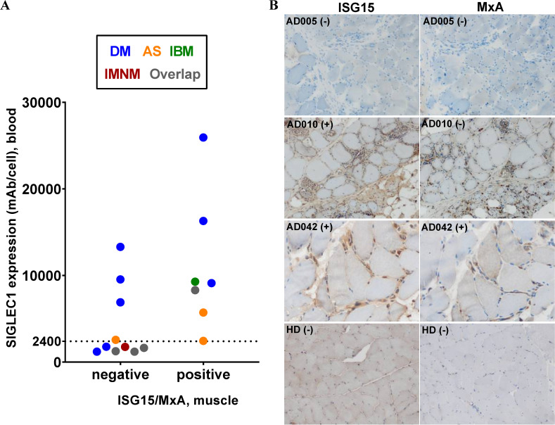
Methods: We performed a retrospective analysis of adult and paediatric patients with the diagnosis of IIM. SIGLEC1 expression was assessed by flow cytometry and was compared with Physician Global Assessment or Childhood Myositis Assessment Scale disease activity scores. Mann Whitney U test and receiver operating characteristic curves were used for cross-sectional data analysis (n=96), two-level mixed-effects linear regression model for longitudinal analyses (n=26, 110 visits). Response to treatment was analysed in 14 patients within 12 months, using Wilcoxon test. SIGLEC1 was compared with interferon-stimulated gene 15/MxA status by immunohistochemical staining of muscle biopsies (n=17).

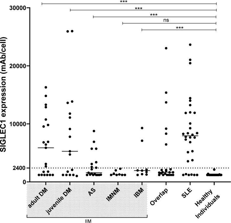
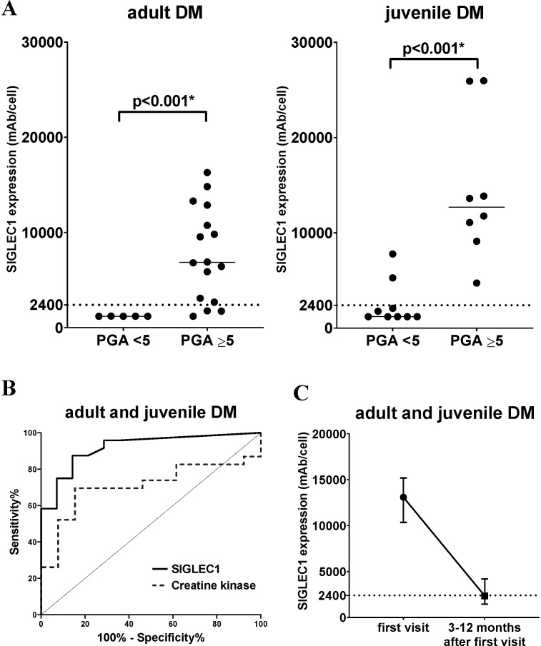
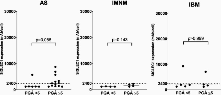
Results: 96 patients with adult (a) and juvenile (j) dermatomyositis (DM, n=38), antisynthetase syndrome (AS, n=19), immune-mediated necrotising myopathy (IMNM, n=8), inclusion body myositis (IBM, n=9) and overlap myositis (n=22) were included. SIGLEC1 distinguished significantly between active and inactive disease with an area under the curve of 0.92 (95% CI 0.83 to 1) in DM and correlated with disease activity longitudinally (aDM: standardised beta=0.54, p<0.001; jDM: standardised beta=-0.70, p<0.001). Response to treatment in DM was associated with a decreasing SIGLEC1 (p<0.01, Wilcoxon test). SIGLEC1 was found upregulated in 8 of 19 patients with AS, 2 of 9 patients with IBM but not in IMNM.

Conclusion: SIGLEC1 is a candidate biomarker to assess type I interferon activity in IIM and proved useful for monitoring disease activity and response to treatment in juvenile and adult DM.

Keywords: autoantibodies; autoimmunity; dermatomyositis; polymyositis.


Conflict of interest statement

Competing interests: None declared.

Figures

Figure 1
Figure 1
SIGLEC1 expression on monocytes of all patients at first visit. Horizontal bars show median values. Mann-Whitney U test was used to compare patients with the following idiopathic inflammatory myopathies (IIM)—adult (n=21) and juvenile (n=17) dermatomyositis (DM), antisynthetase syndrome (AS, n=18), immune-mediated necrotising myopathy (IMNM, n=8), inclusion body myositis (IBM, n=9)—with healthy individuals (n=19); ***p<0.001; ns, not significant; SIGLEC1, sialic acid binding Ig-like lectin 1; SLE, systemic lupus erythematosus.
Figure 2
Figure 2
SIGLEC1 expression in DM patients (A) adult and juvenile DM subgroups (n=21/n=17) are separated by PGA score: PGA <5 (no to moderate disease activity) and PGA ≥5 (moderate to severe disease activity). Horizontal bars show median values; asterisks (*) represent significant results (p<0.05). The Mann-Whitney U test was used to compare groups. (B) Receiver operating characteristic curves for SIGLEC1 and creatine kinase in juvenile and adult DM (n=38). The curves show the ability of each biomarker to distinguish between patients with PGA ≥5 (moderate to severe disease activity) and PGA <5 (no to moderate disease activity): SIGLEC1: AUC=0.92, 95% CI 0.83 to 1; p<0.001; CK: AUC=0.71; 95% CI 0.54 to 0.89; p=0.04 (C) SIGLEC1 expression in adult and juvenile DM patients with a clinically meaningful improvement between the first visit and 3–12 months after the first visit (n=14, p<0.01, Wilcoxon test). The median and IQR for each time point is shown. AUC, area under the curve; DM, dermatomyositis; PGA, physician global assessment; SIGLEC1, sialic acid binding Ig-like lectin 1.
Figure 3
Figure 3
SIGLEC1 expression versus disease activity in AS, IMNM and IBM subgroups, separated by PGA score: PGA <5 (no to moderate disease activity) and PGA ≥5 (moderate to severe disease activity). Horizontal bars show median values. The Mann-Whitney U test was used to compare groups. AS, antisynthetase syndrome; IBM, inclusion body myositis; IMNM, immune-mediated necrotising myopathy; PGA, Physician Global Assessment; SIGLEC1, sialic acid binding Ig-like lectin 1.
Figure 4
Figure 4
SIGLEC1 expression and autoantibody status. SIGLEC1 expression of all IIM patients (n=74) at first visit. Each group represents patients with a positivity for the respective myositis-specific autoantibody (MSA) and (A) PGA ≥5 (moderate to severe disease activity) and (B) PGA <5 (no to moderate disease activity); if present, additional myositis associated antibodies (MAA) are marked by different colours and no MAA is resembled by black. IIM, idiopathic inflammatory myopathies; PGA, Physician Global Assessment; SIGLEC1, sialic acid binding Ig-like lectin 1.
Figure 5
Figure 5
Immunohistochemical staining of type I interferon-inducible proteins (ISG15/MxA) in muscle tissue (A) comparison of SIGLEC1 expression in blood with negative or positive ISG15 and/or MxA status in immunohistochemical muscle biopsy staining (n=17). Colours represent patients with the specified subgroups of IIM and overlap (see legend box). (B) ISG15 and MxA status of three patients and one healthy donor (HD) are shown. Patient AD005 (adult DM, SIGLEC1 in blood 13301 mAb/cell) had negative (−) stains. Patient AD010 (adult IBM, SIGLEC1 in blood 9281 mAb/cell) stained clearly positive (+) for MxA on capillaries and macrophages; regarding the ISG15 status, some macrophages were positive, but myofibers were not. Patient AD042 (adult DM, SIGLEC1 in blood 16295 mAb/cell) was ISG15-positive (more densely stained on sarcolemma than on sarcoplasm) and MxA-positive. AS, antisynthetase syndrome; DM, dermatomyositis; IBM, inclusion body myositis; IMNM, immune-mediated necrotising myopathy; IIM, idiopathic inflammatory myopathies; SIGLEC1, sialic acid binding Ig-like lectin 1.

Similar articles

Cited by

References

    1. Lundberg IE, Tjärnlund A, Bottai M, et al. . 2017 European League against Rheumatism/American College of rheumatology classification criteria for adult and juvenile idiopathic inflammatory myopathies and their major subgroups. Ann Rheum Dis 2017;76:1955–64. 10.1136/annrheumdis-2017-211468 - DOI - PMC - PubMed
    1. Allenbach Y, Benveniste O, Goebel H-H, et al. . Integrated classification of inflammatory myopathies. Neuropathol Appl Neurobiol 2017;43:62–81. 10.1111/nan.12380 - DOI - PubMed
    1. Mariampillai K, Granger B, Amelin D, et al. . Development of a new classification system for idiopathic inflammatory myopathies based on clinical manifestations and myositis-specific autoantibodies. JAMA Neurol 2018;75:1528–37. 10.1001/jamaneurol.2018.2598 - DOI - PMC - PubMed
    1. Tanboon J, Uruha A, Stenzel W, et al. . Where are we moving in the classification of idiopathic inflammatory myopathies? Curr Opin Neurol 2020;33:590–603. 10.1097/WCO.0000000000000855 - DOI - PubMed
    1. Mathur T, Manadan AM, Thiagarajan S, et al. . The utility of serum aldolase in normal creatine kinase dermatomyositis. J Clin Rheumatol 2014;20:47–8. 10.1097/RHU.0000000000000062 - DOI - PubMed

Publication types

Substances