. 2022 Feb 17;12(1):2701.
doi: 10.1038/s41598-022-06773-9.

Inhibition of HIF-1α-AQP4 axis ameliorates brain edema and neurological functional deficits in a rat controlled cortical injury (CCI) model

Affiliations

Inhibition of HIF-1α-AQP4 axis ameliorates brain edema and neurological functional deficits in a rat controlled cortical injury (CCI) model

Ao Xiong et al. Sci Rep. .

Abstract

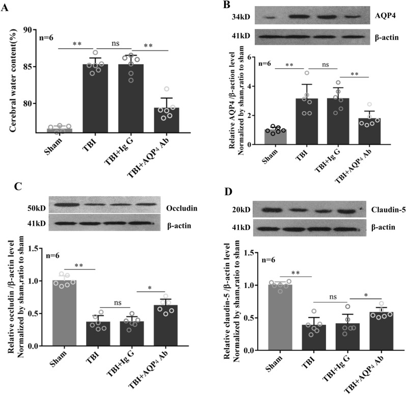
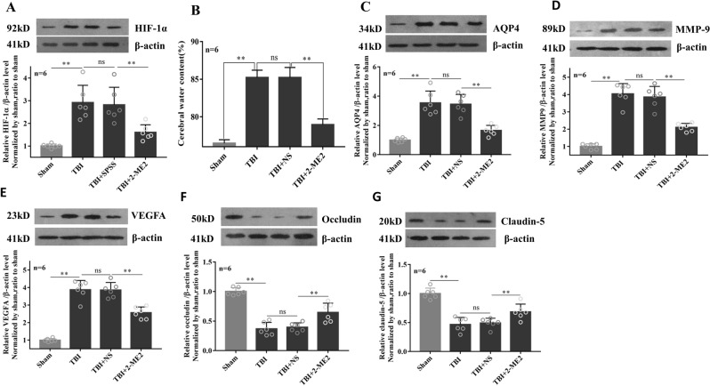
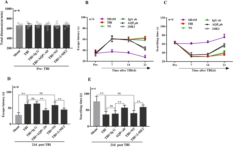
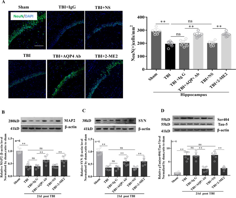
Traumatic brain injury (TBI) is an important cause of death in young adults and children. Till now, the treatment of TBI in the short- and long-term complications is still a challenge. Our previous evidence implied aquaporin 4 (AQP4) and hypoxia inducible factor-1α (HIF-1α) might be potential targets for TBI. In this study, we explored the roles of AQP4 and HIF-1α on brain edema formation, neuronal damage and neurological functional deficits after TBI using the controlled cortical injury (CCI) model. The adult male Sprague Dawley rats were randomly divided into sham and TBI group, the latter group was further divided into neutralized-AQP4 antibody group, 2-methoxyestradiol (2-ME2) group, and their corresponding control, IgG and isotonic saline groups, respectively. Brain edema was examined by water content. Hippocampal neuronal injury was assessed by neuron loss and neuronal skeleton related protein expressions. Spatial learning and memory deficits were evaluated by Morris water maze test and memory-related proteins were detected by western blot. Our data showed that increased AQP4 protein level was closely correlated with severity of brain edema after TBI. Compared with that in the control group, both blockage of AQP4 with neutralized-AQP4 antibody and inhibition of HIF-1α with 2-ME2 for one-time treatment within 30-60 min post TBI significantly ameliorated brain edema on the 1st day post-TBI, and markedly alleviated hippocampal neuron loss and spatial learning and memory deficits on the 21st day post-TBI. In summary, our preliminary study revealed the short-term and long-term benefits of targeting HIF-1α-AQP4 axis after TBI, which may provide new clues for the selection of potential therapeutic targets for TBI in clinical practice.


Conflict of interest statement

The authors declare no competing interests.

Figures

Figure 1
Figure 1
Blockage of AQP4 ameliorated brain edema in the acute phase after TBI. Rats were intravenously injected via the tail vein with 0.25 ml isotonic saline containing neutralized-AQP4 antibody (1 μg/kg of body weight) within 30–60 min after TBI, the control animals were injected with IgG. (A) On the 1st day post TBI, brain edema was examined by brain water content. Protein levels of AQP4 (B), Occludin (C) and Claudin-5 (D) were detected by western blot with β-actin as an internal control. The identical internal control β-actin blots were shared for Occludin and Claudin-5 blots because the detections were performed on the same blot membrane. Data are expressed as mean ± SEM, n = 6 per group. The comparisons among groups were analyzed by one-way ANOVA and the least significant difference (LSD) method. * P < 0.05, between the two groups. ns non-significant.
Figure 2
Figure 2
Inhibition of HIF-1α reduced brain water content and improve blood–brain barrier function. TBI rats were intravenously injected via the tail vein with 0.25 ml isotonic saline containing 2-ME2 (2.5 mg/kg of body weight) within 30–60 min after TBI, the corresponding controls were injected with isotonic saline. On the 1st day after injury, brain water content (B) was determined and protein levels of HIF-1α (A), AQP4 (C), MMP-9 (D), VEGF (E), tight junction protein levels of occluding (F) and claudin-5 (G) were detected by western blot. β-actin was used as loading controls for the total proteins. Data represent mean ± SEM (n = 6 per group). The comparisons among groups were analyzed by one-way ANOVA and the LSD method. Significant differences are shown by asterisks (*P < 0.05). ns non-significant.
Figure 3
Figure 3
Early post-traumatic inhibition of AQP4 and HIF-1α markedly alleviated TBI-induced neurological functional deficits in long term learning and memory. In the water maze test, rats were placed in a large circular pool filled with opaque water and were given the task to swim to a platform that can be either visible or hidden. (A) Before TBI, the swimming distance per minute of rats in each group showed no significant difference. TBI rats were intravenously injected via the tail vein with 0.25 ml isotonic saline containing AQP4 antibodies (1 μg/kg of body weight) or 2-ME2 (2.5 mg/kg of body weight) within 30–60 min after impact, the corresponding controls were injected with IgG and isotonic saline, respectively. The Morris water maze tests were performed and the escape latency (B) and searching time (C) were investigated at the indicated time points, such as shown on the 21st day post-TBI in (D,E). Data represent mean ± SEM, n = 6 per group. The comparisons among groups were analyzed by one-way ANOVA with the LSD method. *P < 0.05, between the two groups. ns non-significant.
Figure 4
Figure 4
Effects of AQP4 and HIF-1α inhibition on neuron loss and the expression levels of neuron structure-related proteins in hippocampus. TBI rats were intravenously injected via the tail vein with 0.25 ml isotonic saline containing AQP4 antibodies (1 μg/kg of body weight) or 2-ME2 (2.5 mg/kg of body weight) within 30–60 min after impact, the corresponding controls were injected with IgG or isotonic saline, respectively. On the 21st day post-TBI, (A) the hippocampal CA1 NeuN-positive neuron numbers of the injured region in each group were calculated (scale bar 100 μm). The hippocampal protein levels of MAP2 (B) and SYN (C) were detected by western blot. (D) The protein expression of pTau (ser404) and Tau-5 was detected in each group. β-actin was used as loading controls for the total proteins. Data represent mean ± SEM, n = 6 per group. The comparisons among groups were analyzed by one-way ANOVA with the LSD method. * P < 0.05, between the two groups. ns non-significant.

Similar articles

Cited by

References

    1. Ilie G, Boak A, Adlaf EM, Asbridge M, Cusimano MD. Prevalence and correlates of traumatic brain injuries among adolescents. JAMA. 2013;309:2550–2552. - PubMed
    1. Korhonen N, Niemi S, Parkkari J, Sievanen H, Kannus P. Incidence of fall-related traumatic brain injuries among older Finnish adults between 1970 and 2011. JAMA. 2013;309:1891–1892. - PubMed
    1. Donkin JJ, Vink R. Mechanisms of cerebral edema in traumatic brain injury: Therapeutic developments. Curr. Opin. Neurol. 2010;23:293–299. - PubMed
    1. Jha RM, Kochanek PM, Simard JM. Pathophysiology and treatment of cerebral edema in traumatic brain injury. Neuropharmacology. 2019;145:230–246. - PMC - PubMed
    1. Winkler EA, Minter D, Yue JK, Manley GT. Cerebral edema in traumatic brain injury: Pathophysiology and prospective therapeutic targets. Neurosurg. Clin. N. Am. 2016;27:473–488. - PubMed

MeSH terms