. 2022 Feb 1:10:e12725.
doi: 10.7717/peerj.12725. eCollection 2022.

Hammerhead flatworms (Platyhelminthes, Geoplanidae, Bipaliinae): mitochondrial genomes and description of two new species from France, Italy, and Mayotte

Affiliations

Hammerhead flatworms (Platyhelminthes, Geoplanidae, Bipaliinae): mitochondrial genomes and description of two new species from France, Italy, and Mayotte

Jean-Lou Justine et al. PeerJ. .

Abstract

Background: New records of alien land planarians are regularly reported worldwide, and some correspond to undescribed species of unknown geographic origin. The description of new species of land planarians (Geoplanidae) should classically be based on both external morphology and histology of anatomical structures, especially the copulatory organs, ideally with the addition of molecular data.

Methods: Here, we describe the morphology and reproductive anatomy of a species previously reported as Diversibipalium "black", and the morphology of a species previously reported as Diversibipalium "blue". Based on next generation sequencing, we obtained the complete mitogenome of five species of Bipaliinae, including these two species.

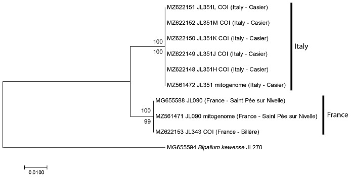
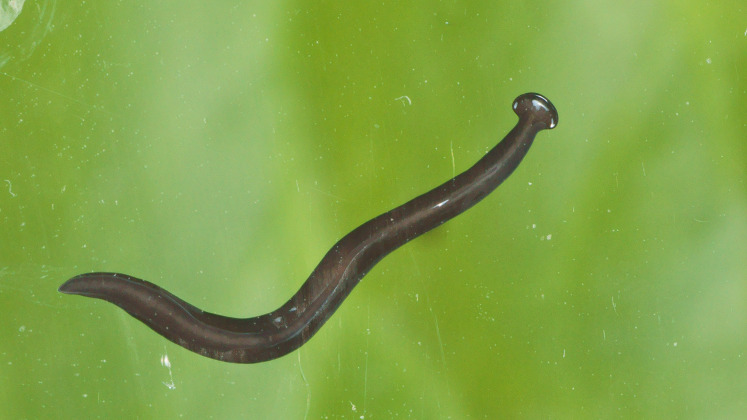
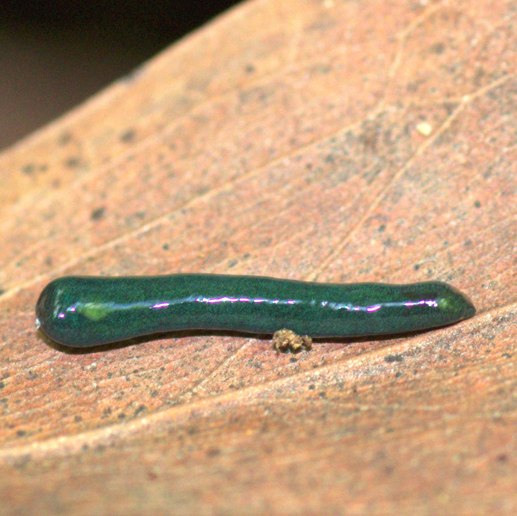
Results: The new species Humbertium covidum n. sp. (syn: Diversibipalium "black" of Justine et al., 2018) is formally described on the basis of morphology, histology and mitogenome, and is assigned to Humbertium on the basis of its reproductive anatomy. The type-locality is Casier, Italy, and other localities are in the Department of Pyrénées-Atlantiques, France; some published or unpublished records suggest that this species might also be present in Russia, China, and Japan. The mitogenomic polymorphism of two geographically distinct specimens (Italy vs France) is described; the cox1 gene displayed 2.25% difference. The new species Diversibipalium mayottensis n. sp. (syn: Diversibipalium "blue" of Justine et al., 2018) is formally described on the basis of external morphology and complete mitogenome and is assigned to Diversibipalium on the basis of an absence of information on its reproductive anatomy. The type- and only known locality is the island of Mayotte in the Mozambique Channel off Africa. Phylogenies of bipaliine geoplanids were constructed on the basis of SSU, LSU, mitochondrial proteins and concatenated sequences of cox1, SSU and LSU. In all four phylogenies, D. mayottensis was the sister-group to all the other bipaliines. With the exception of D. multilineatum which could not be circularised, the complete mitogenomes of B. kewense, B. vagum, B. adventitium, H. covidum and D. mayottensis were colinear. The 16S gene in all bipaliine species was problematic because usual tools were unable to locate its exact position.

Conclusion: Next generation sequencing, which can provide complete mitochondrial genomes as well as traditionally used genes such as SSU, LSU and cox1, is a powerful tool for delineating and describing species of Bipaliinae when the reproductive structure cannot be studied, which is sometimes the case of asexually reproducing invasive species. The unexpected position of the new species D. mayottensis as sister-group to all other Bipaliinae in all phylogenetic analyses suggests that the species could belong to a new genus, yet to be described.

Keywords: Alien invasive species; Barcoding; Citizen science; France; Italy; Land planarians; Mayotte; Mitogenome; Platyhelminthes; Taxonomy.


Conflict of interest statement

Jean-Lou Justine is an Academic Editor of PeerJ.

Figures

Figure 1
Figure 1. Humbertium covidum n. sp. from two populations, tree based on cox1 sequences.
The evolutionary history was inferred using the Maximum Likelihood and the Neighbour-Joining methods; there was a total of 387 positions in the final dataset. All partial cox1 sequences from Italy (six specimens) were identical, as were the three sequences from France, from two localities. Sequences from France and Italy differed by 2.58%. Bootstrap values: above branches, ML; below branches, NJ.
Figure 2
Figure 2. Humbertium covidum n. sp. from Italy, alive.
General dorsal aspect. Photo by Pierre Gros.
Figure 3
Figure 3. Humbertium covidum n. sp. from Italy, alive.
Lateral view showing locomotion and slime trail. Photo by Pierre Gros.
Figure 4
Figure 4. Humbertium covidum n. sp. from Italy, alive.
Individual with raised anterior end showing ventral surface. Photo by Pierre Gros.
Figure 5
Figure 5. Humbertium covidum n. sp. from Italy, alive.
Ventral surface with typical headplate shape. Photo by Pierre Gros.
Figure 6
Figure 6. Humbertium covidum n. sp. from Billère, France, alive.
General dorsal aspect. Photo by Pierre Gros.
Figure 7
Figure 7. Humbertium covidum n. sp. from Billère, France, alive.
Lateral aspect. Photo by Pierre Gros.
Figure 8
Figure 8. Humbertium covidum n. sp. from Billère, France, alive.
Lateral aspect showing extended papillae on headplate. Photo by Pierre Gros.
Figure 9
Figure 9. Humbertium covidum n. sp. from Billère, France, alive.
Individual with raised anterior end. Photo by Pierre Gros.
Figure 10
Figure 10. Humbertium covidum n. sp. from Billère, France, alive.
The flatworm seems to threaten a snail (unidentified species). Photo by Pierre Gros.
Figure 11
Figure 11. Humbertium covidum n. sp. from Saint-Pée-sur-Nivelle, France, preserved.
Specimen MNHN JL090, preserved specimen, dorsal aspect. Showing the partly protruded pharynx. Photo by Jean-Lou Justine. Reproduced from Figure 20 of Justine et al. (2018).
Figure 12
Figure 12. Humbertium covidum n. sp. from Saint-Pée-sur-Nivelle, France, preserved.
Specimen MNHN JL090. Preserved specimen, ventral aspect. The ventral ground colour is grey, with the creeping sole a lighter tone. The pharynx is slightly protruded from the mouth, and the gonopore is evident as a small transverse white slit on the creeping sole some 2 mm below to the mouth. Scale is in mm. Photo by Jean-Lou Justine. Reproduced from Figure 21 of Justine et al. (2018).
Figure 13
Figure 13. Anatomy of Humbertium covidum n. sp., pre-pharyngeal region.
Holotype, specimen MNHN JL351B. Pre-pharyngeal region, transverse section. Arrows indicate the extent of the creeping sole. Photo by Leigh Winsor.
Figure 14
Figure 14. Anatomy of Humbertium covidum n. sp, ventral longitudinal muscular plate.
Holotype, specimen MNHN JL351B. Lateral body showing ventral longitudinal muscular plate. Photo by Leigh Winsor.
Figure 15
Figure 15. Anatomy of Humbertium covidum n. sp., pharynx.
Holotype, specimen MNHN JL351B. Pharynx, sagittal section. Photo by Leigh Winsor.
Figure 16
Figure 16. Morphology of Humbertium covidum n. sp., eye pattern.
Paratype JL 351C. Headplate showing the dorsal and ventral eye patterns in a cleared specimen. The headplate is curled ventrad. Drawing by Leigh Winsor.
Figure 17
Figure 17. Anatomy of Humbertium covidum n. sp., composite drawing of copulatory organs.
Holotype, specimen MNHN JL351B. Composite reconstruction of the copulatory organs, sagittal view. The dashed line in the common atrium indicates the extent of the glandular mesenchyme forming the common genital canal. Anterior: left. Drawing by Leigh Winsor.
Figure 18
Figure 18. Anatomy of Humbertium covidum n. sp., level of gonopore.
Holotype, specimen MNHN JL351B. Copulatory organs at the level of the gonopore, with the female glandular canal entering the common genital canal at the point where it communicates with the common atrium. Anterior: left. Photo by Leigh Winsor.
Figure 19
Figure 19. Anatomy of Humbertium covidum n. sp., putative common genital canal.
Paratype, specimen MNHN JL351C. Glandular mesenchyme of the putative common genital canal on the left side of the body. Anterior: left. Photo by Leigh Winsor.
Figure 20
Figure 20. Anatomy of Humbertium covidum n. sp., common genital canal.
Paratype, specimen MNHN JL351C. The beginning of the slit-like common genital canal. Anterior: left. Photo by Leigh Winsor.
Figure 21
Figure 21. Anatomy of Humbertium covidum n. sp., male atrium.
Paratype, specimen MNHN JL351C. The point where the male atrium is about to open into the common genital canal which has not yet opened into the common atrium. Anterior: left. Photo by Leigh Winsor.
Figure 22
Figure 22. Anatomy of Humbertium covidum n. sp., viscid gland.
Holotype, specimen MNHN JL351B. The viscid gland at the anteriad end of the genital pad below the male organs. Anterior: left. Photo by Leigh Winsor.
Figure 23
Figure 23. Anatomy of Humbertium covidum n. sp., viscid gland and erythrophil glands.
Holotype, specimen MNHN JL351B. The glandular duct of the viscid gland, and erythrophil glands in the atrial crease. Anterior: left. Photo by Leigh Winsor.
Figure 24
Figure 24. Diversibipalium mayottensis n. sp, alive.
Specimen MNHN JL282 from Mayotte, Indian Ocean, dorsal aspect. The headplate of this small planarian is a rusty-brown colour that extends to some irregular patches on the ‘neck.’ The dorsal ground colour is an iridescent blue–green (‘dark turquoise glitter’). Photo by Laurent Charles. Reproduced from Figure 23 in Justine et al. (2018).
Figure 25
Figure 25. Diversibipalium mayottensis n. sp, alive.
Specimen MNHN JL282 from Mayotte, Indian Ocean, dorsal aspect. Same specimen as in Fig. 24. Photo by Laurent Charles. Reproduced from Figure 24 in Justine et al. (2018).
Figure 26
Figure 26. Diversibipalium mayottensis n. sp, alive regenerating specimen.
Dorsal aspect of a regenerating specimen with a damaged anterior end. Specimen MNHN JL280. Under appropriate lighting, the colour of the specimen takes on a beautiful, almost metallic green colour. The iridescence and blue–green colour are lost on fixation, leaving the specimen a dark brown. Photo by Laurent Charles. Reproduced from Figure 25 in Justine et al. (2018).
Figure 27
Figure 27. Diversibipalium mayottensis n. sp, alive regenerating specimen.
Dorsal aspect of a regenerating specimen with a damaged anterior end. Specimen MNHN JL280. A small portion of the brown-pigmented ventral surface with the median pale creeping sole can be seen. Photo by Laurent Charles. Reproduced from Figure 26 in Justine et al. (2018).
Figure 28
Figure 28. Mitogenome of Humbertium covidum n. sp.: genomic map of specimen MNHN JL351.
Specimen from the Italian population in Casier. The mitogenome is 15,540 bp long and contains 12 protein coding genes, two ribosomal RNA genes and 21 transfer RNA genes. The ND3 gene was found with a premature stop codon.
Figure 29
Figure 29. Mitogenome of Humbertium covidum n. sp.: genomic map of specimen MNHN JL090.
Specimen from the French population in Billère (Pyrénées-Atlantiques). The mitogenome is 15,524 bp long and contains 12 protein coding genes, two ribosomal RNA genes and 21 transfer RNA genes. The ND3 gene was found with a premature stop codon.
Figure 30
Figure 30. Mitogenome of Diversibipalium mayottensis n. sp.: genomic map of specimen JL281.
The mitogenome is 15,989 bp long and contains 12 protein coding genes, two ribosomal RNA genes and 22 transfer RNA genes.
Figure 31
Figure 31. Mitogenome of Bipalium vagum: genomic map of specimen JL307.
The mitogenome is 17,149 bp long and contains 12 protein coding genes, two ribosomal RNA genes and 22 transfer RNA genes. The genes cox3, atp6, ND1, ND4L have alternative start codon.
Figure 32
Figure 32. Mitogenome of Bipalium adventitium: genomic map of specimen JL328.
The mitogenome is 15,494 bp long and contains 12 protein coding genes, two ribosomal RNA genes and 21 transfer RNA genes. It was not possible to find a stop codon for the cob gene.
Figure 33
Figure 33. Mitogenome of Diversibipalium multilineatum: genomic map of specimen JL177.
The mitogenome is not complete. The size of the partial genome is 15,660 bp long and contains 12 protein coding genes, two ribosomal RNA genes and 21 transfer RNA genes. The genes ND2 and ND3 have alternative start codon.
Figure 34
Figure 34. Mitogenome of Bipalium kewense: genomic map of specimen JL184A.
The mitogenome is 15,666 bp long and contains 12 protein coding genes, two ribosomal RNA genes and 22 transfer RNA genes.
Figure 35
Figure 35. An alignment of the ‘complete’ 16S genes from all Bipaliinae displayed as a LOGO.
The alignment obtained from seven sequences representing six species shows the presence of a more conserved second part of the gene while the first part appears strongly variable.
Figure 36
Figure 36. Alignments of the ‘complete’ 16S genes from H. covidum JL090 and JL351.
The two specimens are from the French (JL09) and Italian (JL351) populations. The black star indicates the beginning of the most conserved part evidenced by multispecies alignment.
Figure 37
Figure 37. SSU phylogenetic tree of bipaliine geoplanids.
Maximum likelihood phylogenetic tree based on 14 partial SSU genes, using the GTR+I+G model of evolution, with the best tree out of 100 computed for 1,000 bootstrap replications. The tree with the best likelihood is shown (−2,551.353092). ML bootstrap support values on the left. The BI tree had and identical topology, posterior probabilities are indicated on the right as decimal values. Diversibipalium mayottensis n. sp. appears as the sister-group to all other bipaliines. The subfamilies within the Geoplanidae (Rhynchodeminae, Geoplaninae and Bipaliinae) are indicated. Diversibipalium mayottensis branch in bold to show its position as sister-group to all other Bipaliinae.
Figure 38
Figure 38. LSU phylogenetic tree of bipaliine geoplanids.
Maximum likelihood phylogenetic tree based on 20 partial LSU genes, using the GTR+I+G model of evolution, with the best tree out of 100 computed for 1,000 bootstrap replications. The tree with the best likelihood is shown (−4,759.571033). ML bootstrap support values on the left. The BI tree had identical topology; posterior probabilities are indicated on the right as decimal values. The subfamilies within the Geoplanidae (Rhynchodeminae, Geoplaninae and Bipaliinae) are indicated. Diversibipalium mayottensis branch in bold to show its position as sister-group to all other Bipaliinae.
Figure 39
Figure 39. Phylogenetic tree of concatenated mitochondrial proteins of bipaliine geoplanids.
Maximum likelihood phylogenetic tree based on concatenated protein sequences extracted from 19 mitogenomes using the mtART+I+G model, with the best tree out of 100 computed for 1,000 bootstrap replications. The tree with the best likelihood is shown (−4,759.571033). The subfamilies within the Geoplanidae (Rhynchodeminae, Geoplaninae and Bipaliinae) are indicated. Diversibipalium mayottensis branch in bold to show its position as sister-group to all other Bipaliinae.
Figure 40
Figure 40. Three-gene phylogenetic tree of bipaliine geoplanids, based on concatenated cox1, SSU and LSU genes.
Maximum likelihood phylogenetic tree based on 18 concatenated partial sequences of cox1, SSU and LSU, using the GTR+I+G model of evolution, with the best tree out of 100 computed for 1,000 bootstrap replications. The tree with the best likelihood is shown (−24,779.059136). ML bootstrap support values on the left. The BI tree had identical topology; posterior probabilities are indicated on the right as decimal values. The subfamilies within the Geoplanidae (Rhynchodeminae, Geoplaninae and Bipaliinae) are indicated. Diversibipalium mayottensis branch in bold to show its position as sister-group to all other Bipaliinae.

References

    1. Abascal F, Posada D, Zardoya R. MtArt: a new model of amino acid replacement for Arthropoda. Molecular Biology and Evolution. 2007;24(1):1–5. doi: 10.1093/molbev/msl136. - DOI - PubMed
    1. Andújar C, Creedy TJ, Arribas P, López H, Salces-Castellano A, Pérez-Delgado AJ, Vogler AP, Emerson BC. Validated removal of nuclear pseudogenes and sequencing artefacts from mitochondrial metabarcode data. Molecular Ecology Resources. 2021;21(6):1772–1787. doi: 10.1111/1755-0998.13337. - DOI - PubMed
    1. Bankevich A, Nurk S, Antipov D, Gurevich AA, Dvorkin M, Kulikov AS, Lesin VM, Nikolenko SI, Pham S, Prjibelski AD. SPAdes: a new genome assembly algorithm and its applications to single-cell sequencing. Journal of Computational Biology. 2012;19(5):455–477. doi: 10.1089/cmb.2012.0021. - DOI - PMC - PubMed
    1. Bernt M, Donath A, Jühling F, Externbrink F, Florentz C, Fritzsch G, Pütz J, Middendorf M, Stadler PF. MITOS: improved de novo metazoan mitochondrial genome annotation. Molecular Phylogenetics and Evolution. 2013;69(2):313–319. doi: 10.1016/j.ympev.2012.08.023. - DOI - PubMed
    1. Boratyn GM, Schäffer AA, Agarwala R, Altschul SF, Lipman DJ, Madden TL. Domain enhanced lookup time accelerated BLAST. Biology Direct. 2012;7(1):12. doi: 10.1186/1745-6150-7-12. - DOI - PMC - PubMed

Publication types

LinkOut - more resources