Review
. 2022 Feb 1:9:817099.
doi: 10.3389/fcell.2021.817099. eCollection 2021.

IGF Signaling in Intervertebral Disc Health and Disease

Affiliations
Review

IGF Signaling in Intervertebral Disc Health and Disease

Hui Lin et al. Front Cell Dev Biol. .

Abstract

Low back pain (LBP) is a common musculoskeletal symptom, which brings a lot of pain and economic loss to patients. One of the most common causes of LBP is intervertebral disc degeneration (IVDD). However, pathogenesis is still debated, and therapeutic options are limited. Insulin-like growth factor (IGF) signaling pathways play an important role in regulating different cell processes, including proliferation, differentiation, migration, or cell death, which are critical to the homeostasis of tissues and organs. The IGF signaling is crucial in the occurrence and progression of IVDD. The activation of IGF signaling retards IVDD by increasing cell proliferation, promoting extracellular matrix (ECM) synthesis, inhibiting ECM decomposition, and preventing apoptosis and senescence of disc cells. However, abnormal activation of IGF signaling may promote the process of IVDD. IGF signaling is currently considered to have a promising treatment prospect for IVDD. An in-depth understanding of the role of IGF signaling in IVDD may help find a novel approach for IVDD treatment.

Keywords: degeneration; insulin-like growth factor; intervertebral disc; low back pain; nucleus pulposus.


Conflict of interest statement

The authors declare that the research was conducted in the absence of any commercial or financial relationships that could be construed as a potential conflict of interest.

Figures

FIGURE 1
FIGURE 1
Simplified scheme of IGF signaling in the intervertebral disc. With the participation of MMPs, TNFα, or IL-1β, PAPP-A cleaves IGFBP in the extracellular matrix, increasing the amount of bioavailable IGF1 and IGF2. To exert their effects, IGF bind to specific cell surface receptors (IGF1R and IGF2R). IGF1R activation activates various molecular signaling pathways, including the PI3K-Akt and RAS-extracellular signal-regulated kinase (ERK) pathways, to regulate IVD cell death and ECM content. MMP: matrix metalloproteinases; PAPP-A: pregnancy-associated plasma protein-A; IRS-1: insulin-receptor substrate 1; AA: ascorbic acid; SVCT2: sodium-dependent vitamin C transporter 2; PI3K: phosphatidiyl inositol 3 kinase; PIP: phosphatidylinositol-3,4,5-trisphosphate; FoxO1: forkhead box O1; CREB: cAMP-responsive element-binding protein; CA12: carbonic anhydrase 12; MEK: mitogen-activated protein kinase/extracellular signal-regulated kinase; ERK: extracellular signal-regulated kinase.
FIGURE 2
FIGURE 2
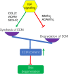
Activation of IGF signaling enhances ECM content by increasing the synthesis of ECM through upregulating the gene expression of COL2, ACAN, SOX-9 and inhibitng the degradation of ECM by downregulating the gene expression of MMPs and ADAMTs to exert protection against IVDD. COL2, Collagen-II; ACAN, Aggrecan.
FIGURE 3
FIGURE 3
Effects of IGF signaling on IVDD initiation and progression. IGF signaling activation inhibits IVDD by promoting ECM synthesis, cell proliferation, and inhibiting inflammatory responses, ECM degradation, and cell apoptosis. However, the abnormal activation of IGF signaling can accelerate the degeneration of IVD by stimulating excessive cell proliferation, increasing vascular and nerve growth, aggravating nutritional deficiency.

Similar articles

Cited by

References

    1. Akyuva Y., Kaplan N., Yilmaz I., Ozbek H., Sirin D. Y., Karaaslan N., et al. (2019). Delivering Growth Factors through a Polymeric Scaffold to Cell Cultures Containing Both Nucleus Pulposus and Annulus Fibrosus. Turk Neurosurg. 29, 180–193. 10.5137/1019-5149.JTN.22672-18.1 - DOI - PubMed
    1. Bergknut N., Smolders L. A., Grinwis G. C. M., Hagman R., Lagerstedt A.-S., Hazewinkel H. A. W., et al. (2013). Intervertebral Disc Degeneration in the Dog. Part 1: Anatomy and Physiology of the Intervertebral Disc and Characteristics of Intervertebral Disc Degeneration. Vet. J. 195, 282–291. 10.1016/j.tvjl.2012.10.024 - DOI - PubMed
    1. Bikle D. D., Tahimic C., Chang W., Wang Y., Philippou A., Barton E. R. (2015). Role of IGF-I Signaling in Muscle Bone Interactions. Bone 80, 79–88. 10.1016/j.bone.2015.04.036 - DOI - PMC - PubMed
    1. Buckwalter J. A. (1995). Aging and Degeneration of the Human Intervertebral Disc. Spine 20, 1307–1314. 10.1097/00007632-199506000-00022 - DOI - PubMed
    1. Chao C.-C., Lee W.-F., Yang W.-H., Lin C.-Y., Han C.-K., Huang Y.-L., et al. (2021). IGFBP-3 Stimulates Human Osteosarcoma Cell Migration by Upregulating VCAM-1 Expression. Life Sci. 265, 118758. 10.1016/j.lfs.2020.118758 - DOI - PubMed