. 2022 Feb 8;4(1):fcac008.
doi: 10.1093/braincomms/fcac008. eCollection 2022.

Aphasia recovery by language training using a brain-computer interface: a proof-of-concept study

Affiliations

Aphasia recovery by language training using a brain-computer interface: a proof-of-concept study

Mariacristina Musso et al. Brain Commun. .

Abstract

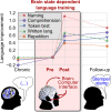
Aphasia, the impairment to understand or produce language, is a frequent disorder after stroke with devastating effects. Conventional speech and language therapy include each formal intervention for improving language and communication abilities. In the chronic stage after stroke, it is effective compared with no treatment, but its effect size is small. We present a new language training approach for the rehabilitation of patients with aphasia based on a brain-computer interface system. The approach exploits its capacity to provide feedback time-locked to a brain state. Thus, it implements the idea that reinforcing an appropriate language processing strategy may induce beneficial brain plasticity. In our approach, patients perform a simple auditory target word detection task whilst their EEG was recorded. The constant decoding of these signals by machine learning models generates an individual and immediate brain-state-dependent feedback. It indicates to patients how well they accomplish the task during a training session, even if they are unable to speak. Results obtained from a proof-of-concept study with 10 stroke patients with mild to severe chronic aphasia (age range: 38-76 years) are remarkable. First, we found that the high-intensity training (30 h, 4 days per week) was feasible, despite a high-word presentation speed and unfavourable stroke-induced EEG signal characteristics. Second, the training induced a sustained recovery of aphasia, which generalized to multiple language aspects beyond the trained task. Specifically, all tested language assessments (Aachen Aphasia Test, Snodgrass & Vanderwart, Communicative Activity Log) showed significant medium to large improvements between pre- and post-training, with a standardized mean difference of 0.63 obtained for the Aachen Aphasia Test, and five patients categorized as non-aphasic at post-training assessment. Third, our data show that these language improvements were accompanied neither by significant changes in attention skills nor non-linguistic skills. Investigating possible modes of action of this brain-computer interface-based language training, neuroimaging data (EEG and resting-state functional MRI) indicates a training-induced faster word processing, a strengthened language network and a rebalancing between the language- and default mode networks.

Keywords: aphasia rehabilitation; brain–computer interface; chronic stroke; language training; neurofeedback training.


Figures

Graphical Abstract
Graphical Abstract
Figure 1
Figure 1
Overlap of the binarized lesions of the 10 patients. Lesions are displayed over sagittal surface (A) and over nine horizontal sections parallel to the AC-PC line (B). Brighter regions indicate a greater degree of overlap of lesions. Images were generated using mricron.
Figure 2
Figure 2
Study protocol for BCI-based language training. (A) Time points (relative to the first training session) of the clinical testings and training sessions that each patient underwent, including an individual familiarization with the paradigm (hours to few sessions per patient), language assessments (AAT, Aachen Aphasia Test; S&V, Snodgrass & Vanderwart naming test; CAL, communicative activity log) and cognitive assessment (TAP, test of attentional performance; digit span, Corsi span, semantic and phonological fluency), see Supplementary Section 2. (B) Structure of a single training session and duration of its components. (C) Set-up of the AMUSE protocol: a subject is placed in the centre of six loudspeakers placed at ear level. In this loudspeaker condition, all auditory stimuli and auditory feedback were presented over these loudspeakers. Within each trial, a 1:1 relation was maintained between the six words and the loudspeakers. Between trials, the target word and the mapping between directions and loudspeakers were pseudo-randomized. In a headphone condition, the patient received sentences, word stimuli and auditory feedback in one mono-channel via headphones such that spatial information could not be exploited. (D) Structure of a single trial, consisting of a ‘get ready’ cue, a sentence presentation, a word sequence presentation, immediate EEG analysis and feedback to the patients. At trial start, the computer played one of six German cueing sentences from an audio file, but the sentence’s last word was missing. Example: ‘Die neue Tonerkartusche steckt schon im … ’ (‘The new cartridge is already in the … ’). During the sentence presentation, patients were asked to listen to and understand it with the goal to infer the target word. Following, a sequence of six different words (all bisyllabic nouns with durations below 300 ms) was played. It took about 32 s (for SOA 350 ms if no dynamic stopping was triggered) and consisted of 15 target words (blue rectangles, Drucker/printer in this example) and 5 × 15 = 75 presentations of the five non-target words (red rectangles). The target and non-target role of a word switched in a pseudo-randomly balanced manner between trials of the same run.
Figure 3
Figure 3
Clear training-induced improvements of language abilities measured by the AAT. (A) Individual changes and groupwise changes (bars with standard deviations) of different language abilities measured by the T-transformed AAT scores. This transformation normalizes the raw AAT scores such that 10 T-transformed AAT points correspond to 1 SD. Significance was assessed by two-sided paired t-tests with the Benjamini–Hochberg correction. The symbol ‘*’ marks P < 0.05 and ‘**’ marks P < 0.01 for the corrected P-values. (B) The average language performance on the group level at four different time points relative to the pre-training performance. Missing data points are annotated and were excluded from the computation of the averages and the statistical tests.
Figure 4
Figure 4
Stronger and earlier word-evoked P300 responses after the training. The plots visualize data obtained by ERP offline analysis for patients (pre- and post-training) and for 20 NACs to indicate how healthy subjects process the word stimuli. The ERP responses were evoked by words played with an SOA of 250 ms from six loudspeakers. (A) The average target and non-target ERP responses for channels Cz and Fz. (B) The spatial distributions of mean target responses within four selected time intervals (in ms relative to stimulus onset): A: (191,240); B: (301,420); C: (421,670); D: (671,800). It can be observed that patients showed P300 and N200 amplitudes lateralized over the right hemisphere before and after the training. However, at post-training, the average ERP time courses and intensities of spatial patterns obtained from patients approximate those of NACs. (C) The average (bars with standard deviation) and individual values (dots) for six different metrics. As a result of the training, the P300 amplitudes have increased, P300 onsets have appeared earlier and target versus non-target classification accuracies have increased. Note that no statistical comparisons have been conducted between data of the NACs and patients. All conducted tests are indicated by black bars. n.s., not significant; AUC, area under the receiver-operating characteristic curve; ‘*’ corresponds to P < 0.05.
Figure 5
Figure 5
Training-induced rebalancing of language- and default mode networks. Pre-/versus post-training changes of rs-fMRI FC are visualized. For every row, the region of interest is indicated by blue-grey colour. The colour bar shows the T-values of a paired t-test where T-values above 3.1 denote a significant change at a family-wise level of P < 0.05 after cluster-extent-based thresholding. The FC of the posterior cingulate cortex (PCC) and the precuneus (Prec) with the left postcentral gyrus increase after training. The PCC also shows an increased FC with the left primary motor cortex (M1), dorsolateral prefrontal cortex (DLPC), middle superior temporal gyri (STGm), frontal orbital and anterior cingulate cortex (ACC). The two hubs of the DMN (PCC and Prec) show a decreased FC with the main hubs of the language network—pars triangularis (F3tri), pars opercularis (F3op) and pars orbitalis (F3orb) of the inferior frontal gyrus (Broca’s region) and the posterior superior temporal gyrus (STGp, Wernicke’s region)—as well as with other hubs of the DMN—anterior precuneus, temporo-parietal junction (TPJ), angular gyrus (AG), infero-temporal cortex (ITG), parietal lobe (PL) and occipital cortex (OC). F3op, F3tri and F3orb show increased FC with each other, and also with ACC and STGp. F3orb/tri/op showed decreased FC with MTG, left DLPC, Prec and M1. Wernicke’s region exhibited increased FC with left PL, ACC and cerebellum, and decreased FC with the OC, Prec, TP, premotor and motor cortex (PMC, M1) and DLPC. Non-significant changes are denoted by ‘ns’. Images were generated using mricron (https://www.nitrc.org/projects/mricron).

Similar articles

Cited by

References

    1. Dickey L, Kagan A, Lindsay MP, Fang J, Rowland A, Black S. Incidence and profile of inpatient stroke-induced aphasia in Ontario, Canada. Arch Phys Med Rehabil. 2010;91(2):196–202. - PubMed
    1. Dalemans RJ, De Witte LP, Beurskens AJ, Van Den Heuvel WJ, Wade DT. An investigation into the social participation of stroke survivors with aphasia. Disabil Rehabil. 2010;32(20):1678–1685. - PubMed
    1. Doucet T, Muller F, Verdun-Esquer C, Debelleix X, Brochard P. Returning to work after a stroke: A retrospective study at the Physical and Rehabilitation Medicine Center ‘La Tour de Gassies’. Ann Phys Rehabil Med. 2012;55(2):112–127. - PubMed
    1. Berthier ML. Poststroke aphasia: Epidemiology, pathophysiology and treatment. Drugs Aging. 2005;22(2):163–182. - PubMed
    1. Brady MC, Kelly H, Godwin J, Enderby P, Campbell P. Speech and language therapy for aphasia following stroke. Cochrane Database Syst Rev. 2016;6(6):CD000425. - PMC - PubMed

LinkOut - more resources