. 2022 Mar 1;149(5):dev199995.
doi: 10.1242/dev.199995. Epub 2022 Mar 11.

Notch signaling enhances bone regeneration in the zebrafish mandible

Affiliations

Notch signaling enhances bone regeneration in the zebrafish mandible

Jessica M Kraus et al. Development. .

Abstract

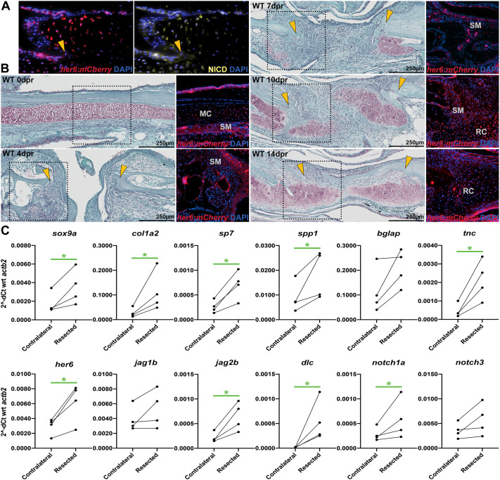
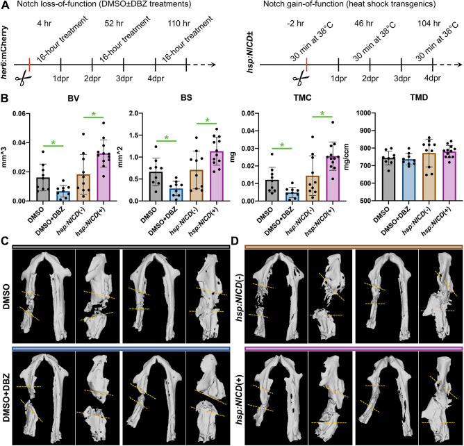
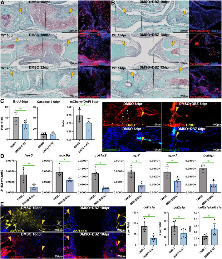
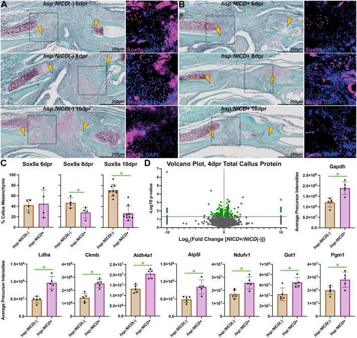
Loss or damage to the mandible caused by trauma, treatment of oral malignancies, and other diseases is treated using bone-grafting techniques that suffer from numerous shortcomings and contraindications. Zebrafish naturally heal large injuries to mandibular bone, offering an opportunity to understand how to boost intrinsic healing potential. Using a novel her6:mCherry Notch reporter, we show that canonical Notch signaling is induced during the initial stages of cartilage callus formation in both mesenchymal cells and chondrocytes following surgical mandibulectomy. We also show that modulation of Notch signaling during the initial post-operative period results in lasting changes to regenerate bone quantity one month later. Pharmacological inhibition of Notch signaling reduces the size of the cartilage callus and delays its conversion into bone, resulting in non-union. Conversely, conditional transgenic activation of Notch signaling accelerates conversion of the cartilage callus into bone, improving bone healing. Given the conserved functions of this pathway in bone repair across vertebrates, we propose that targeted activation of Notch signaling during the early phases of bone healing in mammals may both augment the size of the initial callus and boost its ossification into reparative bone.

Keywords: Bone; Fracture healing; Notch signaling; Osteoblasts; Regeneration; Zebrafish.


Conflict of interest statement

Competing interests The authors declare no competing or financial interests.

Figures

Fig. 1.
Fig. 1.
Notch signaling is active in the regenerating mandible callus. (A) IHC of mCherry and NICD in a 10 dpr wild-type callus demonstrates colocalization of the her6:mCherry transgene with native NICD. (B) Safranin O staining (left column) and mCherry IHC (right column) from 0-32 dpr reveal spatiotemporal context of Notch signaling during mandible regeneration. Dotted boxes in Safranin O images represent approximate source locations and scale of IHC images from adjacent slides. MC, Meckel's chondrocytes; RC, repair chondroblasts; SM, stromal/mesenchymal cells. Yellow arrowheads indicate approximate surgical margins. (C) At 10 dpr, broad upregulation of osteochondral and Notch signaling-associated genes was observed by qPCR within the callus. Each point represents a different pool of least 11 animals, and solid lines connect paired resected versus contralateral (unresected) tissues. *P<0.05 (ratio-paired, two-tailed t-tests).
Fig. 2.
Fig. 2.
Regenerate bone quantity scales with post-operative Notch signaling dose. (A) Complementary approaches were used to assess the role of Notch signaling in mandible regeneration during the initial post-operative period. (B) µCT analyses at 32 dpr demonstrate reduced bone formation following DBZ treatment and enhanced bone formation with hsp:NICD. Notch signaling levels correlate with bone quantity (BV, BS, TMC) but not density (TMD). The four groups represent 9, 9, 10 and 12 animals, from left to right. Each data point represents an individual fish, with bars depicting mean±s.d. *P<0.05 (two-tailed t-tests with Welch's correction). (C,D) Two representative µCT surface renderings are shown for each group to illustrate range, in two orientations offset by 90°: supine, and lateral recumbent with the intact side digitally removed for clarity. Approximate surgical margins are depicted with yellow dashed lines; actual injury margins were carefully delineated slice-by-slice during µCT analysis.
Fig. 3.
Fig. 3.
Inhibition of Notch signaling represses osteochondral callus formation. (A,B) Safranin O staining (left column) and mCherry IHC (right column) from her6:mCherry reporter zebrafish at 10-32 dpr reveal inhibition of initial callus formation and Notch signaling following three sessions of post-operative DBZ treatment (as in Fig. 2A). Dotted boxes in Safranin O images represent approximate source locations and scale of IHC images from adjacent slides. Yellow arrowheads indicate approximate surgical margins. (C) At 6 dpr, DBZ treatment decreased proliferation (as measured by BrdU staining) of mCherry+ cells at the injury margins, whereas apoptosis (measured by caspase-3 IHC) was not changed (n=6 per group). Representative images are shown on the right. Yellow arrowheads indicate approximate surgical margins. (D) At 10 dpr, 5 days following the conclusion of DBZ treatment, lasting alterations in callus osteochondral gene expression were observed. Each point represents a pool of least 11 animals (n=5 pools in each group). (E) ISH for col1a1a and col2a1a reveals fewer callus cells and increased col2a1a/col1a1a ratio following DBZ treatment (n=6 per group). Each data point represents an individual fish, except for qPCR data, for which each point represents a different pool. Bars depict mean±s.d. *P<0.05 (two-tailed t-tests with Welch's correction).
Fig. 4.
Fig. 4.
Overactivation of Notch signaling results in direct, accelerated bone regeneration. (A,B) Safranin O staining (left column) and Sox9a IHC (right column) show a marked trend of decreased cartilage and increased bone from 6-10 dpr in hsp:NICD+ animals relative to heat-shocked hsp:NICD(−) sibling controls (treated as in Fig. 2A). Dotted boxes in Safranin O images represent approximate source locations and scale of IHC images from adjacent slides. Yellow arrowheads indicate approximate surgical margins. (C) Quantification of Sox9a IHC reveals divergent trajectories of Sox9a positivity based on Notch signaling dose. Percentage is calculated by the number of Sox9a+ cells over the total DAPI count per field (n=4 per group at 6 and 8 dpr; n=9 per group at 10 dpr). (D) A volcano plot depicts dysregulated proteins between the hsp:NICD+ and hsp:NICD(−) mandible calluses at 4 dpr. Major and reproducible changes to proteins with known functions in metabolism and osteogenesis were detected, including numerous enzymes associated with aerobic glycolysis (n-4 per group). Green dots in the volcano plot represent significantly dysregulated proteins. In bar graphs, each data point represents an individual fish, with bars depicting mean±s.d. *P<0.05 (two-tailed t-tests with Welch's correction).

Similar articles

Cited by

References

    1. Alyahya, A. and Swennen, G. R. J. (2019). Bone grafting in orthognathic surgery: a systematic review. Int. J. Oral Maxillofac. Surg. 48, 322-331. 10.1016/j.ijom.2018.08.014 - DOI - PubMed
    1. Bales, C. B., Kamath, B. M., Munoz, P. S., Nguyen, A., Piccoli, D. A., Spinner, N. B., Horn, D., Shults, J., Leonard, M. B., Grimberg, A.et al. (2010). Pathologic lower extremity fractures in children with Alagille syndrome. J. Pediatr. Gastroenterol. Nutr. 51, 66-70. 10.1097/MPG.0b013e3181cb9629 - DOI - PMC - PubMed
    1. Barske, L., Askary, A., Zuniga, E., Balczerski, B., Bump, P., Nichols, J. T. and Crump, J. G. (2016). Competition between Jagged-Notch and Endothelin1 signaling selectively restricts cartilage formation in the zebrafish upper face. PLoS Genet. 12, e1005967. 10.1371/journal.pgen.1005967 - DOI - PMC - PubMed
    1. Bergen, D. J. M., Kague, E. and Hammond, C. L. (2019). Zebrafish as an emerging model for osteoporosis: a primary testing platform for screening new osteo-active compounds. Front. Endocrinol. 10, 6. 10.3389/fendo.2019.00006 - DOI - PMC - PubMed
    1. Broussonet, P. M. A. (1789). Memoir on the regeneration of certain parts of the bodies of fishes. The Literary Magazine and British Review 3, 111-113. 10.5962/bhl.title.5761 - DOI

Publication types