. 2022 Mar 15;135(6):jcs259512.
doi: 10.1242/jcs.259512. Epub 2022 Mar 31.

The role of SPAG1 in the assembly of axonemal dyneins in human airway epithelia

Affiliations

The role of SPAG1 in the assembly of axonemal dyneins in human airway epithelia

Amanda J Smith et al. J Cell Sci. .

Abstract

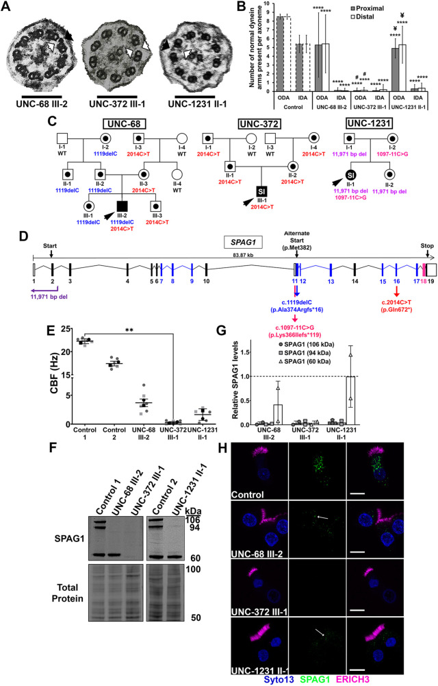
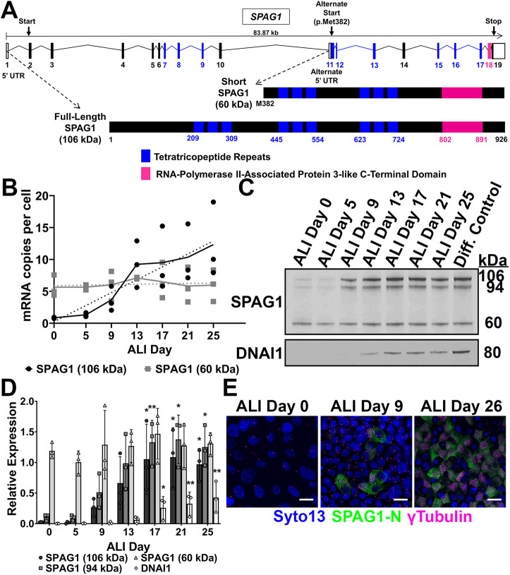
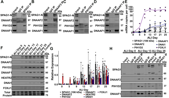
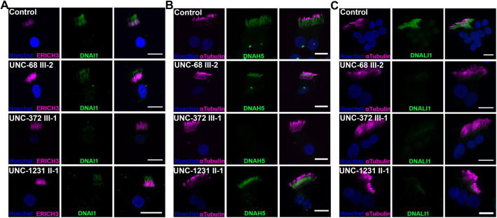
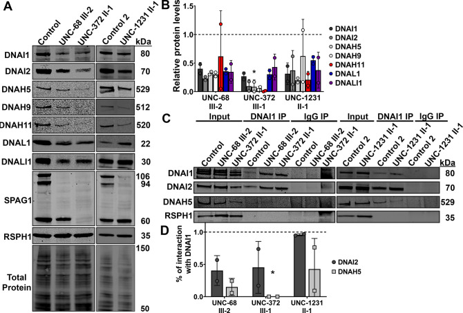
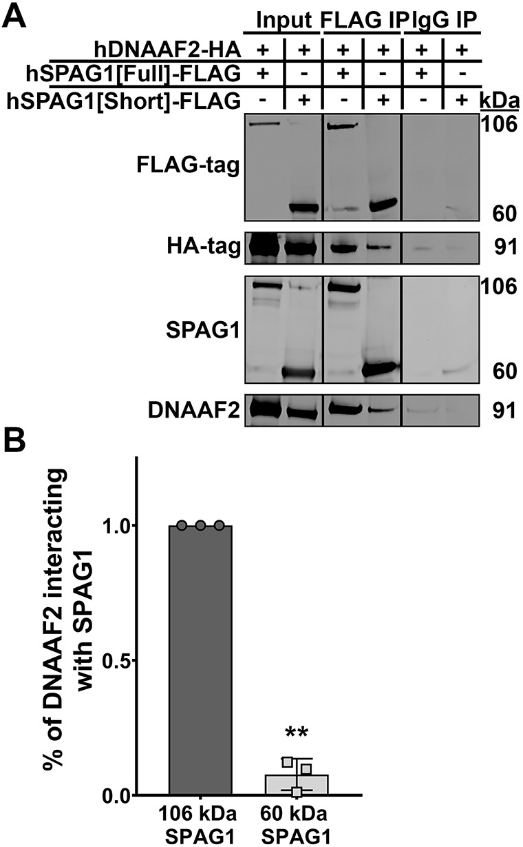
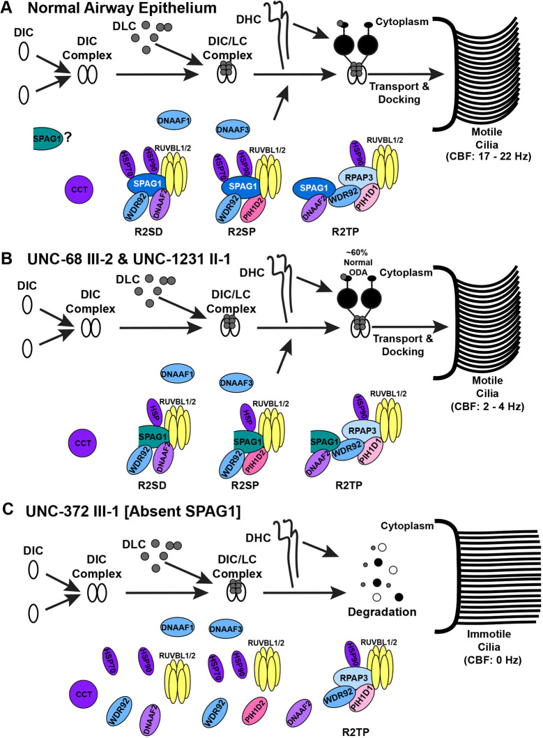
Mutations in SPAG1, a dynein axonemal assembly factor (DNAAF) that facilitates the assembly of dynein arms in the cytoplasm before their transport into the cilium, result in primary ciliary dyskinesia (PCD), a genetically heterogenous disorder characterized by chronic oto-sino-pulmonary disease, infertility and laterality defects. To further elucidate the role of SPAG1 in dynein assembly, we examined its expression, interactions and ciliary defects in control and PCD human airway epithelia. Immunoprecipitations showed that SPAG1 interacts with multiple DNAAFs, dynein chains and canonical components of the R2TP complex. Protein levels of dynein heavy chains (DHCs) and interactions between DHCs and dynein intermediate chains (DICs) were reduced in SPAG1 mutants. We also identified a previously uncharacterized 60 kDa SPAG1 isoform, through examination of PCD subjects with an atypical ultrastructural defect for SPAG1 variants, that can partially compensate for the absence of full-length SPAG1 to assemble a reduced number of outer dynein arms. In summary, our data show that SPAG1 is necessary for axonemal dynein arm assembly by scaffolding R2TP-like complexes composed of several DNAAFs that facilitate the folding and/or binding of the DHCs to the DIC complex.

Keywords: Dynein arm assembly; Motile cilia; Primary ciliary dyskinesia; R2TP complex; SPAG1.


Conflict of interest statement

Competing interests The authors declare no competing or financial interests.

Figures

Fig. 1.
Fig. 1.
Expression of 60-kDa SPAG1 results in an atypical ciliary ultrastructural defect. (A) TEM of nasal cilia cross-sections from probands UNC-68 III-2 (left), UNC-372 III-1 (middle) and UNC-1231 II-1 (right). UNC-372 III-1 has absent IDAs/ODAs (white arrows), whereas UNC-68 III-2 and UNC-1231 II-1 have normal ODAs (black arrows). Scale bars: 150 nm. (B) Quantification of normal ODAs and IDAs present per proximal (gray bars) and distal (white bars) ciliary axoneme examined by TEM. Control values were based on averages of normal ODAs and IDAs scored on TEM of 62 healthy individuals (dashed bars) (Shapiro and Leigh, 2017). UNC-68 III-2, UNC-372 III-1 and UNC-1231 II-1, n=18, 31 and 10 proximal axonemes and 24, 24 and 19 distal axonemes, respectively. ****P<0.0001 compared to Control; #P<0.0001 compared to UNC-68 III-2; ¥P<0.0001 compared to UNC-371 III-1 (ANOVA with Tukey's multiple comparisons). (C) Pedigrees of genetic variants identified in families UNC-68, UNC-372 and UNC-1231. Symbols with black dots indicate carrier status, and black symbols indicate affected individuals with PCD. Probands are designated by black arrowheads, and SI indicates individuals with situs inversus. WT, wild type. (D) Diagram of the SPAG1 gene (NCBI, NG_033834.2). Blue and pink boxes represent the exons that translate into tetratricopeptide domains and the RPAP3-like C-terminal domain, respectively. The positions of all genetic variants found in families UNC-68 (blue and red arrows), UNC-372 (red arrow) and UNC-1231 (purple and magenta arrows) are shown. (E) CBF measurements of HNEC cultures from control and PCD-affected subjects. Six cultures were measured across n=2 independent experiments. **P<0.01 (Kruskal–Wallis with uncorrected Dunn's multiple comparisons). (F) Representative immunoblots for SPAG1, and corresponding total protein stain, on HNEC lysates from normal and PCD-affected individuals. (G) Quantification of immunoblots probing for SPAG1 protein levels. Raw signals were normalized to a total protein stain, then a separate differentiated HBEC control to control for variation across immunoblots, and then the control means were normalized to 1.0 (dashed line). n=2 independent experiments. (H) Representative isolated cell immunofluorescence images of multiciliated HNECs from normal and PCD-affected individuals stained for SPAG1 (green) and ERICH3 (magenta), a cilia marker. SYTO13 (blue) was used to stain the nuclei. Arrows indicate SPAG1 puncta that are still present in UNC-68 III-2 and UNC-1231 II-1 nasal cells. Images are representative of a total of 20 fields across n=2 independent experiments. Scale bars: 10 μm. Data are mean±s.d.
Fig. 2.
Fig. 2.
Different SPAG1 isoforms have distinct expression patterns. (A) Schematic of the canonical and alternate start sites of SPAG1 and the two different SPAG1 isoforms that are produced. The full-length SPAG1 is a 926-amino acid protein (106 kDa) with three TPR domains (blue boxes) and a RPAP3-like C-terminal domain (pink box). The 60-kDa SPAG1 isoform, or the short isoform, starts at the methionine at amino acid 382 of the full-length SPAG1 and produces a 545-amino acid protein (60 kDa) that has only two of the TPR domains and the RPAP3-like C-terminal domain. (B) Quantification of mRNA expression of 106-kDa and 94-kDa (black) or 60-kDa (gray) SPAG1 isoforms in differentiating HBEC cultures using ddPCR. Calreticulin (CALR) was used as a reference gene. Linear regression analysis (dashed lines) determined that the slope of 60-kDa SPAG1 was not significantly non-zero (P=0.603) compared to the slope of 106-kDa and 94-kDa SPAG1 (P<0.001). Data shown are means across timepoints (solid lines) and individual data points; n=3 independent experiments using three distinct cell codes. (C) Representative immunoblot for SPAG1 and DNAI1 on differentiating HBEC lysates. DNAI1 was used as a ciliogenesis control. (D) Quantification of SPAG1 isoforms and DNAI1 protein levels analyzed by immunoblots on differentiating HBEC lysates. Raw signals were normalized to a total protein stain and then a separate differentiated HBEC lysate control. Data are mean±s.d. n=3 independent experiments using three distinct cell codes. *P<0.05, **P<0.01 compared to ALI day 0 (Kruskal–Wallis with uncorrected Dunn's multiple comparisons). (E) Whole-mount immunofluorescence images on differentiating HBEC cultures stained for the N terminus of SPAG1-N (green), γ-tubulin (magenta) to mark basal bodies, and the nuclei (blue). Images are representative of a total of six fields of view across two cultures per timepoint per independent experiment. Scale bars: 10 μm. n=3 independent experiments using three distinct cell codes.
Fig. 3.
Fig. 3.
SPAG1 interacts with other known DNAAFs. (A) Representative immunoblots for SPAG1, DNAAF2, PIH1D2 and DNAAF1 analyzing co-immunoprecipitation samples for endogenous SPAG1 in HBEC cultures. n=3 independent experiments using three distinct cell codes. (B-D) Representative immunoblots for SPAG1, DNAAF2, PIH1D2 and DNAAF1 analyzing reverse co-immunoprecipitation samples for endogenous DNAAF2 (B), PIH1D2 (C) and DNAAF1 (D) in HBEC cultures. n=2 independent experiments using two distinct cell codes. (E) Quantification of mRNA co-expression between SPAG1, FOXJ1, DNAI1, DNAAF1, DNAAF2 and PIH1D2 in differentiating HBEC cultures. FOXJ1 and DNAI1 were used as ciliogenesis controls. Calreticulin (CALR) was used as a reference gene. Data shown are means across timepoints (lines) and individual data points. n=3 independent experiments using three distinct cell codes. (F,G) Representative immunoblots (F) and quantification (G) of protein co-expression of SPAG1, DNAAF2, PIH1D2, DNAAF1, HEATR2, DNAI1 and FOXJ1 in differentiating HBEC culture lysates. DNAI1 and FOXJ1 were used as ciliogenesis controls. Total protein stain was used as a loading control. Raw signals were normalized to a total protein stain and then a separate differentiated HBEC lysate control. Data are mean±s.d. n=3 independent experiments using three distinct cell codes. *P<0.05, **P<0.01 compared to ALI day 0 (Kruskal–Wallis with uncorrected Dunn's multiple comparisons). (H) Representative immunoblots for SPAG1, DNAAF2, PIH1D2 and DNAAF1 in time-course co-immunoprecipitation for endogenous SPAG1 samples. HBEC cultures were lysed at various days (ALI days 0, 13, 18 and 28) throughout differentiation, and lysates were used to immunoprecipitate endogenous SPAG1 and interactors. n=2 independent experiments using two distinct cell codes. IP, immunoprecipitation. FT, flow through. Input lanes represent 5% of the total lysate.
Fig. 4.
Fig. 4.
UNC-372 III-1 has defective ODAs and IDAs, whereas UNC-68 III-2 and UNC-1231 II-1 have normal ODAs. Single-cell immunofluorescence images of cultured multiciliated nasal cells from normal and PCD-affected individuals stained for ODAs, using DNAI1 (A) and DNAH5 (B), and IDAs, using DNALI1 (C) (green). Cilia were labeled with either ERICH3 or acetylated α-tubulin (magenta), and nuclei (blue) were stained using Hoechst 33342. Images are representative of a total of 20 fields of view across n=2 independent experiments. Scale bars: 10 μm.
Fig. 5.
Fig. 5.
SPAG1 facilitates the addition of DHCs to the intermediate chain complex. (A) Immunoblots for various ODA dyneins (DNAI1, DNAI2, DNAH5, DNAH9, DNAH11 and DNAL1), an IDA dynein chain (DNALI1), SPAG1, a cilia control (RSPH1) and total protein stain on the HNEC lysates of normal and PCD-affected individuals. (B) Quantification of dynein chain protein expression measured by immunoblots on the HNEC lysates of normal and PCD-affected individuals. Background-subtracted signals were normalized to a total protein stain and RSPH1, then a separate differentiated HBEC lysate to control for variations across immunoblots, and then the nasal control means were set to 1.0 (dashed line). n=2 independent experiments. *P<0.05 compared to control (Kruskal–Wallis test with uncorrected Dunn's multiple comparisons). (C) Immunoblots for DNAI1, DNAI2, DNAH5 and a cilia control (RSPH1) analyzing co-immunoprecipitation for endogenous DNAI1 samples from HNECs of normal and PCD-affected individuals. (D) Quantification of the amount of DNAI2 and DNAH5 protein interacting with DNAI1 in PCD-affected HNECs compared to normal HNECs. Background-subtracted DNAI2 or DNAH5 signals were divided by DNAI1 signals per each eluate sample, and then the control means were set to 1.0 (dashed line). Data shown are mean±s.d. n=2 independent experiments. *P<0.05 compared to control (Kruskal–Wallis test with uncorrected Dunn's multiple comparisons). IP, immunoprecipitation. Input lanes represent 5% of the total lysate.
Fig. 6.
Fig. 6.
The 60-kDa SPAG1 isoform can still interact with DNAAF2. (A) Immunoblots for SPAG1, DNAAF2, FLAG-tag and HA-tag to analyze co-immunoprecipitations of FLAG-tagged full-length or 60-kDa SPAG1 with HA-tag DNAAF2 in HEK293T cells. IP, immunoprecipitations. Input lanes represent 5% of the total lysate. (B) Quantification of the amount of DNAAF2 interacting with 106-kDa or 60-kDa SPAG1 isoforms. Background-subtracted HA-tag signals were divided by FLAG-tag signals per each sample, and then the 106-kDa SPAG1 ratio was set to 1.0. Data are mean±s.d. n=3 independent experiments. **P<0.01 compared to 106-kDa SPAG1 (two-tailed one-sample t-test).
Fig. 7.
Fig. 7.
Function of full-length SPAG1 and compensation of 60-kDa SPAG1 in axonemal dynein arm assembly. (A) In normal human airway multiciliated cells, full-length SPAG1 (blue with white text) interacts with RUVBL1/2, DNAAF2 or PIH1D2, and WDR92, to form potential R2TP-like complexes that facilitate the folding and binding of the DHCs to the DIC-DLC (DIC/LC) complex. It is also possible that SPAG1 interacts with the canonical R2TP complex through WDR92 to facilitate dynein arm assembly. These R2TP-like complexes can further interact with different chaperones (purple), such as HSP70, HSP90 and CCT, and other DNAAFs (light blue), including DNAAF1 and DNAAF3, to recruit dynein chains to chaperones for formation into dynein arms. The dynein arms are then transported into the ciliary axoneme by intraflagellar transport, leading to motile cilia (CBF, 17-22 Hz). The 60-kDa SPAG1 isoform (teal) could play a role in dynein arm assembly to assemble a subset of ODAs, or it has a cilia-independent function. (B) In cells that are lacking full-length SPAG1 but still express the 60-kDa isoform of SPAG1 (UNC-68 III-2 and UNC-1231 II-1), some assembly of ODAs occurs even though SPAG1 is lacking one of its HSP binding sites. About 60% of normal ODAs leads to some ciliary motility (CBF, 2-4 Hz). (C) If SPAG1 is completely absent in airway epithelium (UNC-372 III-1), these R2TP-like complexes are unlikely to form, and thus, the folding and binding of DHCs to the DIC/LC complex is impaired. The cytoplasmic assembly process of dynein arms is inhibited, and the partially assembled dynein complex is degraded, instead of being transported into the ciliary axoneme. With this, cilia are completely immotile (CBF, 0 Hz).

Similar articles

Cited by

References

    1. Austin-Tse, C., Halbritter, J., Zariwala, M. A., Gilberti, R. M., Gee, H. Y., Hellman, N., Pathak, N., Liu, Y., Panizzi, J. R., Patel-King, R. S.et al. (2013). Zebrafish ciliopathy screen plus human mutational analysis identifies C21orf59 and CCDC65 defects as causing primary ciliary dyskinesia. Am. J. Hum. Genet. 93, 672-686. 10.1016/j.ajhg.2013.08.015 - DOI - PMC - PubMed
    1. Benbahouche, N. E. H., Iliopoulos, I., Török, I., Marhold, J., Henri, J., Kajava, A. V., Farkaš, R., Kempf, T., Schnölzer, M., Meyer, P.et al. (2014). Drosophila Spag is the homolog of RNA polymerase II-associated protein 3 (RPAP3) and recruits the heat shock proteins 70 and 90 (Hsp70 and Hsp90) during the assembly of cellular machineries. J. Biol. Chem. 289, 6236-6247. 10.1074/jbc.M113.499608 - DOI - PMC - PubMed
    1. Biermann, K., Heukamp, L. C., Steger, K., Zhou, H., Franke, F. E., Sonnack, V., Brehm, R., Berg, J., Bastian, P. J. and Müller, S. C. (2007). Genome-wide expression profiling reveals new insights into pathogenesis and progression of testicular germ cell tumors. Cancer Genomics Proteomics 4, 359-367. - PubMed
    1. Bio-Rad. (2018). Droplet DigitalTM PCR Droplet DigitalTM PCR Applications Guide. Bulletin 6407, 1-145.
    1. Blackburn, K., Bustamante-Marin, X., Yin, W., Goshe, M. B. and Ostrowski, L. E. (2017). Quantitative proteomic analysis of human airway cilia identifies previously uncharacterized proteins of high abundance. J. Proteome Res. 16, 1579-1592. 10.1021/acs.jproteome.6b00972 - DOI - PMC - PubMed