. 2022 Apr;13(2):919-931.
doi: 10.1002/jcsm.12937. Epub 2022 Feb 17.

Cell interactome in sarcopenia during aging

Affiliations

Cell interactome in sarcopenia during aging

Laura González-Blanco et al. J Cachexia Sarcopenia Muscle. 2022 Apr.

Abstract

Background: The diversity between the muscle cellular interactome of dependent and independent elderly people is based on the interrelationships established between different cellular mechanisms, and alteration of this balance modulates cellular activity in muscle tissue with important functional implications.

Methods: Thirty patients (85 ± 8 years old, 23% female) scheduled to undergo hip fracture surgery participated in this study. During the surgical procedures, skeletal muscle tissue was obtained from the Vastus lateralis. Two groups of participants were studied based on their Barthel index: 15 functional-independent individuals (100-90) and 15 severely functional-dependent individuals (40-0). The expression of proteins from the most important cellular mechanisms was studied by western blot.

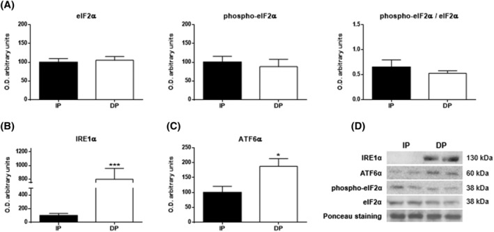
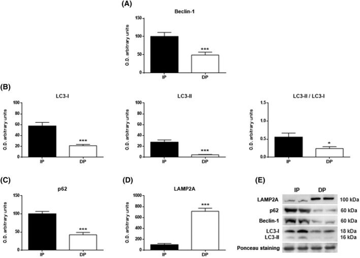
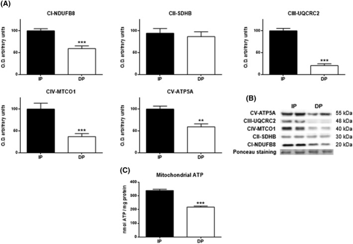
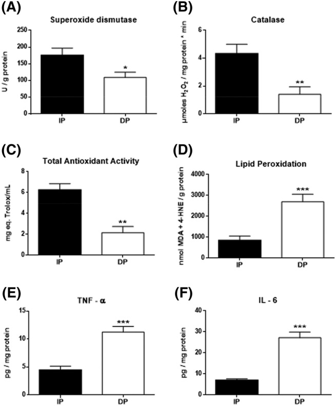
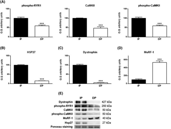
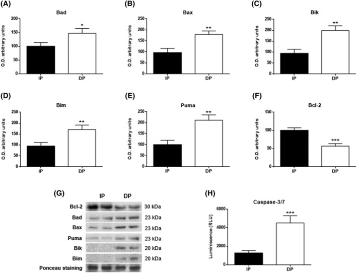
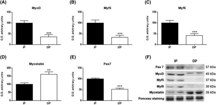
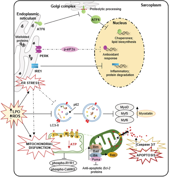
Results: Compared with independent elderly patients, dependent elderly showed an abrupt decrease in the capacity of protein synthesis; this decrease was only partially compensated for at the response to unfolded or misfolded proteins (UPR) level due to the increase in IRE1 (P < 0.001) and ATF6 (P < 0.05), which block autophagy, an essential mechanism for cell survival, by decreasing the expression of Beclin-1, LC3, and p62 (P < 0.001) and the antioxidant response. This lead to increased oxidative damage to lipids (P < 0.001) and that damage was directly associated with the mitochondrial impairment induced by the significant decreases in the I, III, IV, and V mitochondrial complexes (P < 0.01), which drastically reduced the energy capacity of the cell. The essential cellular mechanisms were generally impaired and the triggering of apoptosis was induced, as shown by the significantly elevated levels of most proapoptotic proteins (P < 0.05) and caspase-3/7 (P < 0.001) in dependents. The death of highly damaged cells is not detrimental to organs as long as the regenerative capacity remains unaltered, but in the dependent patients, this ability was also significantly altered, which was revealed by the reduction in the myogenic regulatory factors and satellite cell marker (P < 0.001), and the increase in myostatin (P < 0.01). Due to the severely disturbed cell interactome, the muscle contractile capacity showed significant damage.

Conclusions: Functionally dependent patients exhibited severe alterations in their cellular interactome at the muscle level. Cell apoptosis was caused by a decrease in successful protein synthesis, to which the cellular control systems did not respond adequately; autophagy was simultaneously blocked, the mitochondrion malfunctioned, and as the essential recovery mechanisms failed, these cells could not be replaced, resulting in the muscle being condemned to a loss of mass and functionality.

Keywords: Elderly; Mitochondria; Myogenic regulatory factors; Oxidative stress; Sarcopenia; Unfolded protein response.


Conflict of interest statement

The authors declare no conflict of interest.

Figures

Figure 1
Figure 1
Endoplasmic reticulum stress in the muscle tissues of functional‐independent patients (IP) and functional‐dependent patients (DP). Bar chart shows the quantification of the optical densities (O.D.) of blot bands of (A) eukaryotic initiation factor 2α (eIF2α), phospho‐eIF2α, phospho‐eIF2α/eIF2α, (B) inositol‐requiring enzyme 1α (IRE1α), and (C) activating transcription factor 6α (ATF6α). (D) Representative immunoblots. Ponceau staining was used as a loading control. Data are represented as the mean ± SEM. *P < 0.05; ***P < 0.001.
Figure 2
Figure 2
Autophagy in the muscle tissues of functional‐independent patients (IP) and functional‐dependent patients (DP). Bar chart shows the quantification of the optical densities (O.D.) of blot bands for (A) Beclin‐1, (B) LC3‐I, LC3‐II, LC3‐II/LC3‐I, (C) p62 and (D) LAMP2A. (E) Immunoblots of autophagy markers. Ponceau staining was used as a loading control. Data are represented as the mean ± SEM. *P < 0.05; ***P < 0.001.
Figure 3
Figure 3
Western blot analysis for studying the oxidative phosphorylation profile in muscle tissues of functional‐independent patients (IP) and functional‐dependent patients (DP). Bar chart shows the quantification of the optical densities (O.D.) of blot bands for (A) subunits from the protein complexes of the mitochondrial electron transport chain (NADH dehydrogenase (ubiquitone) 1b subcomplex 8 (NDUFB8) from complex I (CI), iron sulfur subunit (SDHB) from complex II (CII), ubiquinol‐cytochrome c reductase core protein II (UQCRC2) subunit from complex III (CIII), cytochrome c oxidase subunit I (MTCO1) from complex IV (CIV) and ATP synthase subunit α (ATP5A) from complex V (CV). (B) Representative immunoblots. Ponceau staining was used as a loading control. (C) ATP content was evaluated by bioluminescence (expressed as nmol ATP/mg protein. Data are represented as the mean ± SEM. **P < 0.01; ***P < 0.001.
Figure 4
Figure 4
Oxidative stress status and inflammatory response in the muscle of functional‐independent patients (IP) and functional‐dependent patients (DP). (A) Superoxide dismutase (expressed as SOD units/mg of protein), (B) catalase (expressed as μmol H2O2/min mg protein), (C) total antioxidant activity (expressed as mg eq. Trolox/mL), and (E) lipid peroxidation (expressed as MDA + 4‐HNE/g protein). Inflammation levels by the determination of (E) TNF‐α and (F) IL‐6 (expressed as pg/mg protein). Data are represented as the mean ± SEM. *P < 0.05; **P < 0.01; ***P < 0.001.
Figure 5
Figure 5
Skeletal muscle excitation‐contraction coupling mechanism in muscle tissues of functional‐independent patients (IP) and functional‐dependent patients (DP). (A) Levels of phospho‐RYR1, Ca2+/calmodulin‐dependent protein kinase II (CaMKII) and phospho‐CaMKII. (B) Levels of Hsp27, (C) Dystrophin, and (D) MuRF‐1. Bar chart shows the quantification of the optical densities (O.D.). (E) Representative immunoblots. Ponceau staining was used as a loading control. Data are represented as the mean ± SEM. ***P < 0.001.
Figure 6
Figure 6
Apoptosis in muscle tissues of functional‐independent patients (IP) and functional‐dependent patients (DP). Bar chart shows the quantification of the optical densities (O.D.) of blot bands for (A) bad, (B) Bax, (C) Bik, (D) Bim, (E) Puma, and (F) Bcl‐2. (G) Representative immunoblots. (H) Caspase‐3/7 was evaluated by bioluminescence. Ponceau staining was used as a loading control. Data are represented as the mean ± SEM. *P < 0.05; **P < 0.01; ***P < 0.001.
Figure 7
Figure 7
Myogenic differentiation in muscle tissues of functional‐independent patients (IP) and functional‐dependent patients (DP). Bar chart shows the quantification of the optical densities (O.D.) of blot bands for (A) MyoD, (B) Myf5, (C) Myf6, (D) myostatin, and (E) Pax7. (F) Representative immunoblots. Ponceau staining was used as a loading control. Data are represented as the mean ± SEM. **P < 0.01; ***P < 0.001.
Figure 8
Figure 8
Global effect of dependence in the skeletal muscle of elderly people. In the muscle of dependent patients, a concatenation of cellular alterations ends up inducing their death by apoptosis and reducing their regenerative capacity, which could be the main causes of the muscle reduction evident in these subjects. Thus, the abrupt decrease in protein synthesis triggers reticular stress and activation of the response to non‐folded proteins (mainly IRE‐1, ATF‐6) which induces an increase in oxidative damage (LPO) and blockage of autophagy (LC3‐II, p62) which prevents the degradation of damaged proteins while mitochondrial injury reduces cellular energy capacity. This has a negative effect on muscle contraction (phospho‐RYR1, phospho‐CaMKII) and cell proliferation by reducing myogenic regulatory factors (MyoD, Myf5, Myf6) and Pax7, while increasing myostatin. These last alterations will be the cause of the inhibition of muscle regeneration. Simultaneously, the accumulation of cellular damage causes an apoptotic imbalance with an increase in proapoptotic proteins (Bad, Bax, Bik, Bim, Puma) and a decrease in antiapoptotic proteins (Bcl‐2) leading to the death by apoptosis of muscle cells in extreme sarcopenia.

References

    1. Wall BT, Dirks ML, van Loon LJ. Skeletal muscle atrophy during short‐term disuse: implications for age‐related sarcopenia. Ageing Res Rev 2013;12:898–906. - PubMed
    1. Lee SM, Lee SH, Jung Y, Lee Y, Yoon JH, Choi JY, et al. FABP3‐mediated membrane lipid saturation alters fluidity and induces ER stress in skeletal muscle with aging. Nat Commun 2020;11:5661. - PMC - PubMed
    1. Christian CJ , Benian GM. Animal models of sarcopenia. Aging Cell 2020;19:e13223. - PMC - PubMed
    1. Potes Y, Pérez‐Martinez Z, Bermejo‐Millo JC, Rubio‐Gonzalez A, Fernandez‐Fernández M, Bermudez M, et al. Overweight in the elderly induces a switch in energy metabolism that undermines muscle integrity. Aging Dis 2019;10:217–230. - PMC - PubMed
    1. Potes Y, de Luxán‐Delgado B, Rodriguez‐González S, Guimarães MRM, Solano JJ, Fernández‐Fernández M, et al. Overweight in elderly people induces impaired autophagy in skeletal muscle. Free Radic Biol Med 2017;110:31–41. - PubMed

Publication types