. 2022 Mar;11(5):e022991.
doi: 10.1161/JAHA.121.022991. Epub 2022 Feb 18.

Myocardial Cytoskeletal Adaptations in Advanced Kidney Disease

Affiliations

Myocardial Cytoskeletal Adaptations in Advanced Kidney Disease

Arvin Halim et al. J Am Heart Assoc. 2022 Mar.

Erratum in

Abstract

Background The myocardial cytoskeleton functions as the fundamental framework critical for organelle function, bioenergetics and myocardial remodeling. To date, impairment of the myocardial cytoskeleton occurring in the failing heart in patients with advanced chronic kidney disease has been largely undescribed. Methods and Results We conducted a 3-arm cross-sectional cohort study of explanted human heart tissues from patients who are dependent on hemodialysis (n=19), hypertension (n=10) with preserved renal function, and healthy controls (n=21). Left ventricular tissues were subjected to pathologic examination and next-generation RNA sequencing. Mechanistic and interference RNA studies utilizing in vitro human cardiac fibroblast models were performed. Left ventricular tissues from patients undergoing hemodialysis exhibited increased myocardial wall thickness and significantly greater fibrosis compared with hypertension patients (P<0.05) and control (P<0.01). Transcriptomic analysis revealed that the focal adhesion pathway was significantly enriched in hearts from patients undergoing hemodialysis. Hearts from patients undergoing hemodialysis exhibited dysregulated components of the focal adhesion pathway including reduced β-actin (P<0.01), β-tubulin (P<0.01), vimentin (P<0.05), and increased expression of vinculin (P<0.05) compared with controls. Cytoskeletal adaptations in hearts from the hemodialysis group were associated with impaired mitochondrial bioenergetics, including dysregulated mitochondrial dynamics and fusion, and loss of cell survival pathways. Mechanistic studies revealed that cytoskeletal changes can be driven by uremic and metabolic abnormalities of chronic kidney disease, in vitro. Furthermore, focal adhesion kinase silencing via interference RNA suppressed major cytoskeletal proteins synergistically with mineral stressors found in chronic kidney disease in vitro. Conclusions Myocardial failure in advanced chronic kidney disease is characterized by impairment of the cytoskeleton involving disruption of the focal adhesion pathway, mitochondrial failure, and loss of cell survival pathways.

Keywords: cardiovascular; chronic kidney disease; cytoskeleton; dialysis; end stage kidney disease; heart failure; mitochondria.


Figures

Figure 1
Figure 1. The Cardiac Aging in CKD (CAIN) Cohort and Study Protocol.
A total of 109 deceased donor participants have been recruited to‐date in the CAIN cohort. Patients in the hypertension (HTN) group were included based on a documented history of diagnosed hypertension. All donor hearts underwent quality control (QC) screening for RNA purity. RNA quality was determined by nucleic acids absorbance ratio by spectrophotometry and a ratio ≈2.0 was considered as high purity. One patient was excluded due to poor RNA quality. Strict exclusion criteria for each group were used as illustrated. A total of 50 participants were included in the final analysis. CKD, chronic kidney disease; GFR, glomerular filtration rate; LVH, left ventricular hypertrophy.
Figure 2
Figure 2. Cardiac characterization.
A, Gross pathologic characterization. All heart samples underwent gross pathologic examination. Hemodialysis (HD) heart weight in grams (i) and normalized to body surface area (BSA) (ii) was significantly greater in HD and hearts from the hypertension group (HTN). HD and HTN hearts had increased left ventricular wall thickness (iii) expressed in centimeters (cm) and (iv) normalized to BSA. While HD and HTN hearts appeared to have increased left atrial area (v) expressed in cm2, this was not statistically significant following normalization to BSA (vi). Box‐and‐whisker plot denote extremes, interquartile range, and median. Displayed data points include outliers. P value was obtained by ANOVA for 3‐group comparison. Post hoc analysis by Tukey‐Kramer Honestly Significant Differences (HSD)test. B, Masson's trichrome stain of fibrotic changes in HD and HTN heart tissues: Left ventricular tissue sections were stained with Masson’s trichome stain. Left panels: 500 µm scale; Right panels: 100 µm scale of enlarged areas shown in the yellow boxes in the top panel. Left ventricular tissues from HD participants had severe and widespread fibrosis compared with patients with hypertension, as illustrated by the alinine blue color which stains for collagen. Little to no significant fibrosis were found in left ventricular tissue from control patients. C, Quantification of Masson’s trichrome stain of fibrosis in Control, HTN, and HD hearts: Quantification of fibrosis, as indicated by intensity of alinine blue staining, showed that left ventricular tissues from patients undergoing hemodialysis had significantly greater fibrosis compared with HTN and control groups. Box‐and‐whisker plot denote extremes, interquartile range, and median. Displayed data points include outliers. P value was obtained by ANOVA for 3‐group comparison. Post hoc analysis by Tukey‐Kramer HSD. Con indicates control; LA, left atrial; LV, left ventricular.
Figure 3
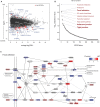
Figure 3. Transcriptomic profiles of heart tissues, ex vivo.
A, RNA‐seq analysis of heart tissues (left ventricular [LV] free wall) comparing hemodialysis (HD) and control groups. Top 26 significantly differentially expressed genes are highlighted in red (false discovery rate [FDR] adjusted P values <0.05). The x‐axis denotes mean log counts per million (log2CPM) and the y‐axis denotes log fold change (log2FC). B, Transcriptomics pathway enrichment analysis. Approximately 300 Kyoto Encyclopedia of Genes and Genomes (KEGG) pathways are aligned in the order of statistical significance. C, Gene expression ratios mapped to the focal adhesion pathway and pseudo‐colored according to the indicated scale. Genes with blank rectangles had insufficient counts. Dotted lines denote indirect effect. CPM indicates counts per million; and FC, fold‐change.
Figure 4
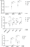
Figure 4. Dysregulation of the cytoskeleton is a cardinal feature of myocardial failure in patients undergoing hemodialysis, ex vivo.
A, Cytoskeletal genes are significantly altered in hemodialysis (HD) hearts. mRNA expression of major cytoskeletal proteins β‐actin, β‐tubulin, vimentin and vinculin as assessed by quantitative polymerase chain reaction (PCR). β‐actin, β‐tubulin, and vimentin was significantly dysregulated in hearts from patients undergoing hemodialysis compared with control. Hypertensive (HTN) hearts had dysregulation of β‐actin and β‐tubulin, but not vimentin compared with control. GAPDH was used as a standard. B, Cytoskeletal protein expressions in left ventricular heart tissues. (i) β‐actin, (ii) β‐tubulin, and (iii) vimentin expression was significantly suppresed in HD hearts. However, only β‐tubulin and vimentin expression was reduced in HTN hearts; (iv) vinculin expression was significantly upregulated in HD hearts but not in HTN hearts. (*P<0.05; **P<0.01).
Figure 5
Figure 5. Disruption of mitochondrial bioenergetics and cell survival pathways occurs in the failing myocardium in patients undergoing hemodialysis, ex vivo.
quantitative polymerase chain reaction was used to assess mRNA levels of genes identified on RNA sequencing related to mitochondrial function, cell survival and apoptosis. A, Mitochondrial function is significantly altered in hearts from HD and patients with HTN: Mitochondrial function, reflected by expression of OPA1 and MFN1, critical genes that regulate mitochondrial division and fusion activities was severely suppressed in both hearts from HD and patients with HTN. B, Pro‐apoptotic pathways are activated in hearts from HD and patients with HTN. In HD hearts, caspase 3, as well as P53, cytochrome C (CYCS) and BAX was upregulated. In HTN hearts, caspase 3 was downregulated but expression of P53, CYCS and BAX was increased. C, Anti‐apoptotic genes are suppressed in HD and HTN hearts. Anti‐apoptotic genes, BCL2 and BAG1 were down‐regulated in both HD and HTN hearts. (*:P<0.05; **:P<0.01).
Figure 6
Figure 6. Cytoskeletal dysregulation can be driven by uremic stress, in vitro.
Primary human cardiac fibroblasts were cultured from the ventricle. Expression of target proteins was assesed by immunoblotting. A, Dysregulation of all major cytoskeletal compartments following treatment with pooled uremic serum (US). Cardiac fibroblasts were treated with 10% pooled human uremic serum or 10% human serum from healthy donors for 48 hours. (i) β‐actin, (ii) β‐tubulin, and (iii) vimentin was significantly suppresed in cardiac fibroblasts from the ventricle following uremic treatment. Vinculin (iv) expression did not significantly change with uremic treatment. B, Dysregulation of all major cytoskeletal compartments following treatment with TNF‐α. Cardiac fibroblasts were treated with TNF‐α at 20 ng/mL for 48 hours. (i) β‐actin, (ii) β‐tubulin, (iii) vimentin, and (iv) vinculin were all suppressed in cardiac fibroblasts from the ventricle following treatment. C, Cytoskeletal dysregulation can be driven by uremic mineral stress. β‐actin (i) was significantly suppressed with either phosphate or calcium treatments, but not with combined phosphate and calcium treatment. β‐tubulin (ii) was significantly suppressed only with phosphate treatment. Combined phosphate and calcium treatment significantly suppressed vimentin (iii) but upregulated vinculin (iv). Primary human cardiac fibroblasts treated for 48 hours with either high phosphate (P: 5 mmol/L β‐glycerolphosphate), high calcium (Ca: 5 mmol/L calcium chloride), or both high calcium and high phosphate (P+Ca, 5 mmol/L+5 mmol/L). Cytoskeletal components were differentially regulated by high calcium and high phosphate. GAPDH was used as a standard. N=6 for all treatments. (*P<0.05, **P<0.01). Con indicates control.
Figure 7
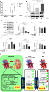
Figure 7. Regulation of the cardiac cytoskeleton by focal adhesion kinase (FAK).
A, Expression of FAK mRNA is significantly upregulated in left ventricular tissues from patients undergoing hemodialysis, ex vivo. Analysis of FAK mRNA expression by quantitative polymerase chain reaction revealed that FAK was significantly upregulated in hearts from patients undergoing hemodialysis. However, FAK mRNA expression was suppressed in hearts from patients with HTN. B, Expression of FAK protein is upregulated in left ventricular tissues from patients undergoing hemodialysis, ex vivo. Protein expression of FAK was significantly upregulated in hearts from patients undergoing hemodialysis as assessed by immunoblotting. FAK protein expression was significantly suppressed in HTN hearts. C, FAK silencing mediates cytoskeletal dysfunction in the presence of mineral stressors. (i) Primary human ventricular cardiac fibroblasts were transfected once with either scrambled or FAK siRNA. Cells were then treated daily with or without mineral stressors (ie P+Ca: 2 mmol/L β‐glycerophosphate with 1 unit/mL alkaline phosphatase, and 2.7 mmol/L calcium chloride) for 5 days. After treatment, target cytoskeletal proteins were assessed by immunoblotting. (ii) Treatment with FAK siRNA significantly reduced full‐length FAK expression at 5 days, and exposure to high calcium and high phosphate further reduced FAK expression. High calcium and high phosphate treatment resulted in detection of 80 kDa N‐terminal FAK fragment, which was decreased in combined FAK siRNA and high mineral stressors treatment. There was no statistical difference in FAK expression between the control and scrambled siRNA groups. (iii) There was no significant difference in β‐actin expression across the groups. (iv) β‐Tubulin was significantly decreased in cardiac fibroblasts exposed to mineral stressors compared with controls, but was not significantly different in the FAK silencing groups. (v) Vimentin was decreased significantly when cardiac fibroblasts were exposed to high calcium and high phosphate, and further decreased in the FAK silenced group exposed to high calcium and high phosphate. (vi) Expression of vinculin followed a similar pattern to vimentin. D, Schematic diagram of ultrastructural adaptations of the failing myocardium in CKD. In the healthy individuals, interactions between the heart and kidney is critical for normal physiological function. Patients with advanced CKD on hemodialysis (HD) exhibited significant cytoskeletal maladaptations that were generally more severe than hypertensive control hearts. HD hearts exhibited a unique pattern of severely reduced β‐actin, β‐tubulin and upregulation of vinculin expression. Cytoskeletal dysregulation in HD and HTN hearts is associated with mitochondrial dysfunction (reduced OPA1 and MFN1 genes responsible for mitochondrial fusion and division) and loss of cell survival pathways. FAK is a cytosolic tyrosine kinase that plays a central role in regulating cytoskeletal integrity. FAK expression is upregulated in HD hearts. This may account for increased expression of anchor protein vinculin observed as a protective response. GFR indicates glomerular filtration rate.

References

    1. Thompson S, James M, Wiebe N, Hemmelgarn B, Manns B, Klarenbach S, Tonelli M, Alberta Kidney Disease N . Cause of death in patients with reduced kidney function. J Am Soc Nephrol. 2015;26:2504–2511. doi: 10.1681/ASN.2014070714 - DOI - PMC - PubMed
    1. Edwards NC, Moody WE, Chue CD, Ferro CJ, Townend JN, Steeds RP. Defining the natural history of uremic cardiomyopathy in chronic kidney disease: the role of cardiovascular magnetic resonance. JACC Cardiovasc Imaging. 2014;7:703–714. doi: 10.1016/j.jcmg.2013.09.025 - DOI - PubMed
    1. Rutherford E, Mark PB. What happens to the heart in chronic kidney disease? J R Coll Physicians Edinb. 2017;47:76–82. doi: 10.4997/JRCPE.2017.117 - DOI - PubMed
    1. Kahn MR, Robbins MJ, Kim MC, Fuster V. Management of cardiovascular disease in patients with kidney disease. Nat Rev Cardiol. 2013;10:261–273. doi: 10.1038/nrcardio.2013.15 - DOI - PubMed
    1. Verbrugge FH, Tang WH, Hazen SL. Protein carbamylation and cardiovascular disease. Kidney Int. 2015;88:474–478. doi: 10.1038/ki.2015.166 - DOI - PMC - PubMed

Publication types