. 2022 Dec;100(8):946-959.
doi: 10.1111/aos.15112. Epub 2022 Feb 18.

Choroidal arteriovenous anastomoses: a hypothesis for the pathogenesis of central serous chorioretinopathy and other pachychoroid disease spectrum abnormalities

Affiliations

Choroidal arteriovenous anastomoses: a hypothesis for the pathogenesis of central serous chorioretinopathy and other pachychoroid disease spectrum abnormalities

Joost Brinks et al. Acta Ophthalmol. 2022 Dec.

Abstract

The pachychoroid disease spectrum (PDS) includes several chorioretinal diseases that share specific choroidal abnormalities. Although their pathophysiological basis is poorly understood, diseases that are part of the PDS have been hypothesized to be the result of venous congestion. Within the PDS, central serous chorioretinopathy is the most common condition associated with vision loss, due to an accumulation of subretinal fluid in the macula. Central serous chorioretinopathy is characterized by distinct risk factors, most notably a high prevalence in males and exposure to corticosteroids. Interestingly, sex differences and corticosteroids are also strongly associated with specific types of arteriovenous anastomoses in the human body, including dural arteriovenous fistula and surgically created arteriovenous shunts. In this manuscript, we assess the potential of such arteriovenous anastomoses in the choroid as a causal mechanism of the PDS. We propose how this may provide a novel unifying concept on the pathophysiological basis of the PDS, and present cases in which this mechanism may play a role.

Keywords: arteriovenous anastomoses; arteriovenous fistula; corticosteroids; pachychoroid disease spectrum; sex differences; venous congestion.


Figures

Fig. 1
Fig. 1
Thirty‐three‐year‐old man with central serous chorioretinopathy showing typical aspects of the pachychoroid disease spectrum. Foveal optical coherence tomography (A) revealed subretinal fluid accumulation. Pachyvessels with apparent attenuation of the overlying choriocapillaris were also observed on optical coherence tomography (B; red arrowheads). Indocyanine green angiography shows a prominent appearance of pachyvessels that appear to adhere to vortex vein quadrants (C; red arrowheads), as well as intervortex vein anastomoses nasal to the macula (C; red arrowheads). Mid‐phase indocyanine green angiography (D) showed hypofluorescent and hyperfluorescent areas with indistinct borders that are typical for diseases that are part of the pachychoroid disease spectrum.
Fig. 2
Fig. 2
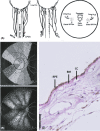
Overview of the anatomy of the choroid. (A) Reproduced with permission from Hayreh (2004). Schematic representation of the branching pattern of medial and lateral posterior ciliary arteries in 2 eyes (illustrations at the left and the middle) and of the site of entry of the various branches of the posterior ciliary arteries, as seen at the back of the eye (illustration at the right). (B) Reproduced with permission from Hayreh (1975). Schematic representation of distribution of choriocapillaris supplied by various temporal short posterior ciliary arteries. (Left) Normal pattern. (Right) Postulated pattern caused by generalized chronic ischaemic disorder of the choroid, with reduction in choriocapillaris most marked in macular region and equatorial choroid. (C) A haematoxylin and eosin staining of a cross section of the human choroid showing the retinal pigment epithelium (RPE), Bruch’s membrane (BM) and the choriocapillaris (CC). In the deeper layers, the lumen of a large vessel and the presence of other cell types resident in the choroid, such as melanocytes, can be observed. Scale bar = 50 μm.
Fig. 3
Fig. 3
Reproduced with permission from Jung et al. (2020). Representative mid‐phase ultra‐widefield indocyanine green angiography images (Optos California; Optos, Inc., Dunfermline, UK) after removal of imaging artefacts from a left eye diagnosed with chronic central serous chorioretinopathy. Vortex vein ampullas are indicated with orange arrowheads.
Fig. 4
Fig. 4
Illustration of the hypothesis presented in this manuscript, in which choroidal arteriovenous anastomoses are proposed to play a central role in the pathogenesis of pachychoroid spectrum diseases. In a normal situation (bottom half of the illustration), circulation of the choroid starts in short posterior ciliary arteries, which branches into smaller arterioles that eventually feed choriocapillaris lobules. The choriocapillaris is drained by collector venules which merge into vortex veins and exit the eye at the ampulla, through the sclera and continue as ophthalmic veins. In the abnormal situation (upper half of the illustration), a choroidal arteriovenous anastomosis is formed, either as a result of a dysregulated physiological anastomosis or due to a primarily pathological fistula. In the case of such an abnormal choroidal arteriovenous anastomosis, shunting of blood flow occurs from arteries directly into choroidal veins, bypassing the capillary bed. Without the dampening effect of the choriocapillaris on blood pressure, choroidal veins will be affected due to the direct arterial filling, which now become pachyvessels. Consequently, additional signs of venous overload emerge. This includes (1) congestion of blood flow from the ‘upstream’ choriocapillaris resulting in vascular hyperpermeability; (2) venous dilatation of other choroidal veins within the same vortex vein quadrant due to congestion of blood flow at the ampulla; and (3) the manifestation of intervortex vein anastomosis which cross watershed zones and shunt blood from overloaded vortex vein quadrants to neighbouring vortex vein quadrants. This can eventually affect the barrier function of the retinal pigment epithelium overlying these choroidal abnormalities, leading to subretinal fluid accumulation.
Fig. 5
Fig. 5
Multimodal imaging of a patient with unilateral central serous chorioretinopathy. Multimodal imaging (A–G) of the left eye of a 56‐year‐old man patient with complaints of central vision loss. The complaints started after a stressful period. Medical history of this patient included hypertension and asthma, for which fluticasone was prescribed. The foveal optical coherence tomography (OCT) scan (A) revealed subretinal fluid (SRF) and a subfoveal choroidal thickness of 364 μm. The OCT also showed pachyvessels with attenuation of the overlying choriocapillaris. Fundus autofluorescence (B) mainly showed mild hypo‐ and hyperautofluorescent changes in the macular region. On mid‐phase fluorescein angiography (C), several ‘hot spots’ of leakage were observed in the macula. The arterial phase of indocyanine green angiography at 18 s (D) revealed filling of several vessels with a large lumen suspect for choroidal veins (blue arrowheads). There appears to be a delayed arterial filling of the nasal side compared with the temporal side (E). Several seconds later, complete vortex vein quadrants containing pachyvessels became visible (F), suggestive for asymmetrical venous filling (blue arrowheads E compared with F). In addition, intervortex vein anastomoses emerge which cross the horizontal watershed zone through the macular region (F). Mid‐phase indocyanine green angiography (G) showed hyperfluorescent areas with indistinct borders, typical for central serous chorioretinopathy.
Fig. 6
Fig. 6
Multimodal imaging of the right eye of a patient with bilateral central serous chorioretinopathy before (A–G) and after (H–N) treatment with half‐dose photodynamic therapy. This 46‐year‐old male patient presented with complaints of central vision loss and micropsia. Medical history included a burn‐out 8 years ago, although there was no reported stress in the period of onset of the complaints. The patient did not use corticosteroids or any other type of medication. The foveal optical coherence tomography (OCT) scan (A) revealed subretinal fluid (SRF) and a subfoveal choroidal thickness of 425 μm. The OCT also showed pachyvessels with attenuation of the overlying choriocapillaris. Fundus autofluorescence (B) mainly showed extensive hyperautofluorescent changes and several spots of hypo‐autofluorescence. The mid‐phase fluorescein angiography (C) revealed extensive windows defects and hyperfluorescent areas representing active leakage. The arterial phase of indocyanine green angiography at 17 s (D) showed filling of two vessels in the macular region suggestive to be of arterial origin. One second later, filling of choroidal veins started (E; blue arrowheads). Several seconds later, choroidal veins reminiscent of pachyvessels became visible (F; blue arrowheads), which appeared to be directly connected to the arteries (F; red arrowheads) that emerged a few seconds earlier (D; red arrowheads). Mid‐phase indocyanine green angiography (G) showed mainly hyperfluorescent changes with indistinct borders, typical for central serous chorioretinopathy, as well as some choroidal folds. The patient received half‐dose photodynamic therapy, and multimodal imaging was performed 6 weeks after this treatment (H–N). A complete resolution of subretinal fluid was observed (H), as well as a reduction in hyperfluorescent abnormalities on both fluorescein angiography (J) and indocyanine green angiography (N). Strikingly, signs of vascular remodelling may be observed on the early phase of indocyanine green angiography (K–L), which appeared to show a notable reduction in some of the pachyvessels after half‐dose photodynamic therapy (M; top blue arrowheads) compared with the imaging before treatment (F; top blue arrowheads), as well as a general decrease in previous abnormal hyperfluorescence (N). Moreover, the degree of filling of the apparent pachyvessels during the arterial phase in the macular region appeared to be diminished after treatment with half‐dose photodynamic therapy (K–M compared with D–F; red arrowheads).

References

    1. Allison DJ & Kennedy A (1991): Peripheral arteriovenous malformations. BMJ: Br Med J 303: 1191–1194. - PMC - PubMed
    1. Amyere M, Revencu N, Helaers R et al. (2017): Germline loss‐of‐function mutations in EPHB4 cause a second form of capillary malformation‐arteriovenous malformation (CM‐AVM2) deregulating RAS‐MAPK signaling. Circulation 136: 1037–1048. - PubMed
    1. Araki T, Ishikawa H, Iwahashi C et al. (2019): Central serous chorioretinopathy with and without steroids: a multicenter survey. PLoS One 14: e0213110. - PMC - PubMed
    1. Baek J, Dansingani KK, Lee JH, Lee WK & Freund KB (2019a): Choroidal morphology in eyes with peripapillary polypoidal choroidal vasculopathy. Retina 39: 1571–1579. - PubMed
    1. Baek J, Kook L & Lee WK (2019b): Choriocapillaris flow impairments in association with pachyvessel in early stages of pachychoroid. Sci Rep 9: 1–6. - PMC - PubMed