

Codelivery of 1α,25-Dihydroxyvitamin D3 and CYP24A1 Inhibitor VID400 by Nanofiber Dressings Promotes Endogenous Antimicrobial Peptide LL-37 Induction
- PMID: 35179903
- PMCID: PMC10214699
- DOI: 10.1021/acs.molpharmaceut.1c00944
Codelivery of 1α,25-Dihydroxyvitamin D3 and CYP24A1 Inhibitor VID400 by Nanofiber Dressings Promotes Endogenous Antimicrobial Peptide LL-37 Induction
Abstract
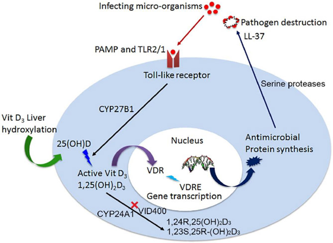
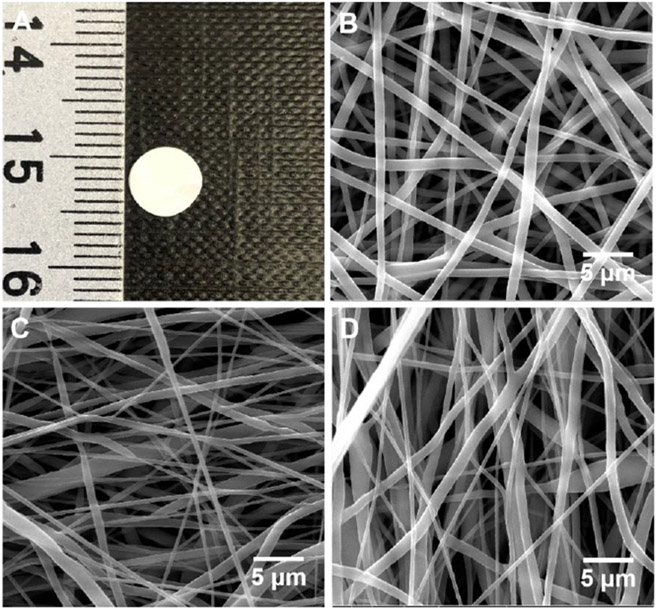
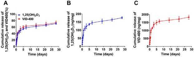
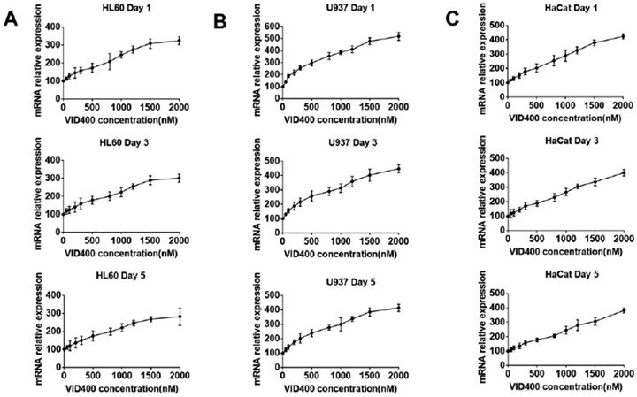
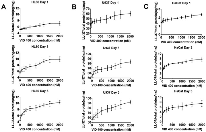
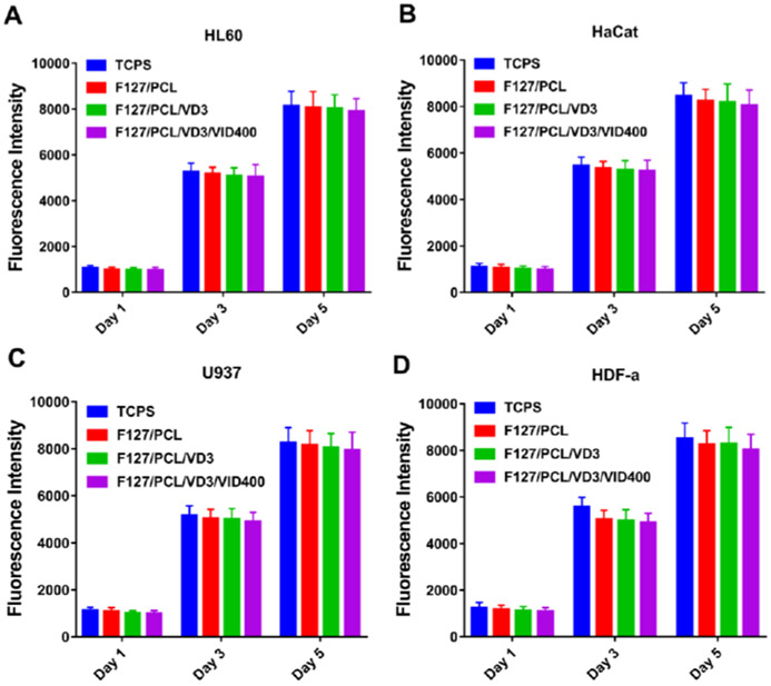
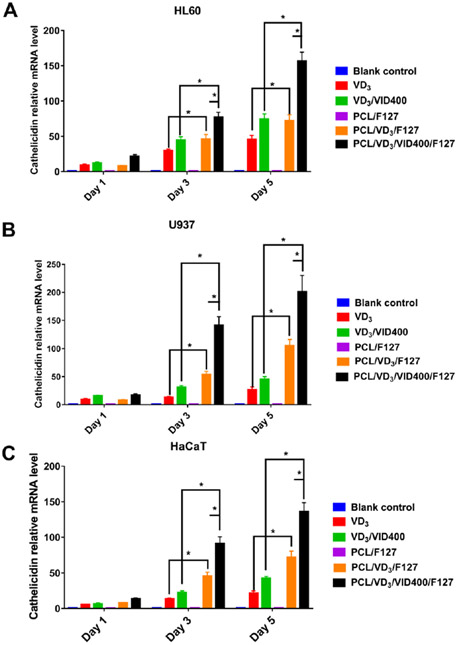
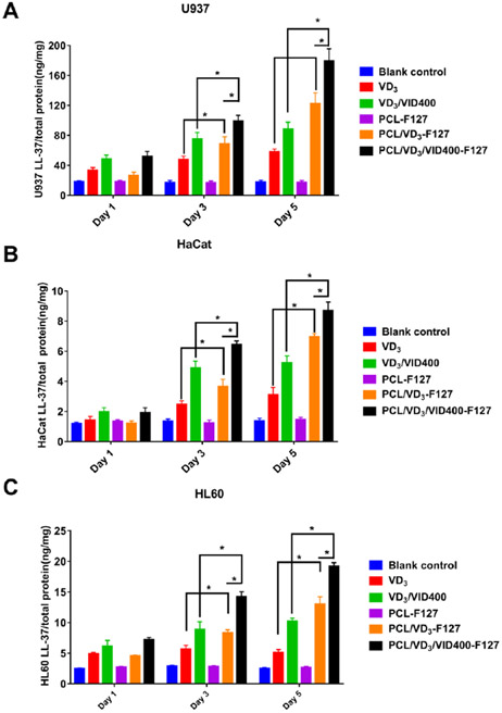
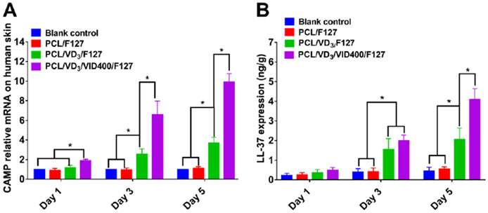
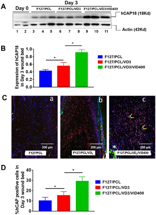
Surgical site infections represent a significant clinical problem. Herein, we report a nanofiber dressing for topical codelivery of immunomodulating compounds including 1α,25-dihydroxyvitamin D3 (1,25(OH)2D3) and VID400, a CYP24A1 inhibitor in a sustained manner, for inducing the expression of the endogenous cathelicidin antimicrobial peptide (CAMP) gene encoding the hCAP18 protein, which is processed into the LL-37 peptide. Nanofiber wound dressings with coencapsulation of 1,25(OH)2D3 and VID400 were generated by electrospinning. Both 1,25(OH)2D3 and VID400 were coencapsulated into nanofibers with loading efficiencies higher than 90% and exhibited a prolonged release from nanofiber membranes longer than 28 days. Incubation with 1,25(OH)2D3/VID400-coencapsulated poly(ϵ-caprolactone) nanofiber membranes greatly induced the hCAP18/LL-37 gene expression in monocytes, neutrophils, and keratinocytes in vitro. Moreover, the administration of 1,25(OH)2D3/VID400-coencapsulated nanofiber membranes dramatically promoted the hCAP18/LL-37 expression in dermal wounds created in both human CAMP transgenic mice and human skin tissues. The 1,25(OH)2D3- and VID400-coencapsulated nanofiber dressings enhanced innate immunity via the more effective induction of antimicrobial peptide than the free drug alone or 1,25(OH)2D3-loaded nanofibers. Together, 1,25(OH)2D3/VID400-embedded nanofiber dressings presented in this study show potential in preventing surgical site infections.
Keywords: 1α,25-dihydroxyvitamin D3; CYP24A1 inhibitor; antimicrobial peptide LL-37; codelivery; nanofiber dressings.
Figures
Similar articles
-
1α,25-dihydroxyvitamin D3-eluting nanofibrous dressings induce endogenous antimicrobial peptide expression.Nanomedicine (Lond). 2018 Jun;13(12):1417-1432. doi: 10.2217/nnm-2018-0011. Epub 2018 Jul 4. Nanomedicine (Lond). 2018. PMID: 29972648 Free PMC article.
-
Altered pharmacokinetics of 1alpha,25-dihydroxyvitamin D3 and 25-hydroxyvitamin D3 in the blood and tissues of the 25-hydroxyvitamin D-24-hydroxylase (Cyp24a1) null mouse.Endocrinology. 2005 Feb;146(2):825-34. doi: 10.1210/en.2004-1116. Epub 2004 Oct 21. Endocrinology. 2005. PMID: 15498883
-
Metabolic stability of 3-epi-1α,25-dihydroxyvitamin D3 over 1 α 25-dihydroxyvitamin D3: metabolism and molecular docking studies using rat CYP24A1.J Cell Biochem. 2013 Oct;114(10):2293-305. doi: 10.1002/jcb.24576. J Cell Biochem. 2013. PMID: 23606409
-
CYP24A1 as a potential target for cancer therapy.Anticancer Agents Med Chem. 2014 Jan;14(1):97-108. doi: 10.2174/18715206113139990307. Anticancer Agents Med Chem. 2014. PMID: 23869781 Review.
-
Metabolism of vitamin D3 by cytochromes P450.Front Biosci. 2005 Jan 1;10:119-34. doi: 10.2741/1514. Print 2005 Jan 1. Front Biosci. 2005. PMID: 15574355 Review.
Cited by
-
Engineered Exosomes Containing Cathelicidin/LL-37 Exhibit Multiple Biological Functions.Adv Healthc Mater. 2022 Oct;11(20):e2200849. doi: 10.1002/adhm.202200849. Epub 2022 Aug 12. Adv Healthc Mater. 2022. PMID: 35930707 Free PMC article.
-
Cholecalciferol Exhibits no Antibacterial Effect on Staphylococcus aureus and Escherichia coli: An in vitro Study.Recent Adv Antiinfect Drug Discov. 2024;19(4):315-321. doi: 10.2174/0127724344277290231211051800. Recent Adv Antiinfect Drug Discov. 2024. PMID: 38275070
-
Induction of Endogenous Antimicrobial Peptides to Prevent or Treat Oral Infection and Inflammation.Antibiotics (Basel). 2023 Feb 9;12(2):361. doi: 10.3390/antibiotics12020361. Antibiotics (Basel). 2023. PMID: 36830272 Free PMC article. Review.
References
-
- Wenzel RP Minimizing surgical-site infections. N. Engl. J. Med 2010, 362, 75. - PubMed
-
- Gottrup F. Prevention of surgical-wound infections, N. Engl. J. Med 2000, 342, 202–204. - PubMed
-
- Berríos-Torres SI; Umscheid CA; Bratzler DW; Leas B; Stone EC; Kelz RR; Reinke CE; Morgan S; Solomkin JS; Mazuski JE; Dellinger EP; Itani KMF; Berbari EF; Segreti J; Parvizi J; Blanchard J; Allen G; Kluytmans JAJW; Donlan R; Schecter WP; Healthcare Infection Control Practice Advisory Committee. Centers for disease control and prevention guideline for the prevention of surgical site infection, JAMA Surg. 2017, 152, 784–791. - PubMed
-
- Shiroky J; Lillie E; Muaddi H; Sevigny M; Choi WJ; Karanicolas PJ The impact of negative pressure wound therapy for closed surgical incisions on surgical site infection: a systematic review and meta-analysis. Surgery 2020, 167, 1001–1009. - PubMed
-
- Eagye KJ; Kim A; Laohavaleeson S; Kuti JL; Nicolau DP Surgical site infection: does inadequate antibiotic therapy affect patient outcomes?. Surg. Infect. (Larchmt) 2009, 10, 323–331. - PubMed
Publication types
MeSH terms
Substances
Grants and funding
LinkOut - more resources
Full Text Sources
