Clinical Trial
. 2022 Mar 3;126(8):1336-1344.
doi: 10.1021/acs.jpca.1c09968. Epub 2022 Feb 18.

Interaction with a Biomolecule Facilitates the Formation of the Function-Determining Long-Lived Triplet State in a Ruthenium Complex for Photodynamic Therapy

Affiliations
Clinical Trial

Interaction with a Biomolecule Facilitates the Formation of the Function-Determining Long-Lived Triplet State in a Ruthenium Complex for Photodynamic Therapy

Avinash Chettri et al. J Phys Chem A. .

Abstract

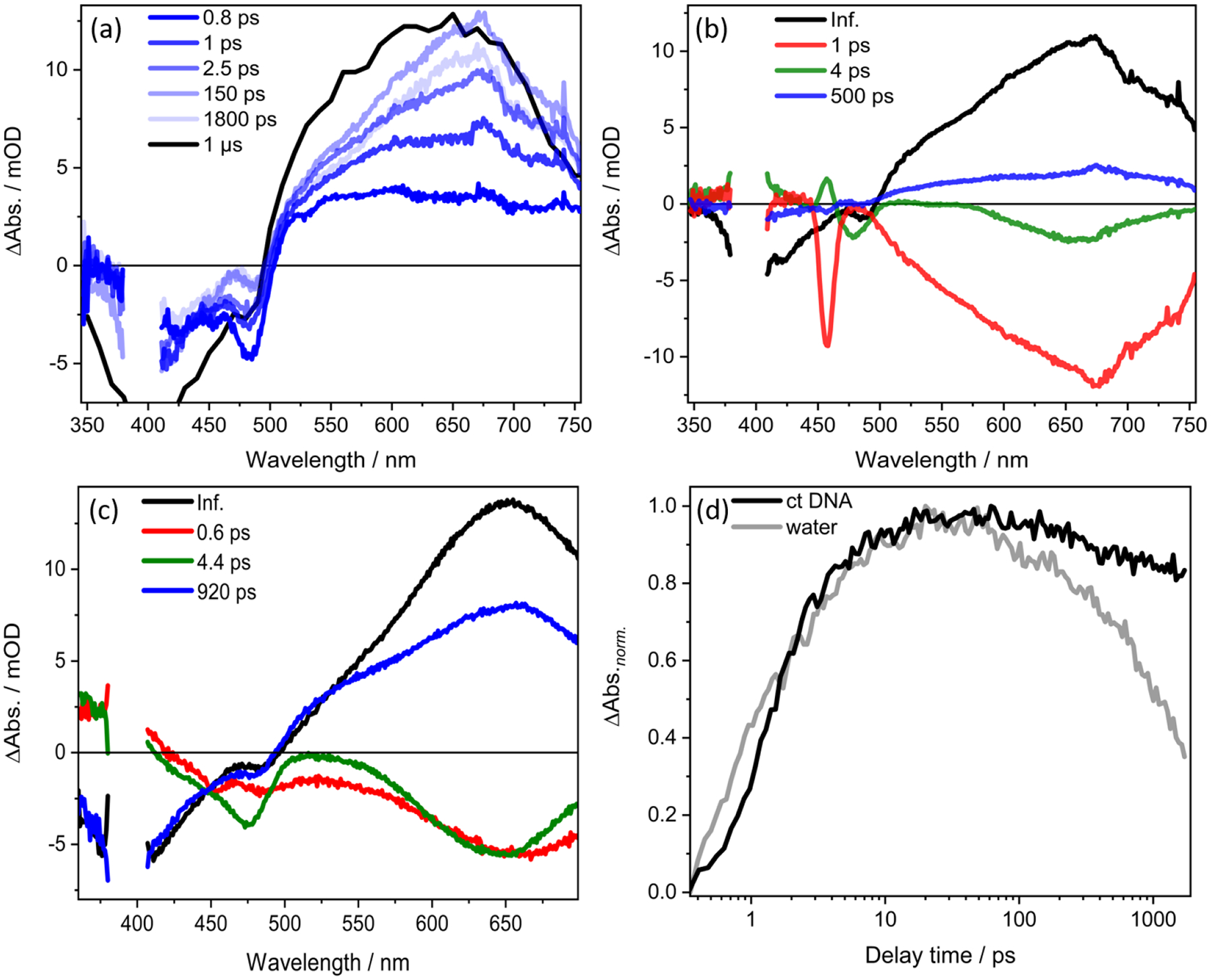
TLD1433 is the first ruthenium (Ru)-based photodynamic therapy (PDT) agent to advance to clinical trials and is currently in a phase II study for treating nonmuscle bladder cancer with PDT. Herein, we present a photophysical study of TLD1433 and its derivative TLD1633 using complex, biologically relevant solvents to elucidate the excited-state properties that are key for biological activity. The complexes incorporate an imidazo [4,5-f][1,10]phenanthroline (IP) ligand appended to α-ter- or quaterthiophene, respectively, where TLD1433 = [Ru(4,4'-dmb)2(IP-3T)]Cl2 and TLD1633 = [Ru(4,4'-dmb)2(IP-4T)]Cl2 (4,4'-dmb = 4,4'-dimethyl-2,2'-bipyridine; 3T = α-terthiophene; 4T = α-quaterthiophene). Time-resolved transient absorption experiments demonstrate that the excited-state dynamics of the complexes change upon interaction with biological macromolecules (e.g., DNA). In this case, the accessibility of the lowest-energy triplet intraligand charge-transfer (3ILCT) state (T1) is increased at the expense of a higher-lying 3ILCT state. We attribute this behavior to the increased rigidity of the ligand framework upon binding to DNA, which prolongs the lifetime of the T1 state. This lowest-lying state is primarily responsible for O2 sensitization and hence photoinduced cytotoxicity. Therefore, to gain a realistic picture of the excited-state kinetics that underlie the photoinduced function of the complexes, it is necessary to interrogate their photophysical dynamics in the presence of biological targets once they are known.


Conflict of interest statement

S.A.M. has a potential research conflict of interest due to a financial interest with Theralase Technologies, Inc. and PhotoDynamic, Inc. A management plan has been created to preserve objectivity in research in accordance with UTA policy

Figures

Figure 1.
Figure 1.
Chemical structures of (a) TLD1433, [Ru(4,4’-dmb)2(IP-3T)]2+ (Ru-ip-3T) and (b) TLD1633, [Ru(4,4’-dmb)2(IP-4T)]2+ (Ru-ip-4T). 4,4’-dmb=4,4’-dimethyl-2,2’-bipyridine; IP=imidazo [4,5-f][9,10] phenanthroline; 3T=α-terthiophene; 4T=α-quaterthiophene.
Figure 2.
Figure 2.
(a) Steady state UV-vis absorption spectra of Ru-ip-3T in water, ct DNA, and lysate of SK-MEL-28 cells. (b) Emission of Ru-ip-3T in water and ct DNA at excitation wavelength of 450 nm. (c) ns-TA of Ru-ip-3T in ct DNA recorded at selected delay times upon excitation at 410 nm. (d) Ground state recovery lifetimes (τTA) associated with T1 state of Ru-ip-3T in water, ct DNA and lysates of SK-MEL-28 cell under aerated conditions. A [DNA]bp/[complex] of 5:1 was used for all measurements in ct DNA solutions. Both ct DNA and SK-MEL-28 lysate solutions were prepared in Tris-HCl / NaCl (5 mM / 50 mM, pH 7.4) buffer. The kinetics from 630–680 nm were averaged to obtain the resultant kinetics and fitted to obtain the corresponding lifetimes. Note: For data analysis in Figure 2c, the contribution by emission is subtracted from the experimental data in the spectral range between 630 and 680 nm, thereby highlighting the features of the excited-state absorption spectrum (rather than the transient absorption spectrum, which of course contains information on emission). Experimentally, a transient absorption measurement is performed first. In order to account for the contribution by emission from this differential absorption kinetics, an additional measurement is conducted exclusively in only ‘pump on’ mode. This process allows us to separately collect the emission kinetics which is calibrated accordingly to ΔAbs. unit and subtracted from the differential absorption kinetics.
Figure 3.
Figure 3.
(a) Femtosecond TA spectra with 400-nm excitation of Ru-ip-3T in ct DNA solution. The spectrum at 1 μs is extracted from the ns-TA spectrum of Ru-ip-3T in ct DNA solution with contribution from emission subtracted from the overall spectra. (b) DAS for Ru-ip-3T in ct DNA solution. The intense band at at 460 nm in the 1-ps spectrum is due to Raman scattering by water. (c) DAS for Ru-ip-3T in water. (d) Transient absorption kinetics recorded at 660 nm of Ru-ip-3T in ct DNA solution versus water following excitation at 400 nm. Data for Ru-ip-3T in water were reported elsewhere.
Figure 4.
Figure 4.
(a) Femtosecond TA spectra for Ru-ip-4T in ct DNA solution with 400-nm excitation. The spectrum at 1 μs is extracted from the ns-TA spectrum for Ru-ip-4T in ct-DNA solution with contribution from emission subtracted from the overall spectra. (b) DAS of Ru-ip-4T in ct DNA solution. The intense band at 460 nm in the 1-ps spectrum is due to Raman scattering by water. (c) DAS for Ru-ip-4T in water. (d) Transient absorption kinetics recorded at 700 nm of Ru-ip-4T in ct DNA solution versus water recorded upon excitation at 400 nm. Data for Ru-ip-4T in water was reported elsewhere.
Figure 5.
Figure 5.
Jablonski diagram to describe the relaxation dynamics of Ru-ip-3T and Ru-ip-4T in biologically complex solvents with 400 nm excitation. The solid bold larger arrow in black indicates that the preferential population of T1 state increases on moving from water to ct DNA solution. The ct DNA solution was 5:1 [DNA]bp/[complex].

Similar articles

Cited by

References

    1. Bonnett R Chemical Aspects of Photodynamic Therapy; CRC Press: London, 2000.
    1. Photodynamic Therapy: Basic Principles and Clinical Applications; Henderson BW, Dougherty TJ, Eds.; CRC Press: New York, 1992
    1. Handbook of Photomedicine, 1st ed.; Hamblin MR, Huang Y, Eds.; CRC Press: Boca Raton, FL, 2013.
    1. Hamblin MR; Mroz P Advances in Photodynamic Therapy: Basic, Translational, and Clinical; Engineering in Medicine and Biology; Artech House: Norwood, MA, 2008.
    1. Photodynamic Medicine: From Bench to Clinic, 1st ed.; Kostron H, Hasan T, Eds.; Royal Society of Chemistry: Cambridge, U.K., 2016.

Publication types